Comparative Study
. 2007 Feb 27;104(9):3502-7.
doi: 10.1073/pnas.0610095104. Epub 2007 Feb 20.

SseL, a Salmonella deubiquitinase required for macrophage killing and virulence

Affiliations
Comparative Study

SseL, a Salmonella deubiquitinase required for macrophage killing and virulence

Anne Rytkönen et al. Proc Natl Acad Sci U S A. .

Abstract

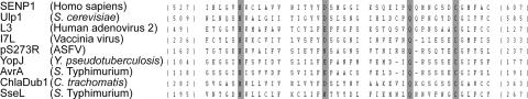
Expression of the Salmonella enterica serovar Typhimurium pathogenicity island 2 (SPI-2) type III secretion system is controlled by the two-component regulatory system SsrA-SsrB. We used a transcriptomic approach to help define the SsrA-SsrB regulon. We identified a gene encoding an uncharacterized effector (SseL) whose translocation into host cells depends on the SPI-2 secretion system. SseL has similarities to cysteine proteases with deubiquitinating activity. A GST-SseL fusion protein specifically hydrolyzed mono- and polyubiquitin substrates in vitro with a preference for K63-linked ubiquitin chains. Ubiquitin-modified proteins accumulated in macrophages infected with Salmonella sseL mutant strains but to a lesser extent when infected with bacteria expressing active protein, demonstrating that SseL functions as a deubiquitinase in vivo. Salmonella sseL mutant strains did not show a replication defect or induce altered levels of cytokine production upon infection of macrophages but were defective for a delayed cytotoxic effect and were attenuated for virulence in mice.


Conflict of interest statement

The authors declare no conflict of interest.

Figures

Fig. 1.
Fig. 1.
Amino acid alignment of SseL with a selection of cysteine proteases from clan CE. The conserved catalytic residues (H, D/E/N, Q, and C) are highlighted in gray. ASFV indicates African swine fever virus.
Fig. 2.
Fig. 2.
Enzymatic activity of GST-fusion proteins. (A) Purified proteins or isopeptidase T (a Ub-specific protease) were incubated with Ub-AMC, and their activity was determined by release of fluorescent AMC. (B) Michaelis–Menten plot to determine kinetic constants for GST-SseL (Km = 1.5 μM; Vmax = 2354 pM s−1). Cleavage of branched K48- (C) and K63- (D) linked multiUb chains by GST-SseL. Purified proteins (5 μM) were incubated with substrate with (+) or without (−) inhibitor (Ub aldehyde) for 1 h at 37°C and fractioned by SDS/PAGE. Proteins were transferred to PVDF membrane and labeled with an anti-Ub antibody (Zymed). Purified proteins of increasing concentration (0–5.0 μM) were incubated for 30 min at 37°C with equimolar amounts of either K48- (E) or K63- (F) linked multiUb chains and subjected to SDS/PAGE and immunoblotted with an anti-Ub antibody (Zymed). Bands correspond to different Ub oligomers (indicated on the left). GST-SseL in each sample was visualized by Coomassie blue staining.
Fig. 3.
Fig. 3.
Ubiquitination of proteins in infected macrophages. (A Left) J774 cells were infected with the ΔsseL mutant strain expressing either SseL-2HA or SseLC262A-2HA. At different time points, cells were lysed and proteins subjected to SDS/PAGE, followed by transfer to PVDF membranes and labeling with an anti-Ub antibody (Zymed). The blot was then stripped and reprobed with anti-tubulin and anti-HA antibodies to provide a loading control for host proteins and to demonstrate equivalent expression of epitope-tagged SseL in cells infected with the two strains. (A Right) J774 cells were uninfected or infected for 16 h with wild-type, ΔsseL or ΔsseL mutant strain expressing either SseL-2HA or SseLC262A-2HA. Cell lysates were analyzed as described above, without anti-HA immunoblot. (B) Coimmune precipitation of HA-tagged proteins from macrophages infected for 16 h with the same strains as in A Left). Macrophage lysates were incubated with anti-HA antibody. Antibody-bound proteins were recovered by using protein G-coated beads and immunoblotted (Left) with an anti-HA antibody. HC and LC indicate heavy and light antibody chains. HA labeling indicates equivalent recovery from the two samples. Recovered fractions were probed with antibodies recognizing polyUb (FK1) or both monoUb and polyUb (FK2) (Right). Positions of size markers are indicated on the left of blots.
Fig. 4.
Fig. 4.
SseL is required for a delayed cytotoxic effect in infected macrophages. (A) TNF release from infected macrophages. J774 cells were infected for different periods of time with the wild-type or ΔsseL strain, and supernatants were recovered and analyzed for TNF by sandwich ELISA. TNF release from macrophages exposed to LPS (1 μg/ml) for 16 h was used as a positive control. (B) Cytotoxicity assay of macrophages after infection with various bacterial strains. Cells were infected for 10 or 20 h and then incubated with PI for 20 min and analyzed by flow cytometry. The levels of PI labeling of uninfected cells were subtracted from those of infected cells at the two time points. In each case, the percentages of PI-positive cells are from a total of 20,000 cells. Samples were analyzed in triplicate, and the standard errors of the mean are shown. The results shown are representative of four independent experiments. (C) Macrophages were infected as in B with the wild-type or ΔsseL strain carrying a GFP-expressing plasmid. At 20 h after uptake, 20,000 GFP-positive macrophages were analyzed, and the number of PI-positive cells was counted. Asterisks indicate P < 0.05.

References

    1. Rathman M, Barker LP, Falkow S. Infect Immun. 1997;65:1475–1485. - PMC - PubMed
    1. Buchmeier NA, Heffron F. Infect Immun. 1991;59:2232–2238. - PMC - PubMed
    1. Waterman SR, Holden DW. Cell Microbiol. 2003;5:501–511. - PubMed
    1. Cirillo DM, Valdivia RH, Monack DM, Falkow S. Mol Microbiol. 1998;30:175–188. - PubMed
    1. Geddes K, Worley M, Niemann G, Heffron F. Infect Immun. 2005;73:6260–6271. - PMC - PubMed

Publication types

MeSH terms

LinkOut - more resources