Developmental hearing loss eliminates long-term potentiation in the auditory cortex
- PMID: 17360680
- PMCID: PMC1805556
- DOI: 10.1073/pnas.0607177104
Developmental hearing loss eliminates long-term potentiation in the auditory cortex
Abstract
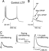
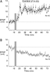
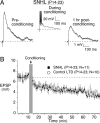
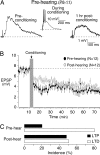
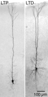
Severe hearing loss during early development is associated with deficits in speech and language acquisition. Although functional studies have shown a deafness-induced alteration of synaptic strength, it is not known whether long-term synaptic plasticity depends on auditory experience. In this study, sensorineural hearing loss (SNHL) was induced surgically in developing gerbils at postnatal day 10, and excitatory synaptic plasticity was examined subsequently in a brain slice preparation that preserves the thalamorecipient auditory cortex. Extracellular stimuli were applied at layer 6 (L6), whereas evoked excitatory synaptic potentials (EPSPs) were recorded from L5 neurons by using a whole-cell current clamp configuration. In control neurons, the conditioning stimulation of L6 significantly altered EPSP amplitude for at least 1 h. Approximately half of neurons displayed long-term potentiation (LTP), whereas the other half displayed long-term depression (LTD). In contrast, SNHL neurons displayed only LTD after the conditioning stimulation of L6. Finally, the vast majority of neurons recorded from control prehearing animals (postnatal days 9-11) displayed LTD after L6 stimulation. Thus, normal auditory experience may be essential for the maturation of synaptic plasticity mechanisms.
Conflict of interest statement
The authors declare no conflict of interest.
Figures



Similar articles
-
Hearing loss raises excitability in the auditory cortex.J Neurosci. 2005 Apr 13;25(15):3908-18. doi: 10.1523/JNEUROSCI.5169-04.2005. J Neurosci. 2005. PMID: 15829643 Free PMC article.
-
Continuous white noise exposure during and after auditory critical period differentially alters bidirectional thalamocortical plasticity in rat auditory cortex in vivo.Eur J Neurosci. 2007 Nov;26(9):2576-84. doi: 10.1111/j.1460-9568.2007.05857.x. Epub 2007 Oct 26. Eur J Neurosci. 2007. PMID: 17970743
-
Conductive hearing loss disrupts synaptic and spike adaptation in developing auditory cortex.J Neurosci. 2007 Aug 29;27(35):9417-26. doi: 10.1523/JNEUROSCI.1992-07.2007. J Neurosci. 2007. PMID: 17728455 Free PMC article.
-
Developmental plasticity of auditory cortical inhibitory synapses.Hear Res. 2011 Sep;279(1-2):140-8. doi: 10.1016/j.heares.2011.03.015. Epub 2011 Apr 2. Hear Res. 2011. PMID: 21463668 Free PMC article. Review.
-
Synaptic plasticity at thalamocortical synapses in developing rat somatosensory cortex: LTP, LTD, and silent synapses.J Neurobiol. 1999 Oct;41(1):92-101. J Neurobiol. 1999. PMID: 10504196 Review.
Cited by
-
Gamma-Band Modulation in Parietal Area as the Electroencephalographic Signature for Performance in Auditory-Verbal Working Memory: An Exploratory Pilot Study in Hearing and Unilateral Cochlear Implant Children.Brain Sci. 2022 Sep 25;12(10):1291. doi: 10.3390/brainsci12101291. Brain Sci. 2022. PMID: 36291225 Free PMC article.
-
[Molecular biological aspects of neuroplasticity: approaches for treating tinnitus and hearing disorders].HNO. 2010 Oct;58(10):973-82. doi: 10.1007/s00106-010-2177-8. HNO. 2010. PMID: 20811868 German.
-
Cognitive decline in acoustic neuroma patients: An investigation based on resting-state functional magnetic resonance imaging and voxel-based morphometry.Front Psychiatry. 2022 Aug 1;13:968859. doi: 10.3389/fpsyt.2022.968859. eCollection 2022. Front Psychiatry. 2022. PMID: 35978844 Free PMC article.
-
Subplate neurons: crucial regulators of cortical development and plasticity.Front Neuroanat. 2009 Aug 20;3:16. doi: 10.3389/neuro.05.016.2009. eCollection 2009. Front Neuroanat. 2009. PMID: 19738926 Free PMC article.
-
Developmental hearing loss impedes auditory task learning and performance in gerbils.Hear Res. 2017 Apr;347:3-10. doi: 10.1016/j.heares.2016.07.020. Epub 2016 Oct 13. Hear Res. 2017. PMID: 27746215 Free PMC article.
References
Publication types
MeSH terms
Grants and funding
LinkOut - more resources
Full Text Sources

