

Male fetal germ cells are targets for androgens that physiologically inhibit their proliferation
- PMID: 17360691
- PMCID: PMC1805536
- DOI: 10.1073/pnas.0611421104
Male fetal germ cells are targets for androgens that physiologically inhibit their proliferation
Abstract
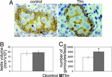
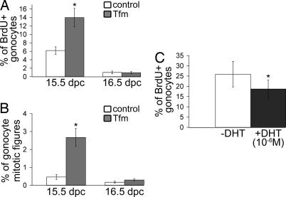
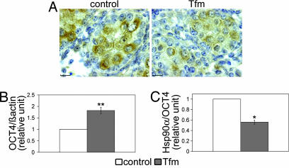
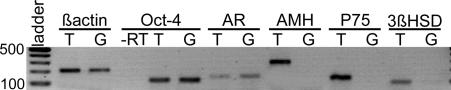
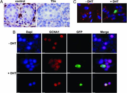
In adulthood, the action of androgens on seminiferous tubules is essential for full quantitatively normal spermatogenesis and fertility. In contrast, their role in the fetal testis, and particularly in fetal germ cell development, remains largely unknown. Using testicular feminized (Tfm) mice, we investigated the effects of a lack of functional androgen receptor (AR) on fetal germ cells, also named gonocytes. We demonstrated that endogenous androgens/AR physiologically control normal gonocyte proliferation. We observed an increase in the number of gonocytes at 17.5 days postconception resulting from an increase in proliferative activity in Tfm mice. In a reciprocal manner, gonocyte proliferation is decreased by the addition of DHT in fetal testis organotypic culture. Furthermore, the AR coregulator Hsp90alpha (mRNA and protein) specifically expressed in gonocytes was down-regulated in Tfm mice at 15.5 days postconception. To investigate whether these effects could result from direct action of androgens on gonocytes, we collected pure gonocyte preparations and detected AR transcripts therein. We used an original model harboring a reporter gene that specifically reflects AR activity by androgens and clearly demonstrated the presence of a functional AR protein in fetal germ cells. These data provide in vivo and in vitro evidence of a new control of endogenous androgens on gonocytes identified as direct target cells for androgens. Finally, our results focus on a new pathway in the fetal testis during the embryonic period, which is the most sensitive to antiandrogenic endocrine disruptors.
Conflict of interest statement
The authors declare no conflict of interest.
Figures
Similar articles
-
Methoxyacetic acid disregulation of androgen receptor and androgen-binding protein expression in adult rat testis.Biol Reprod. 2003 Apr;68(4):1437-46. doi: 10.1095/biolreprod.102.004937. Epub 2002 Nov 27. Biol Reprod. 2003. PMID: 12606434
-
TGFbeta signaling in male germ cells regulates gonocyte quiescence and fertility in mice.Dev Biol. 2010 Jun 1;342(1):74-84. doi: 10.1016/j.ydbio.2010.03.007. Epub 2010 Mar 24. Dev Biol. 2010. PMID: 20346356
-
Prenatal estrogen exposure differentially affects estrogen receptor-associated proteins in rat testis gonocytes.Biol Reprod. 2004 Nov;71(5):1652-64. doi: 10.1095/biolreprod.104.030205. Epub 2004 Jun 30. Biol Reprod. 2004. PMID: 15229138
-
Gonocytes, the forgotten cells of the germ cell lineage.Birth Defects Res C Embryo Today. 2009 Mar;87(1):1-26. doi: 10.1002/bdrc.20142. Birth Defects Res C Embryo Today. 2009. PMID: 19306346 Review.
-
Gonocytes, from the fifties to the present: is there a reason to change the name?Biol Reprod. 2013 Aug 29;89(2):46. doi: 10.1095/biolreprod.113.110544. Print 2013 Aug. Biol Reprod. 2013. PMID: 23843237 Review.
Cited by
-
The neural androgen receptor: a therapeutic target for myelin repair in chronic demyelination.Brain. 2013 Jan;136(Pt 1):132-46. doi: 10.1093/brain/aws284. Brain. 2013. PMID: 23365095 Free PMC article.
-
Role of the testis interstitial compartment in spermatogonial stem cell function.Reproduction. 2017 Apr;153(4):R151-R162. doi: 10.1530/REP-16-0588. Epub 2017 Jan 23. Reproduction. 2017. PMID: 28115580 Free PMC article. Review.
-
Role of prostate cancer stem-like cells in the development of antiandrogen resistance.Cancer Drug Resist. 2022 Jun 1;5(2):459-471. doi: 10.20517/cdr.2022.07. eCollection 2022. Cancer Drug Resist. 2022. PMID: 35800367 Free PMC article. Review.
-
Expression of P-450 aromatase, estrogen receptor α and β, and α-inhibin in the fetal baboon testis after estrogen suppression during the second half of gestation.Endocrine. 2011 Feb;39(1):75-82. doi: 10.1007/s12020-010-9414-5. Endocrine. 2011. PMID: 21061091 Free PMC article.
-
Heterogenous effect of androgen receptor CAG tract length on testicular germ cell tumor risk: shorter repeats associated with seminoma but not other histologic types.Carcinogenesis. 2011 Aug;32(8):1238-43. doi: 10.1093/carcin/bgr104. Epub 2011 Jun 3. Carcinogenesis. 2011. PMID: 21642359 Free PMC article.
References
-
- Jost A, Vigier B, Prepin J, Perchellet JP. Recent Prog Horm Res. 1973;29:1–41. - PubMed
-
- McPhaul MJ. Mol Cell Endocrinol. 2002;198:61–67. - PubMed
-
- O'Shaughnessy PJ, Johnston H, Willerton L, Baker PJ. J Cell Sci. 2002;115:3491–3496. - PubMed
-
- Young CY, Johnson MP, Prescott JL, Tindall DJ. Endocrinology. 1989;124:771–775. - PubMed
Publication types
MeSH terms
Substances
LinkOut - more resources
Full Text Sources
Molecular Biology Databases
Research Materials
