Comparative Study
. 2007 Feb 27;104(9):3639-44.
doi: 10.1073/pnas.0607703104. Epub 2007 Feb 20.

Arabidopsis plasma membrane protein crucial for Ca2+ influx and touch sensing in roots

Affiliations
Comparative Study

Arabidopsis plasma membrane protein crucial for Ca2+ influx and touch sensing in roots

Yuko Nakagawa et al. Proc Natl Acad Sci U S A. .

Abstract

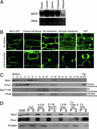
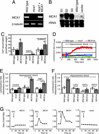
Plants can sense and respond to mechanical stimuli, like animals. An early mechanism of mechanosensing and response is speculated to be governed by as-yet-unidentified sensory complexes containing a Ca(2+)-permeable, stretch-activated (SA) channel. However, the components or regulators of such complexes are poorly understood at the molecular level in plants. Here, we report the molecular identification of a plasma membrane protein (designated Mca1) that correlates Ca(2+) influx with mechanosensing in Arabidopsis thaliana. MCA1 cDNA was cloned by the functional complementation of lethality of a yeast mid1 mutant lacking a putative Ca(2+)-permeable SA channel component. Mca1 was localized to the yeast plasma membrane as an integral membrane protein and mediated Ca(2+) influx. Mca1 also increased [Ca(2+)](cyt) upon plasma membrane distortion in Arabidopsis. The growth of MCA1-overexpressing plants was impaired in a high-calcium but not a low-calcium medium. The primary roots of mca1-null plants failed to penetrate a harder agar medium from a softer one. These observations demonstrate that Mca1 plays a crucial role in a Ca(2+)-permeable SA channel system that leads to mechanosensing in Arabidopsis. We anticipate our findings to be a starting point for a deeper understanding of the molecular mechanisms of mechanotransduction in plants.


Conflict of interest statement

The authors declare no conflict of interest.

Figures

Fig. 1.
Fig. 1.
Function in yeast cells and structural features of Mca1 and Mca2. (A) Complementation of lethality of the yeast mid1 mutant by MCA1. Cell viability of the mid1 mutant strain bearing pFL61-MCA1 or empty vector pFL61 and wild-type strain bearing pFL61 was determined after being exposed to the mating pheromone α-factor. Data are means ± SD from three independent experiments. ∗, P < 0.01 versus mid1/pFL61. (B) Ca2+ uptake activity. Exponentially growing cells of the above strains were suspended in Hepes/Tris buffer containing 74 kBq/ml 45CaCl2 (74 MBq/nmol) and aliquots of the suspension were taken at 1-min intervals over 8 min and filtered through a Millipore filter (type HA; 0.45 μm). The radioactivity retained on the filters was counted. Data are means ± SD from three independent experiments. ∗, P < 0.01 versus mid1/pFL61. (C) Genomic organization of the MCA1 and MCA2 genes. Boxes represent exons and their black areas show the ORF. T-DNA (drawn to an arbitrary size) is inserted into the site 28 bp upstream of the fourth exon, producing an mca1-null allele. (D) Multiple amino acid sequence alignment of the Mca1, Mca2, and rice OsMca1 with Clustal W (version 1.8). Amino acid sequence identity (and similarity) between Mca1 and Mca2 is 72.7% (89.4%), that between Mca1 and OsMca1 is 65.0% (90.3%), and that between Mca2 and OsMca1 is 57.2% (86.4%). Asterisk indicates identical amino acid; colon indicates amino acid with strong similarity; dot indicates amino acid with weak similarity. The ARPK domain (for amino-terminal domain of rice putative protein kinases) is boxed. The line under each sequence shows a potential transmembrane segment (TM) predicted by TopPred (19). The lines above the Mca1 sequence represent an EF-hand-like structure, a coiled-coil region, and a C-terminal, cysteine-rich region similar to the PLAC8 motif found in plant and animal proteins of unknown function. (E) Schema of Mca1 and the hydropathy profile of Mca1. The bars indicate the position of the potential transmembrane segments (TM) described in D. (F) MCA1 has the ability to increase Ca2+ accumulation even in the mid1 cch1 double mutant. MCA1 cDNA on a plasmid was expressed under the control of the TDH3 promoter in each yeast mutant (mid1, cch1, or mid1 cch1) as well as the parental strain (MID1 CCH1). The MID1 gene was expressed from the plasmid YEpMID1 (25). Exponentially growing cells were incubated for 2 h in the low Ca2+ medium SD.Ca100 (15) containing 185 kBq/ml 45CaCl2 (1.8 kBq/nmol) and aliquots of the culture were taken and filtered through a Millipore filter (type HA; 0.45 μm). ∗, P < 0.05 versus vector in each mutant.
Fig. 2.
Fig. 2.
Expression of MCA1. (A) Northern blotting of the MCA1 transcripts. Total RNA was isolated from roots, leaves, stems, flowers, and siliques of mature Arabidopsis plants and subjected to Northern blotting. rRNA was used for internal controls for the amount of RNA loaded. (B) GFP fluorescence images suggesting the localization of Mca1-GFP in the plasma membrane of root cells. The upper row represents intact roots and the bottom row those treated with 0.8 M mannitol for at least 10 min. Note that mannitol induced plasmolysis. The sample and membrane marker proteins used are as follows: Mca1-GFP, a GFP fusion to the C terminus of the full length Mca1 protein; Plasma membrane, a GFP fusion to the plasma membrane channel protein PIP2A expressed in line Q8 (27); ER membrane, a GFP fusion to an endoplasmic reticulum membrane protein expressed in line Q4 (27); Vacuolar membrane, a GFP fusion to the vacuolar membrane channel protein delta-TIP expressed in line Q5 (27); and cytoplasmic GFP. (Scale bars, 20 μm.) (C) Membrane fractionation by sucrose density gradient centrifugation and localization of Mca1 expressed in yeast. Pma1, plasma membrane H+-ATPase; Sec71-HA, an endoplasmic reticulum membrane protein tagged with hemagglutinin antigen (HA); Pho8, a vacuolar membrane protein. (D) Mca1 is an integral membrane protein in yeast. Note that Mca1 is not solubilized with NaCl, Na2CO3, and urea, all of which are known to solubilize peripheral membrane proteins. P, pellet after centrifugation at 100,000 × g for 1 h, containing membranes; S, supernatant.
Fig. 3.
Fig. 3.
Mca1 protein enhances Ca2+ uptake activity in planta. (A) RT-PCR showing no detectable production of MCA1 transcripts in the mca1-null line. β-tubulin is a control. (B) Northern blotting, showing MCA1 mRNA levels in MCA1ox (S1, S2, and S3), wild-type, and vector-bearing wild-type lines. rRNA was used for control experiments. (C) Overexpression of Mca1 increases Ca2+ uptake in intact roots. The roots were incubated with 45CaCl2 for 20 min and washed five times with a washing solution containing LaCl3, which displaces 45Ca2+ nonspecifically bound to the cell wall (33). The uptake is inhibited by 1 mM Gd3+, but not by 20 μM verapamil. ∗, P < 0.01 versus wild type. (D) Hypoosmotic shock-induced [Ca2+]cyt changes, as revealed in aequorin luminescence. Wild-type, mca1-null, and MCA1ox seedlings with aequorin in MS medium (400 μl) were subjected to hypoosmotic shock with the addition of H2O (200 μl). Representative data are shown for each seedling. (E) Summary of hypoosmotic shock- and TNP-induced [Ca2+]cyt changes. As for hypoosmotic shock, experimental conditions were the same as those in D. For TNP stimulation and control experiments, 0.3 mM TNP (200 μl) and MS medium (200 μl) were added to the seedlings in MS medium (400 μl), respectively. The average of five independent experiments is shown for each sample. ∗, P < 0.05 versus each control; ∗∗, P < 0.005 versus wild type in each treatment. (F) Effect of channel blockers and a Ca2+ chelator on the hypoosmotic shock-induced [Ca2+]cyt increase. Ten minutes before hypoosmotic shock, 1 mM La3+, 1 mM Gd3+, or 5 mM EGTA (calcium chelator) was added to the medium. ∗, P < 0.005 versus wild-type and reagent-treated samples. (G) Stretch-activated Ca2+ response in MCA1-expressing and mock-transfected CHO cells. Cells cultured on elastic silicone membranes were loaded with 1 μM fura-2/AM and subjected to a uniaxial stretch pulse (10, 20, or 30% of the length for 1 s at room temperature). Fura-2 fluorescence intensities at excitation wavelengths of 340 and 380 nm were acquired and the ratio (F340/F380) was calculated. An increase in the ratio donates a [Ca2+]cyt increase. (Right) Mock-transfected cells. (Center and Left) MCA1-expressing cells.
Fig. 4.
Fig. 4.
Phenotypes of mca1-null and MCA1ox lines. (A) A seedling of the MCA1ox line S3, showing browning of the hypocotyl. (Scale bars, 2.0 mm.) (B) An MCA1ox plant (line S1), showing a phenotype with no petals, shrunken siliques, short stems, and small rosettes. (White bars, 1.5 mm; black bars, 1.0 cm.) (C) Ca2+-dependent growth phenotypes of MCA1ox seedlings (line S1). Note that the ordinary MS medium contains 3.0 mM CaCl2. On this medium, MCA1ox seedlings exhibited growth deficiency. (Scale bar, 1.0 cm.) (D) Failure to penetrate the lower, harder agar (1.6%) medium of the primary roots of the mca1-null mutant. Seeds of various lines were placed on the surface of the upper, softer agar (0.8%) medium of the two-phase-agar medium, allowed to grow for 9 days, and then photographed. (Left) Wild type (side view). (Upper Center) mca1-null (side view). (Lower Center) mca1-null (oblique upper view), showing the root tips were coiled at the interface of the two agar media. (Right) mca1-null mutant complemented with wild-type MCA1. Arrowheads represent the interface of the two agar media. (Scale bar, 1.0 cm.) (E) Quantitative representation of the results shown in D. Number of seedlings examined: Wild type, n = 150; mca1-null, n = 150; mca1-null + MCA1, n = 80. ∗, P < 0.01 versus wild type and mca1-null + MCA1.

Similar articles

Cited by

References

    1. Darwin C. The Power of Movement in Plants. London: John Murray; 1880.
    1. Trewavas A, Knight M. Plant Mol Biol. 1994;26:1329–1341. - PubMed
    1. Braam J. New Phytol. 2005;165:373–389. - PubMed
    1. Knight MR, Campbell AK, Smith SM, Trewavas AJ. Nature. 1991;352:524–526. - PubMed
    1. Legué V, Blancaflor E, Wymer C, Perbal G, Fantin D, Gilroy S. Plant Physiol. 1997;114:789–800. - PMC - PubMed

Publication types

MeSH terms