Comparative Study
. 2007 Jun;81(11):5685-95.
doi: 10.1128/JVI.02574-06. Epub 2007 Mar 14.

Old World and clade C New World arenaviruses mimic the molecular mechanism of receptor recognition used by alpha-dystroglycan's host-derived ligands

Affiliations
Comparative Study

Old World and clade C New World arenaviruses mimic the molecular mechanism of receptor recognition used by alpha-dystroglycan's host-derived ligands

Jillian M Rojek et al. J Virol. 2007 Jun.

Abstract

alpha-Dystroglycan (DG) is an important cellular receptor for extracellular matrix (ECM) proteins and also serves as the receptor for Old World arenaviruses Lassa fever virus (LFV) and lymphocytic choriomeningitis virus (LCMV) and clade C New World arenaviruses. In the host cell, alpha-DG is subject to a remarkably complex pattern of O glycosylation that is crucial for its interactions with ECM proteins. Two of these unusual sugar modifications, protein O mannosylation and glycan modifications involving the putative glycosyltransferase LARGE, have recently been implicated in arenavirus binding. Considering the complexity of alpha-DG O glycosylation, our present study was aimed at the identification of the specific O-linked glycans on alpha-DG that are recognized by arenaviruses. As previously shown for LCMV, we found that protein O mannosylation of alpha-DG is crucial for the binding of arenaviruses of distinct phylogenetic origins, including LFV, Mobala virus, and clade C New World arenaviruses. In contrast to the highly conserved requirement for O mannosylation, more generic O glycans present on alpha-DG are dispensable for arenavirus binding. Despite the critical role of O-mannosyl glycans for arenavirus binding under normal conditions, the overexpression of LARGE in cells deficient in O mannosylation resulted in highly glycosylated alpha-DG that was functional as a receptor for arenaviruses. Thus, modifications by LARGE but not O-mannosyl glycans themselves are most likely the crucial structures recognized by arenaviruses. Together, the data demonstrate that arenaviruses recognize the same highly conserved O-glycan structures on alpha-DG involved in ECM protein binding, indicating a strikingly similar mechanism of receptor recognition by pathogen- and host-derived ligands.


Figures

FIG. 1.
FIG. 1.
Recombinant retroviral vectors pseudotyped with arenavirus GPs. (A) The packaging cell line GP2-293 (BD Biosciences) stably transfected with MLV Gag and Pol was cotransfected with a plasmid containing the packable MLV genome pLZRs-Luc-gfp (52) carrying a luciferase and a GFP reporter and an expression plasmid for the heterologous GP (pC-AVGP). Retroviral pseudotypes were released into the cell supernatant. LTR, long terminal repeat. (B) Binding of mAb 83.6 to pseudotypes. Equal amounts of concentrated pseudotypes containing the GPs of LFV, LCMV cl-13, AMA, and VSV were immobilized in microtiter plates and probed with mAb 83.6. Primary antibodies were detected with HRP-conjugated anti-mouse IgG in a color reaction using the substrate ABTS. The optical density (OD) at 405 nm was measured using an enzyme-linked immunosorbent assay reader (n = 3 ± standard deviation [SD]). (C) Infection of cells with LFV and LCMV pseudotypes is dependent on α-DG. DG-deficient (DG−/−) and DG+/− mouse ES cells (ESC) cultured in 96-well plates were infected with retroviral pseudotypes of LFV, LCMV cl-13, AMA, or VSV or pseudotypes containing no GP (−) at an MOI of 10. Infection was assessed by Bright-Glo luciferase assay after 48 h (n = 3 ± SD). (D) Blocking of arenavirus pseudotype (PS) infection by inactivated viruses. HEK293 cells cultured in 96-well plates were blocked with inactivated LFV, LCMV, AMA, or PAR at the indicated ratios of virus particles per cell. After blocking for 2 h at 4°C, cells were infected with the indicated pseudotypes at an MOI of 1, and infection was assessed after 24 h by Steady-Glo luciferase assay (n = 3 ± SD).
FIG. 2.
FIG. 2.
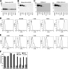
Receptor binding and infection of human cells with LFV and LCMV depend on the functional glycosylation of α-DG. (A) Detection of β-DG in human cell lines. Equal amounts of total cell protein were separated by sodium dodecyl sulfate-polyacrylamide gel electrophoresis (SDS-PAGE), blotted onto nitrocellulose, and probed with anti-β-DG mAb 8D5 in Western blots using an HRP-conjugated secondary antibody and ECL for detection. Molecular masses are indicated. (B) Detection of glycosylated α-DG. Equal amounts of total membrane proteins were separated and probed in Western blots with mAb IIH6 using an HRP-conjugated secondary antibody and ECL. (C) VOPBA. Membrane proteins from B were probed with 107 PFU/ml LCMV cl-13 and inactivated LFV. Bound virus was detected with anti-arenavirus GP2 mAb 83.6 using an HRP-conjugated secondary antibody and ECL. (D) Detection of functionally glycosylated α-DG at the cell surface of human cell lines by flow cytometry. Live, nonpermeabilized cells were stained with mAb IIH6, combined with a PE-labeled secondary antibody, and analyzed by flow cytometry using a FACSCalibur flow cytometer. Data were acquired and analyzed using Cell Quest and FloJo software packages. In histograms, the y axis represents cell numbers, and the x axis represents PE fluorescence intensity. The solid line indicates primary and secondary antibodies, and the broken line indicates secondary antibody only. (E) Detection of α-DG core protein at the cell surface of T- and B-cell lines. Cells were subjected to cell surface staining with the polyclonal antibody GT20ADG, which recognizes the core protein of α-DG independent of functional O glycosylation. Bound primary antibody was detected with a PE-conjugated anti-goat IgG secondary antibody and flow cytometry as in D. The solid line indicates primary and secondary antibodies, and the broken line indicates secondary antibody only. (F) Infection of cells with retroviral pseudotypes. Cells cultured in 96-well plates were infected with LFV, LCMV, and VSV pseudotypes and with pseudotypes without GP (no GP) at MOIs of 1 (HEK293, A549, Huh7, and HUVEC) and 10 (T- and B-cell lines). After 48 h, infection was quantified by luciferase assay, and luminescence was expressed as the increase (n-fold) over uninfected cells (n = 3 ± SD).
FIG. 3.
FIG. 3.
Protein O mannosylation is critical for α-DG's function as a receptor for Old World and clade C New World arenaviruses. (A) Detection of α-DG core protein. CHOK1, Lec35.1, and Lec15.2 cells were subjected to cell surface biotinylation using the reagent N-hydroxysuccinimide-X-biotin. After the quenching of the reaction, cells were lysed, and cleared lysates were subjected to IP with anti-β-DG mAb 8D5 or an isotype control. IPs were separated by SDS-PAGE, blotted onto nitrocellulose, and probed with streptavidin-HRP using ECL detection. The positions of α-DG and β-DG are indicated. (B) Detection of functionally glycosylated α-DG. Membrane lysates of CHOK1, Lec35.1, and Lec15.2 cells were subjected to Western blot analysis using mAb 8D5 (β-DG) and mAb IIH6. (C) Detection of functionally glycosylated α-DG on CHOK1, Lec35.1, and Lec15.2 cells by flow cytometry. Live, nonpermeabilized cells were stained with mAb IIH6, combined with a PE-labeled secondary antibody, and analyzed by flow cytometry as described in the legend of Fig. 2D. In the histograms, the y axis represents cell numbers, and the x axis represents PE fluorescence intensity. The solid line indicates primary and secondary antibodies, and the broken line indicates secondary antibody only. (D) Laminin and virus binding to α-DG from CHOK1 and Lec15.2 cells. α-DG was isolated from CHOK1 and Lec15.2 cells by WGA affinity purification, and eluted proteins were probed for the presence of β-DG in Western blots. Binding to laminin was assessed by LOA using 10 μg/ml mouse laminin-1, a polyclonal anti-laminin antibody, and ECL for detection. For VOPBA, duplicate samples were probed with 107 PFU/ml of LCMV cl-13 and inactivated LFV, Mobala virus, and Oliveros virus as described in the legend of Fig. 1C. (E) Infection of Lec15.2 and wild-type CHOK1 cells with retroviral pseudotypes of LFV, LCMV, and VSV. CHOK1 and LEC15.2 cells cultured in 96-well plates were infected with the indicated pseudotypes (MOI of 1), and infection was quantified by a Steady-Glo luciferase assay after 48 h. Luminescence was expressed as the increase (n-fold) over uninfected cells (n = 3 ± SD). (F) Infection of Lec15.2 cells and wild-type CHOK1 cells with LCMV cl-13 and LCMV WE22. CHOK1 and LEC15.2 cells cultured in 24 well plates were infected with LCMV cl-13 and WE22 (MOI of 1). After 16 h, cells were detached, and infection was quantified by intracellular staining with anti-LCMV NP mAb 113 combined with an FITC-labeled secondary antibody and analyzed by flow cytometry as in C (n = 3 ± SD).
FIG. 4.
FIG. 4.
Generic O-glycans of α-DG are dispensable for virus binding. (A) Enzymatic removal of the sialylated core 1 O-glycans (Galβ1-3GalNAc-S/T) and the SiaAα2-3Galβ1-4 parts of α-DG's O-mannosyl glycans (SiaAα2-3Galβ1-4GlcNAcβ1-2Man). (B) Verification of glycan removal. Purified α-DG was treated with glycosidases as described above (A), blotted onto nitrocellulose, and probed with either mAb IIH6 or the indicated lectins. Peanut agglutinin (PNA) is specific for Galβ(1-3)GalNAc, Erythrina cristagalli agglutinin (ECA) recognizes Galβ(1-4)GlcNAc, and Griffonia simplicifolia lectin II (GsII) detects terminal α- or β-linked GlcNAc and α- or β-linked GalNAc. (C) Binding of laminin, LFV, and LCMV. Nitrocellulose transfers of glycosidase-treated α-DG were subjected to LOA and VOPBA as described in the legend of Fig. 3D.
FIG. 5.
FIG. 5.
LARGE-derived glycan modifications but not O-mannosyl glycans are the crucial structures recognized by arenaviruses. (A) Binding of laminin and viruses to α-DG derived from Lec15.2 cells and wild-type CHO cells with or without overexpression of LARGE. O-mannosylation-deficient Lec15.2 and wild-type CHO cells were transfected with recombinant AdVs expressing LARGE or EGFP. After 48 h, membrane lysates were subjected to WGA affinity purification, and eluted proteins were separated by SDS-PAGE and probed with mAb 8D5 to β-DG in Western blots as described in the legend of Fig. 2A. α-DG was tested for the binding of laminin, LFV, Mobala virus, Oliveros virus, and LCMV as described in the legend of Fig. 3D. (B) Rescue of LFV and LCMV infection in Lec15.2 cells. Lec15.2 and wild-type CHO cells were plated in 96-well plates and infected with AdV-LARGE or AdV-EGFP. After 24 h, cells were infected with pseudotypes of LFV, LCMV, and VSV (MOI of 1), and infection was assessed after 48 h as described in the legend of Fig. 2F.
FIG. 6.
FIG. 6.
Infection of LFV and LCMV is not dependent on GAGs. (A) GAG-deficient psgΑ-745 cells and wild-type CHOK1 cells were infected with LFV, LCMV, and VSV pseudotypes at an MOI of 1 or with pseudotypes without GP (no GP). Infection was quantified after 48 h by luciferase assay as described in the legend of Fig. 2F (n = 3 ± SD). (B) Blocking of LFV infection with HS and heparin. Pseudotypes were preincubated with the indicated concentrations of HS and heparin for 1 h on ice and then added to monolayers of Vero cells (MOI of 1). Infection was quantified as described above (A) (n = 3 ± SD). (C) Heparin blocks binding of laminin-1 to α-DG. One microgram of mouse Engelbreth-Holm-Swarm sarcoma laminin-1 per milliliter was preincubated with the indicated concentrations of heparin and then added to purified rabbit skeletal muscle α-DG immobilized in microtiter plates. Bound laminin was detected with a polyclonal anti-laminin-1 antibody, followed by an HRP-conjugated secondary antibody and a color reaction using the substrate ABTS (n = 3 ± SD).

Similar articles

Cited by

References

    1. Ahmed, R., A. Salmi, L. D. Butler, J. M. Chiller, and M. B. Oldstone. 1984. Selection of genetic variants of lymphocytic choriomeningitis virus in spleens of persistently infected mice. Role in suppression of cytotoxic T lymphocyte response and viral persistence. J. Exp. Med. 160:521-540. - PMC - PubMed
    1. Andac, Z., T. Sasaki, K. Mann, A. Brancaccio, R. Deutzmann, and R. Timpl. 1999. Analysis of heparin, alpha-dystroglycan and sulfatide binding to the G domain of the laminin alpha1 chain by site-directed mutagenesis. J. Mol. Biol. 287:253-264. - PubMed
    1. Barresi, R., and K. P. Campbell. 2006. Dystroglycan: from biosynthesis to pathogenesis of human disease. J. Cell Sci. 119:199-207. - PubMed
    1. Barresi, R., D. E. Michele, M. Kanagawa, H. A. Harper, S. A. Dovicio, J. S. Satz, S. A. Moore, W. Zhang, H. Schachter, J. P. Dumanski, and K. P. Campbell. 2004. LARGE can functionally bypass alpha-dystroglycan glycosylation defects in distinct congenital muscular dystrophy. Nat. Med. 7:696-703. - PubMed
    1. Borrow, P., and M. B. A. Oldstone. 1992. Characterization of lymphocytic choriomeningitis virus-binding protein(s): a candidate cellular receptor for the virus. J. Virol. 66:7270-7281. - PMC - PubMed

Publication types

MeSH terms

LinkOut - more resources