. 2007 Jun;81(11):6099-105.
doi: 10.1128/JVI.02195-06. Epub 2007 Mar 14.

Importance of calcium-binding site 2 in simian virus 40 infection

Affiliations

Importance of calcium-binding site 2 in simian virus 40 infection

Peggy P Li et al. J Virol. 2007 Jun.

Abstract

The exposure of molecular signals for simian virus 40 (SV40) cell entry and nuclear entry has been postulated to involve calcium coordination at two sites on the capsid made of Vp1. The role of calcium-binding site 2 in SV40 infection was examined by analyzing four single mutants of site 2, the Glu160Lys, Glu160Arg, Glu157Lys (E157K), and Glu157Arg mutants, and an E157K-E330K combination mutant. The last three mutants were nonviable. All mutants replicated viral DNA normally, and all except the last two produced particles containing all three capsid proteins and viral DNA. The defect of the site 1-site 2 E157K-E330K double mutant implies that at least one of the sites is required for particle assembly in vivo. The nonviable E157K particles, about 10% larger in diameter than the wild type, were able to enter cells but did not lead to T-antigen expression. Cell-internalized E157K DNA effectively coimmunoprecipitated with anti-Vp1 antibody, but little of the DNA did so with anti-Vp3 antibody, and none was detected in anti-importin immunoprecipitate. Yet, a substantial amount of Vp3 was present in anti-Vp1 immune complexes, suggesting that internalized E157K particles are ineffective at exposing Vp3. Our data show that E157K mutant infection is blocked at a stage prior to the interaction of the Vp3 nuclear localization signal with importins, consistent with a role for calcium-binding site 2 in postentry steps leading to the nuclear import of the infecting SV40.


Figures

FIG. 1.
FIG. 1.
Calcium-binding sites: location on capsid and function in early infection events. (A and B) Locations of calcium-binding sites on the SV40 capsid. The capsid is represented as an α-carbon chain trace of component Vp1 molecules (17, 30), onto which the approximate positions of calcium site 1 (Ca 1) and calcium site 2 (Ca 2) are marked. A front view (A) and a cross-sectional view (B) are presented. (C) Proposed functions of calcium-binding sites in SV40 infection. The major events leading to productive infection by a wild-type virion (Wt) in connection with calcium exchanges at site 1 and site 2 are schematically diagramed. For simplicity, only a small, arbitrarily chosen number of all possible sites 1 or sites 2 is illustrated on each particle. This model portrays a scenario in which some of the calcium ions coordinated at the sites are released at specific stages of infection. Cell entry of the virus is achieved through interaction with a cell surface receptor (Y-shaped molecule on the plasma membrane) and internalization into a membrane-enclosed compartment. Nuclear entry of the virus is achieved through escape from a membrane compartment and exposure of the Vp3 NLS (zigzag lines) for interaction with importins α and β (α/β), leading to import through the nuclear pore complex (NPC). The particles of the E330K (illustrated as lacking functional site 1; cell-binding defective), E157A-E160A and E216K (lacking functional site 2; prematurely dissociating), and E157K (lacking functional site 2; Vp3 exposure defective) mutants are placed at the respective blocked stages of infection. (D) Summary of expected calcium-binding site characteristics for wild-type and mutant particles. The number of calcium ions that can be coordinated by all the sites 1 or sites 2 in one capsid is presumed to be 360 (maximum [Max] of 360) if all site 1 or site 2 side chains are intact. The predicted presence (denoted by a +, as opposed to − for the absence) of a salt bridge at site 1 or site 2 for the E330K or E157K mutant, respectively, is expected to eliminate calcium coordination at the respective mutated site (zero calcium ions at site 1 or 2). The E157A-E160A mutant is expected to coordinate few or no calcium ions at its site 2 as a result of weakened affinity of the site for the ion (few/weak).
FIG. 2.
FIG. 2.
DNA replication, capsid protein expression, and particle formation by mutants. (A) DNA replication and Vp1 accumulation. (Upper panel) Viral DNA was extracted by the Hirt method (10) from 106 CV-1 cells at 72 h posttransfection (15) with the indicated wild-type or mutant NO-SV40 DNA, digested with KpnI and DpnI, resolved by agarose gel electrophoresis, and stained with ethidium bromide. A single low-molecular-weight species at 5.2 kbp was detected for each sample as shown. (Even though the NO-SV40 DNAs used for transfections were equimolar mixtures with pBR322 DNA derived from the processing of NO-pSV40 precursors [11], no additional fragments corresponding to pBR322 were detectable in the Hirt extracts of transfected cells following KpnI-DpnI or BamHI digestion.) (Lower panel) A 2.5 × 10⁴-cell sample per wild-type or mutant DNA from the same transfection experiment was analyzed by anti-Vp1 Western blotting, as described before (14). (B) Subcellular localization of capsid proteins. Cells grown on coverslips were transfected with each indicated NO-SV40 DNA, cultured for 48 h, fixed, doubly stained with guinea pig anti-Vp1 (a, c, e, g, i, k, and m) or rabbit anti-Vp3 (b, d, f, h, j, L, and n), and then doubly stained with rhodamine-labeled (a, c, e, g, i, k, and m) or fluorescein-labeled (b, d, f, h, j, L, and n) secondary antibodies as described before (14). The same cells are shown in panels a and b, c and d, e and f, g and h, i and j, k and L, and m and n. (C to G) Particle formation. Lysates were prepared from cells at 72 h posttransfection (15) with wild-type (C), E157K (D), E157R (E), E160K (F), or E157K-E330K (G) NO-SV40 DNA, treated with DNase I, and sedimented through a 5 to 32% sucrose gradient, as described previously (15). The total number of transfected cells used for the analysis is indicated below each mutant name. Of the 17 fractions collected from the bottom of the gradient, 2/3 was analyzed for viral DNA by Southern blotting (15), and 1/40 was analyzed for Vp1 by Western blotting. The profile for the NO-SV40-E160R mutant was highly similar to that of the E160K mutant shown in panel F. The arrowhead indicates the peak position of viral DNA and Vp1 presence for the wild-type sample in panel C. Short lines point to the species of viral DNA visible in each sample.
FIG. 3.
FIG. 3.
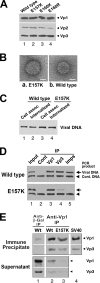
Capsid composition, EM, and infection processes of mutant particles. (A) Capsid protein composition. Peak sucrose fractions for wild-type and various mutant particles, prepared similarly to those for Fig. 2C through G, were pooled and probed for the composition of capsid proteins by anti-Vp1 (upper panel) or anti-Vp3 (lower panel) Western blot analysis. Bands corresponding to Vp1, Vp2, and Vp3 are marked. (B) Electron micrograph of E157K particles. E157K mutant particles (a) or wild-type SV40 virions (b) were purified as for panel A from 4 × 10⁸ to 10 × 108 viral DNA-transfected cells and further purified through a second round of sucrose sedimentation. Three microliters of the E157K mutant or wild-type particles, containing about 1.6 × 10⁷ or 6.6 × 10⁷ particles per μl, respectively, was applied to a glow-discharged carbon-coated copper grid and allowed to settle for 20 s. The excess solution was removed by blotting with a filter paper, and the grid was washed once with 3 μl of water and then stained with a drop of 2% uranyl acetate solution for 15 s. The excess staining was removed by blotting, and the grid was air dried. Tobacco mosaic virus (TMV) was included with the sample as a calibration standard. (The TMV-containing images were windowed in 1,024- by 1,024-pixel images, from which the power spectrum of the images was calculated to reveal the 23-Å layer lines. The half-distance between two layer lines was measured in pixels and used to estimate the pixel size.) The specimens were observed with a JEOL 1230 electron microscope. The mean diameter of the SV40 virions is 47.5 ± 1.9 nm (n = 233), and that of the E157K particles is 52.0 ± 4.0 nm (n = 250). Bar, 20 nm. (C) Cell attachment and internalization by E157K particles. Similar to the previously described procedure (16, 21), cells grown in 100-mm dishes were infected for 6 h, at 1,000 particles per cell, with peak sucrose fractions containing wild-type or E157K particles. One set of infected cells was harvested by scraping (Cell assoc), and another set was harvested by trypsin treatment (Internalized). Viral DNA was extracted by the Hirt method (10) from 2 × 10⁶ of each type of infected cells, linearized by BamHI digestion, resolved by agarose gel electrophoresis, and detected by ethidium bromide staining. (D) Association of internalized E157K DNA with capsid proteins and importins. The state of the internalized E157K mutant particles was assessed by immunoprecipitation essentially as described before (16, 21). Briefly, the cytoplasmic fraction was prepared from cells at 6 h postinfection with wild-type particles (upper panel) or E157K mutant particles (lower panel). Aliquots of the cytoplasmic fractions, equivalent to 2 × 10⁶ infected cells, were reacted with anti-mouse immunoglobulin G (IgG) (lane 2, Cont), anti-Vp1 (lane 3, Vp1), affinity-purified anti-Vp3 (lane 4, Vp3), or a mixture of anti-importin-α and anti-importin-β antibodies (lane 5, Imps). The coimmunoprecipitated (IP) viral DNA was purified from the immune complexes in the presence of 20 to 50 pg of the NO-pSV40Δ NcoI control DNA and detected via semiquantitative PCR. The expected amplification of product of the NO-SV40 genome is 2.2 kbp (arrow, Viral DNA), whereas that of the control DNA is 1.7 kbp (arrowhead, Cont. DNA). For comparison, the viral DNA content of the input cytoplasmic lysate, equivalent to 4 × 10⁵ infected cells (lane 1), was similarly purified in the presence of the control DNA and detected by PCR. (E) Association of Vp3 with Vp1 following E157K particle internalization. The cytoplasmic fraction was extracted from 1 × 10⁸ wild-type (Wt, lanes 1 and 2) or 2 × 10⁸ E157K mutant (lane 3) particle-infected cells at 6 h postinfection and reacted with anti-β-galactosidase (Anti-β-Gal) or anti-Vp1 antibody. The immune complexes were collected via reaction with TrueBlot anti-rabbit Ig beads from eBioscience (San Diego, CA). Both the immunoprecipitates (upper panel) and the supernatants remaining after the bead reactions (lower panel) were probed by anti-Vp1 and anti-Vp3 Western blot analyses similar to those described before (14), except affinity-purified anti-Vp3 IgG was used as the primary antibody for the latter blot analysis and TrueBlot horseradish peroxidase anti-rabbit IgG from eBioscience was used as the secondary antibody for both blot analyses. Purified SV40 virions were included in lane 4 as a Western blot analysis control. Bands corresponding to Vp1 and Vp3 are marked.

Similar articles

Cited by

References

    1. Anderson, H. A., Y. Chen, and L. C. Norkin. 1996. Bound simian virus 40 translocates to caveolin-enriched membrane domains, and its entry is inhibited by drugs that selectively disrupt caveolae. Mol. Biol. Cell 7:1825-1834. - PMC - PubMed
    1. Brady, J. N., V. D. Winston, and R. A. Consigli. 1977. Dissociation of polyoma virus by the chelation of calcium ions found associated with purified virions. J. Virol. 23:717-724. - PMC - PubMed
    1. Chang, D., C. Y. Fung, W. C. Ou, P. C. Chao, S. Y. Li, M. Wang, Y. L. Huang, T. Y. Tzeng, and R. T. Tsai. 1997. Self-assembly of the JC virus major capsid protein, VP1, expressed in insect cells. J. Gen. Virol. 78:1435-1439. - PubMed
    1. Chen, P. L., M. Wang, W. C. Ou, C. K. Lii, L. S. Chen, and D. Chang. 2001. Disulfide bonds stabilize JC virus capsid-like structure by protecting calcium ions from chelation. FEBS Lett. 500:109-113. - PubMed
    1. Christiansen, G., T. Landers, J. Griffith, and P. Berg. 1977. Characterization of components released by alkali disruption of simian virus 40. J. Virol. 21:1079-1084. - PMC - PubMed

Publication types

MeSH terms

LinkOut - more resources