. 2007 Jun;81(11):5669-84.
doi: 10.1128/JVI.02350-06. Epub 2007 Mar 14.

Tyrosine 3 of poliovirus terminal peptide VPg(3B) has an essential function in RNA replication in the context of its precursor protein, 3AB

Affiliations

Tyrosine 3 of poliovirus terminal peptide VPg(3B) has an essential function in RNA replication in the context of its precursor protein, 3AB

Ying Liu et al. J Virol. 2007 Jun.

Abstract

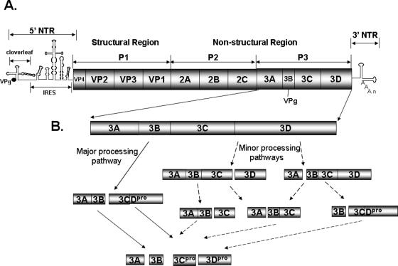
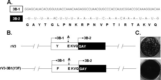
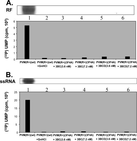
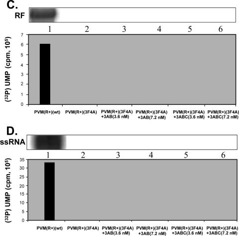
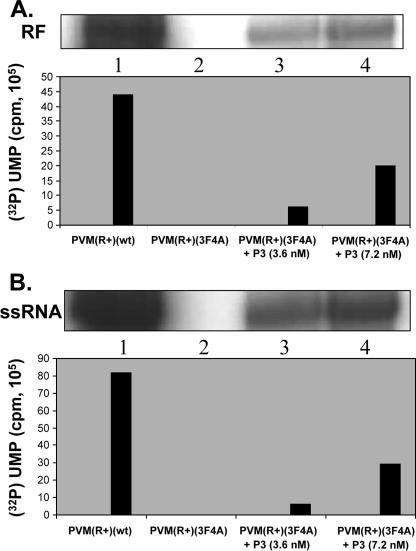
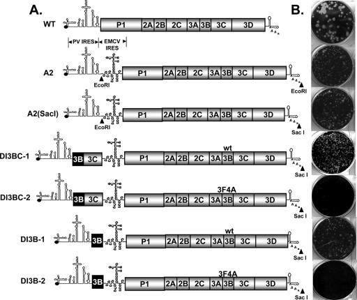
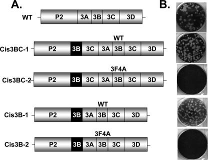
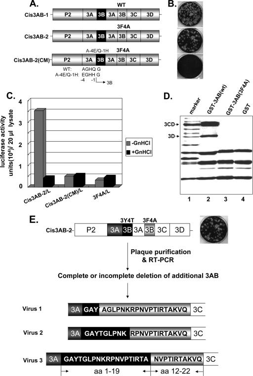
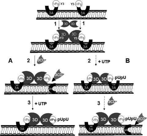
Poliovirus (PV) VPg is a genome-linked protein that is essential for the initiation of viral RNA replication. It has been well established that RNA replication is initiated when a molecule of UMP is covalently linked to the hydroxyl group of a tyrosine (Y3) in VPg by the viral RNA polymerase 3D(pol), but it is not yet known whether the substrate for uridylylation in vivo is the free peptide itself or one of its precursors. The aim of this study was to use complementation analyses to obtain information about the true in vivo substrate for uridylylation by 3D(pol). Previously, it was shown that a VPg mutant, in which tyrosine 3 and threonine 4 were replaced by phenylalanine and alanine (3F4A), respectively, was nonviable. We have now tested whether wild-type forms of proteins 3B, 3BC, 3BCD, 3AB, 3ABC, and P3 provided either in trans or in cis could rescue the replication defect of the VPg(3F4A) mutations in the PV polyprotein. Our results showed that proteins 3B, 3BC, 3BCD, and P3 were unable to complement the RNA replication defect in dicistronic PV or dicistronic luciferase replicons in vivo. However, cotranslation of the P3 precursor protein allowed rescue of RNA replication of the VPg(3F4A) mutant in an in vitro cell-free translation-RNA replication system, but only poor complementation was observed when 3BC, 3AB, 3BCD, or 3ABC proteins were cotranslated in the same assay. Interestingly, only protein 3AB but not 3B and 3BC, when provided in cis by insertion of a wild-type 3AB coding sequence between the P2 and P3 domains of the polyprotein, supported the replication of the mutated genome in vivo. Elimination of cleavage between 3A and 3B in the complementing 3AB protein, however, led to a complete lack of RNA replication. Our results suggest that (i) VPg has to be delivered to the replication complex in the form of a large protein precursor (P3) to be fully functional in replication; (ii) the replication complex formed during PV replication in vivo is essentially inaccessible to proteins provided in trans, even if the complementing protein is translated from a different cistron of the same RNA genome; (iii) 3AB is the most likely precursor of VPg; and (iv) Y3 of VPg has an essential function in RNA replication in the context of both VPg and 3AB.


Figures

FIG. 1.
FIG. 1.
The genomic structure of PV RNA and processing of the P3 domain of the polyprotein. (A) The viral RNA contains a long 5′ NTR, a single ORF, a short 3′ NTR, and a poly(A) tail. The 5′ NTR, which is covalently linked to the terminal protein VPg, consists of a cloverleaf like structure and the IRES. (B) Proteolytic processing of the P3 domain of the polyprotein (35, 36). The major (solid lines) and two minor (broken lines) processing pathways are illustrated. The segments of the polyprotein are not shown according to actual sizes.
FIG. 2.
FIG. 2.
A free N terminus in VPg is required for RNA replication. (A) Nucleotide and amino acid sequences of wt 3B (3B-1, white) and heterologous 3B (3B-2, black) are shown. Note that the amino acid sequences of 3B-1 and 3B-2 are identical, but the nucleotide sequences are different to avoid homologous recombination. Both 3B-1 and 3B-2 have the 10th residue K of wt VPg changed to R, which has been shown to have no effect in viral RNA replication (14). (B) Schematic representation of constructs rV3 and rV3-3B1(Y3F). The two tandemly arranged 3B regions are shown by rectangles. The wt cleavage site AKVQ/G between the two 3B sequences was changed to EKVQ/G in rV3. rV3-3B1(Y3F) contains the same cleavage site mutation and in addition an Y3F change in 3B-1. (C) RNA transcripts of the cDNAs were transfected into HeLa cells and the plaque phenotypes of the resulting viruses were determined by plaque assay (see Materials and Methods).
FIG. 3.
FIG. 3.
Inability of wt 3BC, 3BCD, 3AB or 3ABC to complement viral RNA synthesis of VPg(3F4A) in in vitro translation-RNA replication reactions. (A) 32P-labeled minus-strand RNA synthesis, measured as RF, was carried out as described in Materials and Methods. The autoradiography of the reaction products is shown, and the quantitation of the data is displayed below. (B) 32P-labeled plus-strand ssRNA synthesis was measured as described in Materials and Methods. (C) 32P-labeled minus-strand RNA synthesis, measured as RF, was carried out as described in Materials and Methods. (D) 32P-labeled plus-strand ssRNA synthesis was measured as described in Materials and Methods. (R+) indicates the presence of a ribozyme at the 5′ terminus of the original transcripts (see text).
FIG. 3.
FIG. 3.
Inability of wt 3BC, 3BCD, 3AB or 3ABC to complement viral RNA synthesis of VPg(3F4A) in in vitro translation-RNA replication reactions. (A) 32P-labeled minus-strand RNA synthesis, measured as RF, was carried out as described in Materials and Methods. The autoradiography of the reaction products is shown, and the quantitation of the data is displayed below. (B) 32P-labeled plus-strand ssRNA synthesis was measured as described in Materials and Methods. (C) 32P-labeled minus-strand RNA synthesis, measured as RF, was carried out as described in Materials and Methods. (D) 32P-labeled plus-strand ssRNA synthesis was measured as described in Materials and Methods. (R+) indicates the presence of a ribozyme at the 5′ terminus of the original transcripts (see text).
FIG. 4.
FIG. 4.
Efficient complementation of viral RNA synthesis of VPg(3F4A) by wt P3 in in vitro translation-RNA replication reactions. (A) 32P-labeled minus-strand RNA synthesis, measured as RF, was carried out as described in Materials and Methods. (B) 32P-labeled plus-strand ssRNA synthesis was measured as described in Materials and Methods. (R+) indicates the presence of a ribozyme at the 5′ terminus of the original transcripts (see text).
FIG. 5.
FIG. 5.
Lack of complementation of VPg(3F4A) by wt 3B and 3BC in dc PV. (A) Schematic representation of genomic structures of dc PV used in this study. An EcoRI restriction site was introduced between the PV and EMCV IRESes for the insertion of the coding sequences of the potential complementing proteins. The heterologous 3Bs, or 3Bs in the context of 3BC between the PV and EMCV IRESes, are shown in black. The VPg located within the PV complete polyprotein contained either wt or mutant (3F4A) amino acid sequences. (B) RNA transcripts of the cDNAs were transfected into HeLa cells and the plaque phenotypes of the resulting viruses were determined by plaque assay (see Materials and Methods).
FIG. 6.
FIG. 6.
Lack of complementation of VPg(3F4A) by wt 3BCD and P3 in dc luciferase replicons. (A) Schematic representation of the genomic structures of dc luciferase replicons used in this study. The P1 domain of the PV genome was replaced with the coding sequences of the luciferase gene. The endogenous VPg in the PV genome was either wt or mutant (3F4A). P3 or 3BCD coding sequences were inserted between the PV and the EMCV IRESes, as indicated. (B) Luciferase activities of dc luciferase replicons as a measure of complementation efficiency. The lane marked GnHCl represents the luciferase activity from transfection of replicon RNAs in the presence of 2 mM guanidine HCl. Luciferase assays were performed as described in Materials and Methods. Data are means of results from at least three independent experiments.
FIG. 7.
FIG. 7.
Lack of rescue of VPg(3F4A) by wt 3B and 3BC in cis. (A) Schematic representation of the PV genomic structures in which the coding sequences of the potential complementing proteins (3B or 3BC) were inserted between the P2 and P3 domains of the polyprotein. Heterologous 3B is shown in black. The endogenous VPg contained either wt or mutant (3F4A) sequences. (B) RNA transcripts of the cDNAs were transfected into HeLa cells and the plaque phenotypes of the resulting viruses were determined by plaque assay (see Materials and Methods).
FIG. 8.
FIG. 8.
Y3 of VPg is required for replication both in the context of 3AB and of free VPg. (A) Rescue of VPg(3F4A) in cis by wt 3AB. A schematic representation of constructs in which 3AB is inserted between the P2 and P3 domains of the polyprotein is shown. The endogenous VPg contained either wt or mutant (3F4A) sequences. Construct Cis3AB-2(CM) contained the 3F4A mutations and two mutations (A-4E/Q-1H) in the 3Cpro-specific cleavage signal between the 3A and 3B domains of the potential complementing 3AB protein. wt and mutant (A-4E/Q-1N) cleavage site sequences are shown below. (B) Plaque assays of the resulting viruses. Plaque assays were carried out as described in Materials and Methods. (C) Luciferase activities of constructs, containing the extra copy of 3AB, as a measure of replication efficiency. The lane marked GnHCl represents the luciferase activity of a replicon from cells transfected in the presence of 2 mM guanidine HCl. Data are means of results from at least three independent experiments. (D) GST pull-down assay as a measure of interaction between 3AB and 3Dpol/3CDpro. The assay was carried out with purified GST-3AB(wt), GST-3AB(3F4A), and GST proteins, as described in Materials and Methods. (E) Genetic variants derived from the virus containing the extra copy of 3AB in cis. Viruses isolated from transfection with Cis3AB-2 RNAs were plaque purified, and the RNAs were isolated, reverse transcribed, and sequenced.
FIG. 9.
FIG. 9.
Two possible models for the initiation of PV RNA replication. In both models (A and B) the first step is the interaction of two molecules of membrane-bound 3AB with a dimer of 3Dpol. One molecule of 3AB serves a structural function and anchors the dimer of 3Dpol molecules to the membranes during the uridylylation reaction. In model A (shown by solid lines), the second molecule of 3AB is cleaved by 3CDpro to yield membrane-bound 3A and VPg (step 2) which is uridylylated by 3Dpol to yield VPgpUpU (step 3). In model B (shown by broken lines), the second molecule of 3AB is uridylylated by 3Dpol to yield 3ABpUpU (step 2). This precursor is cleaved by 3CDpro to 3A and VPgpUpU (step 3). The uridylylated VPg made in either pathway A or B is proposed to serve as primer for the elongation of RNA chains by 3Dpol. Our models do not distinguish between the priming of plus and minus RNA strands.

Similar articles

Cited by

References

    1. Agut, H., K. M. Kean, O. Fichot, J. Morasco, J. B. Flanegan, and M. Girard. 1989. A point mutation in the poliovirus polymerase gene determines a complementable temperature-sensitive defect of RNA replication. Virology 168:302-311. - PubMed
    1. Alexander, L., H. H. Lu, and E. Wimmer. 1994. Poliovirus containing picornavirus type 1 and/or type 2 internal ribosomal entry site elements: genetic hybrids and expression of a foreign gene. Proc. Natl. Acad. Sci. USA 91:1406-1410. - PMC - PubMed
    1. Ambros, V., and D. Baltimore. 1978. Protein is linked to the 5′ end of poliovirus RNA by a phosphodiester linkage to tyrosine. J. Biol. Chem. 253:5263-5266. - PubMed
    1. Andino, R., G. E. Rieckhof, P. L. Achacoso, and D. Baltimore. 1993. Poliovirus RNA synthesis utilizes an RNP complex formed around the 5′-end of viral RNA. EMBO J. 12:3587-3598. - PMC - PubMed
    1. Banerjee, R., and A. Dasgupta. 2001. Interaction of picornavirus 2C polypeptide with the viral negative-strand RNA. J. Gen. Virol. 82:2621-2627. - PubMed

Publication types