. 2007 Apr;134(8):1499-508.
doi: 10.1242/dev.02829. Epub 2007 Mar 14.

Lack of the murine homeobox gene Hesx1 leads to a posterior transformation of the anterior forebrain

Affiliations

Lack of the murine homeobox gene Hesx1 leads to a posterior transformation of the anterior forebrain

Cynthia L Andoniadou et al. Development. 2007 Apr.

Abstract

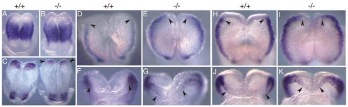
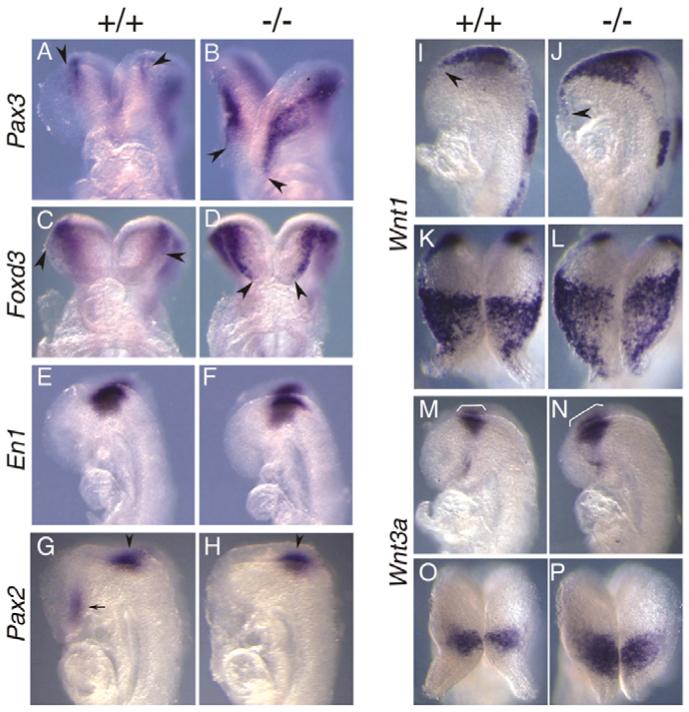
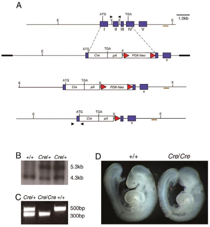
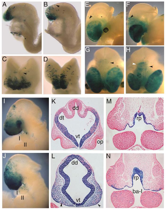
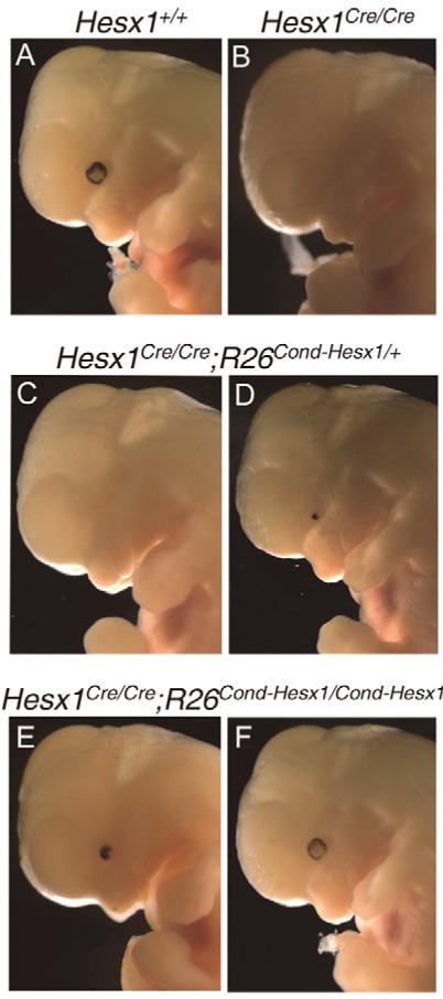
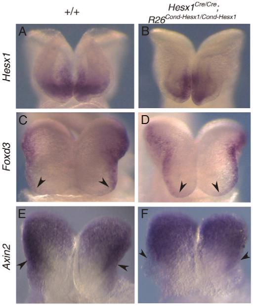
The homeobox gene Hesx1 is an essential repressor that is required within the anterior neural plate for normal forebrain development in mouse and humans. Combining genetic cell labelling and marker analyses, we demonstrate that the absence of Hesx1 leads to a posterior transformation of the anterior forebrain (AFB) during mouse development. Our data suggest that the mechanism underlying this transformation is the ectopic activation of Wnt/beta-catenin signalling within the Hesx1 expression domain in the AFB. When ectopically expressed in the developing mouse embryo, Hesx1 alone cannot alter the normal fate of posterior neural tissue. However, conditional expression of Hesx1 within the AFB can rescue the forebrain defects observed in the Hesx1 mutants. The results presented here provide new insights into the function of Hesx1 in forebrain formation.


Figures

Fig. 1
Fig. 1. Posterior transformation of the AFB in Hesx1-/- mutants at early somite stages
(A,B) Dorsal view of the Pax2 expression domain in wild type (A) and Hesx1-/- mutant (B) at presomitic stages. No significant differences are observable. (C) Dorsal view of the Pax6 expression domain in wild type (left) and Hesx1-/- mutant (right) at the 0- to 1-somite stage. Note the reduction in Pax6 expression in the anterior NP (arrowheads). (D-G) Dorsal (D,E) and frontal (F,G) views of Pax3 expression domain in wild type (D,F) and Hesx1-/- mutant (E,G) at the 2- to 3-somite stage. Pax3 expression domain is anteriorised in the Hesx1-/- mutant (E,G). Arrowheads indicate the rostral limit of Pax3 expression. (H-K) Dorsal (H,I) and frontal (J,K) views of Foxd3 expression domain in wild type (H,J) and Hesx1-/- mutant (I,K) at the 2- to 3-somite stage. Foxd3 expression domain is anteriorised in the Hesx1-/- mutant (I,K). Arrowheads indicate the rostral limit of Foxd3 expression. Anterior is to the top in A-E,H,I.
Fig. 2
Fig. 2. Wnt/β-catenin pathway is activated in the AFB of Hesx1-/- mutants at early somite stages
(A,B) Rostral view of the Wnt1 expression domain in wild type (A) and Hesx1-/- mutant (B) at the 3- to 4-somite stage. Wnt1 transcripts do not reach the rostral tip of the NP in wild-type embryos, but do so in Hesx1-/- mutants (arrowhead). (C-F) Frontal (C,D) and lateral (E,F) views of the Fgf8 expression domain in wild type (C,E) and Hesx1-/- mutant (D,F) at the 4- to 5-somite stage. Fgf8 expression in the anterior neural ridge (ANR) is reduced in the Hesx1-/- mutant. Arrowheads in C,D indicate the posterior limit of the Fgf8 expression domain in the ANR. (G,H) Expression of the Wnt/β-catenin target Axin2 in wild type (G) and Hesx1-/- mutant (H) at the 3- to 4-somite stage. Dorsal view, anterior is to the bottom. Note the expansion of the Axin2 expression domain in the Hesx1-/- mutant (H). Arrowheads indicate the rostral limit of Axin2 expression. (I-N) Dorsal (I-L) and lateral (M,N) views of the expression domain of the Wnt/β-catenin target Sp5 in wild type (I,K,M) and Hesx1-/- mutant (J,L,N) at the 5- to 6-somite (I,J) and the 2- to 3-somite (K-N) stages. Anterior is to the bottom (I-L) or to the left (M,N). Note the expansion of Sp5 expression throughout the anterior NP in the Hesx1 mutant (J,L,N). Only the most rostromedial region of the NP is free of Sp5 transcripts (arrowheads indicate the rostral limit of Sp5 expression).
Fig. 3
Fig. 3. Rostral expansion of dorsal diencephalic markers in Hesx1-/- mutants at the 8- to 10-somite stage
(A-D) Frontal views of the Pax3 (A,B) and Foxd3 (C,D) expression domains in wild type (A,C) and Hesx1-/- mutants (B,D). Both expression domains are anteriorised in the Hesx1-/- mutants. Arrowheads indicate the rostral limit of Pax3 and Foxd3 expression. (E,F) Lateral view of the En1 expression domain in wild type (E) and Hesx1-/- mutant (F). En1 expression domain around the midbrain-hindbrain region is normal in Hesx1-/- mutants when compared with wild-type littermates. (G,H) Lateral view of the Pax2 expression domain in wild type (G) and Hesx1-/- mutant (H). Pax2 expression around the midbrain-hindbrain boundary is normal (arrowheads), but there is no Pax2 expression in the AFB of the Hesx1-/- mutant (arrow in G). (I,J) Lateral view of the Wnt1 expression domain in wild type (I) and Hesx1-/- mutant (J). In the mutant, Wnt1 expression is expanded rostrally until the tip of the NP (arrowhead). (K,L) Dorsal view of the embryos depicted in I,J with anterior to the bottom. (M,N) Lateral view of the Wnt3a expression domain in wild type (M) and Hesx1-/- mutant (N). Note the rostral expansion of Wnt3a expression in the prospective dorsal diencephalon (bracket). (O,P) Dorsal view of the embryos depicted in M and N with anterior to the bottom.
Fig. 4
Fig. 4. Generation of the Hesx1-Cre targeted allele
(A) Top to bottom: structure of the murine Hesx1 wild-type locus, Hesx1-Cre targeting vector and Hesx1-Cre allele prior to and after flipase excision of the neo cassette. The map of the targeting vector shows the replacement of the Hesx1 coding region by a cassette containing the Cre recombinase gene and the neomycin resistance gene under the PGK promoter (PGK-Neo) flanked by Frt sites (red triangles). (B) Southern blot hybridisation of DNA samples from wild type (+/+) and two heterozygous (Cre/+) ES cell clones cut with EcoRI and hybridised with an external probe (brown line in A). (C) Representative example of PCR genotyping of DNA samples from wild-type, Hesx1Cre/+ and Hesx1Cre/Cre embryos. (D) Wild-type embryo (left) and Hesx1Cre/Cre mutant showing lack of AFB tissue (telencephalon and eye).
Fig. 5
Fig. 5. Descendants of Hesx1-expressing cells in the anterior NP colonise posterior regions of the neural tube and the first branchial arch
All embryos were X-Gal stained. (A,B) Higher numbers of lacZ-expressing cells reach more posterior regions of the NP in the Hesx1Cre/-;R26Cond-lacZ/+ mutant (B, arrowhead) as compared with a Hesx1Cre/+;R26Cond-lacZ/+ embryo (A) at the 6- to 7-somite stage. (C,D) This is accentuated in an 8- to 10-somite Hesx1Cre/-;R26Cond-lacZ/+ mutant (D) as compared with an Hesx1Cre/+;R26Cond-lacZ/+ embryo (C). Arrowhead in C indicates the boundary between anterior and posterior forebrain. (E-H) Lateral (E,F) and dorsal (G,H) views of a Hesx1Cre/+;R26Cond-lacZ/+ embryo (E,G) and a Hesx1Cre/-;R26Cond-lacZ/+ mutant (F,H). The brain was partially dissected to improve X-Gal staining. lacZ-expressing cells barely colonise the posterior forebrain of the Hesx1Cre/+;R26Cond-lacZ/+ embryo (arrowheads in E,G), but massively populate this region in the Hesx1Cre/-;R26Cond-lacZ/+ mutant (black arrowheads in F,H). Note that the Hesx1Cre/-;R26Cond-lacZ/+ mutant depicted in F,H shows asymmetric telencephalic development with a very small right telencephalic vesicle. This is concomitant with a more pronounced degree of colonisation of lacZ-expressing cells on the right side of the posterior forebrain (white arrowhead). (I,J) Hesx1Cre/+;R26Cond-lacZ/+ embryo (I) and Hesx1Cre/-;R26Cond-lacZ/+ mutant (J) at 9.5 dpc. Note the presence of lacZ-expressing cells within the first branchial arch in the Hesx1Cre/-;R26Cond-lacZ/+ mutant only (J). (K-N) Frontal sections of a Hesx1Cre/+;R26Cond-lacZ/+ embryo (K,M) and a Hesx1Cre/-;R26Cond-lacZ/+ mutant (L,N) at 10.5 dpc. Many more lacZ-expressing cells are localised in the frontonasal mass (arrowheads in L) of the Hesx1Cre/-;R26Cond-lacZ/+ mutant embryos as compared with the Hesx1Cre/+;R26Cond-lacZ/+ embryo (K). lacZ-expressing cells within the first branchial arch are only observed in the Hesx1Cre/-;R26Cond-lacZ/+ embryo. ba-I, first branchial arch; dd, dorsal diencephalon; dt, dorsal telencephalon; op, olfactory placode; rp, Rathke’s pouch; vt, ventral telencephalon.
Fig. 6
Fig. 6. Genetic rescue of the forebrain defects in the Hesx1-deficient embryos
(A,B) 12.5-dpc wild-type (A) and Hesx1Cre/Cre (B) embryos. Note the small telencephalic vesicle and the absence of the eye in the mutant embryo (B). (C,D) 12.5-dpc Hesx1Cre/Cre;R26Cond-Hesx1/+ compound embryos. Note the significant rescue of telencephalic development, but very little (D) or no rescue (C) of the eye defects. (E,F) 12.5-dpc Hesx1Cre/Cre;R26Cond-Hesx1/ Cond-Hesx1 compound embryos showing significant rescue of both telencephalon and eye development.
Fig. 7
Fig. 7. AFB patterning is improved in Hesx1Cre/Cre;RosaCond-Hesx1/Cond-Hesx1 compound embryos
(A,B) Frontal view of the Hesx1 expression domain in wild type (A) and in a Hesx1Cre/Cre;RosaCond-Hesx1/Cond-Hesx1 compound embryo (B), showing comparable levels of expression. (C,D) Dorsal view of Foxd3 expression domain in wild type (C) and in a Hesx1Cre/Cre;RosaCond-Hesx1/Cond-Hesx1 compound embryo (D). Arrowheads indicate the region of the rostral NP where Foxd3 is not expressed. (E,F) Frontal view of Axin2 expression domain in wild type (E) and in a Hesx1Cre/Cre;RosaCond-Hesx1/Cond-Hesx1 compound embryo (F). Arrowheads indicate the rostral limit of Axin2 expression. Note the presence of AFB tissue that is devoid of Axin2 transcripts in E and F.

References

    1. Brickman JM, Clements M, Tyrell R, McNay D, Woods K, Warner J, Stewart A, Beddington RS, Dattani MT. Molecular effects of novel mutations in Hesx1/HESX1 associated with human pituitary disorders. Development. 2001;128:5189–5199. - PubMed
    1. Carvalho LR, Woods KS, Mendonca BB, Marcal N, Zamparini AL, Stifani S, Brickman JM, Arnhold IJ, Dattani MT. A homozygous mutation in HESX1 is associated with evolving hypopituitarism due to impaired repressor-corepressor interaction. J. Clin. Invest. 2003;112:1192–1201. - PMC - PubMed
    1. Couly GF, Le Douarin NM. Mapping of the early neural primordium in quail-chick chimeras. II. The prosencephalic neural plate and neural folds: implications for the genesis of cephalic human congenital abnormalities. Dev. Biol. 1987;120:198–214. - PubMed
    1. Creuzet S, Couly G, Le Douarin NM. Patterning the neural crest derivatives during development of the vertebrate head: insights from avian studies. J. Anat. 2005;207:447–459. - PMC - PubMed
    1. Crossley PH, Martin GR. The mouse Fgf8 gene encodes a family of polypeptides and is expressed in regions that direct outgrowth and patterning in the developing embryo. Development. 1995;121:439–451. - PubMed

Publication types

MeSH terms