Comparative Study
. 2007 Mar 14;27(11):2727-33.
doi: 10.1523/JNEUROSCI.3758-06.2007.

Reelin depletion in the entorhinal cortex of human amyloid precursor protein transgenic mice and humans with Alzheimer's disease

Affiliations
Comparative Study

Reelin depletion in the entorhinal cortex of human amyloid precursor protein transgenic mice and humans with Alzheimer's disease

Jeannie Chin et al. J Neurosci. .

Abstract

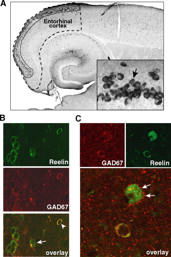
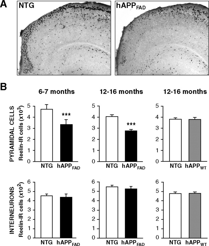
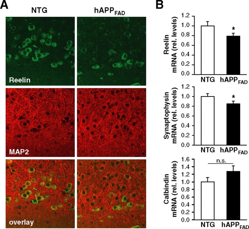
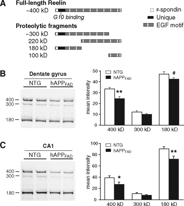
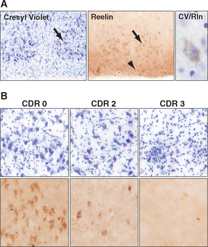
Reelin regulates nervous system development and modulates synaptic plasticity in the adult brain. Several findings suggest that alterations in Reelin signaling may contribute to neuronal dysfunction associated with Alzheimer's disease (AD). Cell surface receptors for Reelin, including integrins and very-low-density lipoprotein receptor/apolipoprotein E2 receptor, may be targets of amyloid-beta (Abeta) peptides presumed to play key roles in the pathogenesis of AD. Reelin also regulates the extent of tau phosphorylation. Finally, increased amounts of Reelin fragments have been found in CSF from AD patients, suggesting altered processing of Reelin. We therefore hypothesized that Reelin levels might be altered in the brains of human amyloid precursor protein (hAPP) transgenic mice, particularly in brain regions vulnerable to AD such as hippocampus and entorhinal cortex. Compared with nontransgenic controls, hAPP mice had significantly fewer Reelin-expressing pyramidal cells in the entorhinal cortex, the major population of glutamatergic neurons expressing Reelin in the brain. Western blot analysis of the hippocampus, which receives projections from the entorhinal cortex, revealed significant reductions in Reelin levels. In contrast, the number of Reelin-expressing GABAergic interneurons was not altered in either the entorhinal cortex or the hippocampus. Thus, neuronal expression of hAPP/Abeta is sufficient to reduce Reelin expression in a specific population of entorhinal cortical pyramidal neurons in vivo. Underscoring the relevance of these findings, we found qualitatively similar reductions of Reelin-expressing pyramidal neurons in the entorhinal cortex of AD brains. We conclude that alterations in Reelin processing or signaling may be involved in AD-related neuronal dysfunction.


Figures

Figure 1.
Figure 1.
Two populations of neurons in the mouse entorhinal cortex express Reelin. A, Low-magnification view of a horizontal brain section from a nontransgenic mouse immunostained for Reelin. The stippled line delineates the entorhinal cortex and the solid line the population of pyramidal neurons in layer II that express Reelin. Inset, High-magnification view of layer II of the entorhinal cortex. Arrow points to a Reelin-expressing pyramidal neuron. B, C, Double immunostaining for Reelin (green) and for GAD67 (red), a marker of GABAergic neurons. Overlay in B demonstrates that Reelin is expressed by glutamatergic pyramidal neurons (arrow) and by GABAergic interneurons (arrowhead). Overlay in C illustrates the presence of GAD67-positive terminals (arrows) surrounding the cell body of a Reelin-positive pyramidal cell.
Figure 2.
Figure 2.
The number of Reelin-expressing pyramidal cells in the entorhinal cortex is decreased in hAPPFAD mice. A, Horizontal sections from nontransgenic (NTG) controls and hAPPFAD mice illustrating Reelin immunoreactivity. B, Reelin-positive cells were counted in every 10th section throughout the dorsoventral extent of the entorhinal cortex. hAPPFAD mice (black bars) had a significant reduction in Reelin-IR pyramidal cells, but not interneurons, at 6–7 (left) and 12–16 (middle) months of age (n = 10–14 mice per genotype and age). In contrast, hAPPWT mice (gray bars; n = 7–8 mice per genotype) did not differ from nontransgenic controls in Reelin-IR pyramidal cells or interneurons (right). ***p < 0.001 versus nontransgenic.
Figure 3.
Figure 3.
hAPPFAD mice have decreased levels of Reelin and synaptophysin mRNAs in the entorhinal cortex but no overt neuronal loss. A, Immunostaining of the entorhinal cortex for Reelin (green) and MAP2 (red) demonstrates comparable overall MAP2 staining and fewer double-labeled pyramidal cells in hAPPFAD mice than in nontransgenic (NTG) controls. B, Quantitative fluorogenic RT-PCR analysis of entorhinal cortex lysates demonstrates decreased Reelin mRNA levels (top) in hAPPFAD mice relative to nontransgenic controls. Although mRNA levels of other synaptically localized proteins such as synaptophysin were also decreased (middle), not all proteins that can influence synaptic functions were decreased (bottom). rel., Relative. *p ≤ 0.05.
Figure 4.
Figure 4.
Reelin levels are decreased in the hippocampus of hAPPFAD mice. A, Schematic illustrating full-length Reelin and the fragments produced by extracellular cleavage. Monoclonal antibody G10, used for both immunostaining and Western blot analyses, binds to Reelin where indicated. B, C, Western blot analysis of protein lysates from dentate gyrus (B) or CA1 (C) demonstrates a significant reduction in Reelin levels in hAPPFAD mice (n = 10 mice per genotype). Equal protein, determined by Bradford assay, was loaded in each lane. #p = 0.06; *p < 0.05; **p < 0.01 versus nontransgenic (NTG).
Figure 5.
Figure 5.
Reelin-expressing pyramidal neurons in the entorhinal cortex are decreased in AD. A, Sections from a control patient illustrating the typical distribution of pyramidal neurons in layer II of the human entorhinal cortex. Cresyl violet (CV) staining is shown on the left, Reelin (Rln) immunoreactivity in the middle, and a pyramidal neuron stained with both cresyl violet and anti-Reelin antibody on the right. Arrows point to pyramidal neurons in layer II; arrowhead points to interneurons in layer I. B, Sections from humans without dementia (CDR 0), or with moderate AD (CDR 2), or severe AD (CDR 3) were stained with cresyl violet (top) or anti-Reelin antibody (bottom).

Similar articles

Cited by

References

    1. Beall MJ, Lewis DA. Heterogeneity of layer II neurons in human entorhinal cortex. J Comp Neurol. 1992;321:241–266. - PubMed
    1. Beffert U, Weeber EJ, Durudas A, Qiu S, Masiulis I, Sweatt JD, Li WP, Adelmann G, Frotscher M, Hammer RE, Herz J. Modulation of synaptic plasticity and memory by Reelin involves differential splicing of the lipoprotein receptor Apoer2. Neuron. 2005;47:567–579. - PubMed
    1. Blennow K, de Leon MJ, Zetterberg H. Alzheimer's disease. Lancet. 2006;368:387–403. - PubMed
    1. Botella-López A, Burgaya F, Gavin R, Garcia-Ayllon MS, Gomez-Tortosa E, Pena-Casanova J, Urena JM, Del Rio JA, Blesa R, Soriano E, Saez-Valero J. Reelin expression and glycosylation patterns are altered in Alzheimer's disease. Proc Natl Acad Sci USA. 2006;103:5573–5578. - PMC - PubMed
    1. Buckner RL. Memory and executive function in aging and AD: multiple factors that cause decline and reserve factors that compensate. Neuron. 2004;44:195–208. - PubMed

Publication types

MeSH terms