Comparative Study
. 2007 Mar 14;27(11):2734-43.
doi: 10.1523/JNEUROSCI.3849-06.2007.

Acute psychosocial stress reduces cell survival in adult hippocampal neurogenesis without altering proliferation

Affiliations
Comparative Study

Acute psychosocial stress reduces cell survival in adult hippocampal neurogenesis without altering proliferation

Rosanne M Thomas et al. J Neurosci. .

Abstract

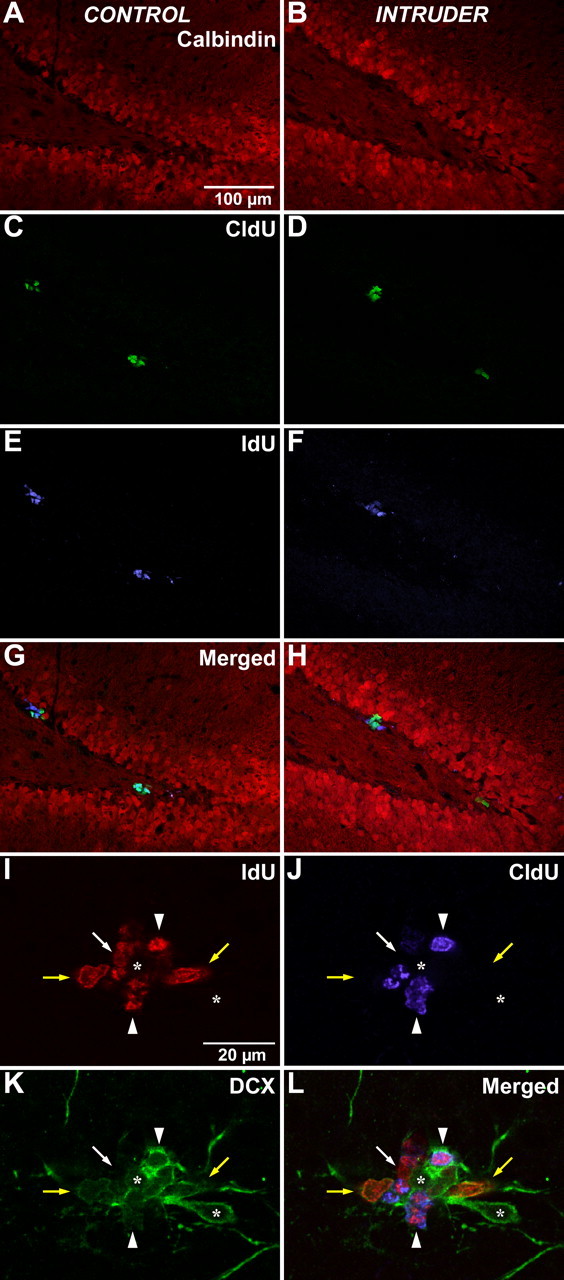
Factors modulating neurogenesis may contribute to the pathophysiology of affective disorders such as major depression. Environmental stressors in animal models have been proposed to alter neurogenesis, suggesting a mechanism for this contribution. The effect of an acute psychosocial stressor on either proliferation or survival (immediate, short term, and long term) was examined along with subsequent neuronal differentiation in the hippocampus of adult male Sprague Dawley rats. Subjects were exposed to a widely used social dominance paradigm that elicits behavioral and physiological responses to an acute psychosocial stressor. This social dominance paradigm may mimic human relational stress more realistically than laboratory stressors and provides a socially relevant model. We found that exposure to an acute psychosocial stressor at the time of cell generation resulted in a decreased number of newly generated cells in the hippocampus. By using sequential thymidine analog administration to provide temporal discrimination of DNA replication, we showed that short-term survival but not initial proliferation or immediate survival was altered in response to stress. Furthermore, we determined that stress experienced subsequent to proliferation also diminished long-term survival of cells. Thus, an acute episode of a social stress produces long-lasting effects on the incorporation of new hippocampal neurons by reducing their survival.


Figures

Figure 1.
Figure 1.
Design of the study. A, Adult neurogenesis in the hippocampal dentate gyrus consists of a process beginning with DNA replication during proliferation of uncommitted progenitor–stem cells that subsequently undergo lineage commitment. Not all newly generated cells survive, and it is the combination of survival and lineage commitment that determines the extent to which new neurons are added to the hippocampus. This study evaluated the modulation of stress on several stages of this continuum of neurogenesis. B, To discriminate the effect of stress on cellular proliferation and immediate survival, two different thymidine analogs, IdU and CldU, were sequentially delivered because they can be separately detected by immunohistochemistry. IdU was administered first to provide a measure of immediate survival, and the episode of acute stress coincided with administration of CldU to evaluate stress modulation of proliferation when assessed the next day (day 4). C, To assess the stress effect on short-term survival, a cohort of proliferating cells was identified by incorporation of another thymidine analog, BrdU, coincident with an episode of acute stress, and evaluated for short-term survival after 1 week (day 10). D, To further discriminate between proliferation and survival, a cohort of newly generated cells was identified by BrdU administration and subjected to acute stress 1 week after birth and evaluated for long-term survival after 4 weeks (day 28). Arrows indicate thymidine analog delivery (two times daily); triangles indicate time of tissue collection.
Figure 2.
Figure 2.
Paradigm for acute psychosocial stress. A, A young naive male rat (intruder; marked with the black X) was introduced into a stable colony of two rats whose female companion had just been removed. B, During initial exploration, the colony rats became aggressive, and the intruder rat (black X) exhibited submissive behavior. C, D, The aggression continued, with the intruder rat being pinned down and bitten.
Figure 3.
Figure 3.
Immunohistochemical detection of halogenated thymidine analogs. Animals that had been administered IdU for 2 d followed by CldU coincident with experiencing the episode of stress were evaluated 1 d later (Fig. 1 B, experiment 1). A, B, Sections from control (A) or intruder (B) rats were stained with calbindin (red) to identify the granule cell layer of the dentate gyrus. C, D, Detection of CldU (green) identified cells that were undergoing DNA replication on the day of the psychosocial stress episode. E, F, Detection of IdU (blue) identified cells that had been generated immediately before the stress episode. Examination of the merged image permits additional detection of cells that coexpress both IdU and CldU, indicating cells that have reentered the cell cycle during the presence of these two thymidine analogs. There is no apparent difference between control rats (A, C, E, G) and intruder rats (B, D, F, H). Scale bar: (in A) A–H, 100 μm. In addition, coexpression of IdU (I, red) and CldU (J, blue) with DCX (K, green) in the subgranular zone of the dentate gyrus was assessed to evaluate the phenotypic distribution of newly generated cells with this marker of early neuronal linage commitment. L, A single focal plane from a sequential series of confocal micrographs from an intruder animal is shown merged. The separation of fluorescence emission in I–K reveals that some IdU-positive cells coexpress DCX (yellow arrows) and some do not (white arrow). Some IdU-positive cells also incorporate CldU (arrowheads), indicating that they had just reentered the cell cycle. In this example, the IdU+/CldU+ cells coexpress DCX, although dual-labeled cells and IdU−/CldU+ cells can be observed that do not coexpress DCX (data not shown). Not all DCX-positive cells in the subgranular zone are engaged in DNA replication (asterisks). Scale bar: (in I) I–L, 20 μm.
Figure 4.
Figure 4.
Short-term survival of newly generated cells is reduced by stress. A, B, Histological evaluation of BrdU-positive cells in animals examined 1 week after the stress episode (Fig. 1 C, experiment 2) revealed a robust population of BrdU-positive cells in the dentate gyrus of control animals (A), but an obvious reduction in cell number for intruder rats (B). C–F, Higher magnification of the boxed region shows BrdU-positive cells at various focal planes. Some cells present at one focal plane (C, D, arrows) are not observed at a deeper focal plane when other BrdU-positive cells come into focus (E, F, arrowheads). The three-dimensional distribution of cells within the section thickness shows the need for stereological quantitation. Scale bars: A, B, 250 μm; C–F, 25 μm.
Figure 5.
Figure 5.
Neuronal differentiation of newly generated cells. Shown is the immunohistochemical detection of BrdU-positive cells in animals that had received BrdU 1 week before experiencing the single episode of psychosocial stress and that were examined at 4 weeks to evaluate their long-term survival (Fig. 1 D, experiment 3). A, B, Cells generated during the delivery of BrdU (green) before the stress episode could be observed in the dentate gyrus of both control and intruder animals. C–F, In both cases, the BrdU-positive cells frequently coexpressed the mature neuronal marker NeuN (red), but only very rarely was any colocalization observed with the mature astrocytic marker S100β (blue). Colocalization was determined on the basis of three-dimensional analysis of coexpression within the same cell imaged by using sequential focal planes obtained by confocal microscopy. Scale bar: (in A) A–F, 50 μm.

Similar articles

Cited by

References

    1. Arango V, Underwood MD, Boldrini M, Tamir H, Kassir SA, Hsiung S, Chen JJ, Mann JJ. Serotonin 1A receptors, serotonin transporter binding and serotonin transporter mRNA expression in the brainstem of depressed suicide victims. Neuropsychopharmacology. 2001;25:892–903. - PubMed
    1. Benninghoff J, Schmitt A, Mossner R, Lesch KP. When cells become depressed: focus on neural stem cells in novel treatment strategies against depression. J Neural Transm. 2002;9:947–962. - PubMed
    1. Blanchard RJ, McKittrick CR, Blanchard DC. Animal models of social stress: effects on behavior and brain neurochemical systems. Physiol Behav. 2001;73:261–271. - PubMed
    1. Cameron HA, McKay RD. Restoring production of hippocampal neurons in old age. Nat Neurosci. 1999;2:894–897. - PubMed
    1. Cameron HA, Hazel TG, McKay RD. Regulation of neurogenesis by growth factors and neurotransmitters. J Neurobiol. 1998;36:287–306. - PubMed

Publication types