Comparative Study
. 2007 Mar 14;27(11):2781-7.
doi: 10.1523/JNEUROSCI.4372-06.2007.

The prefrontal cortex as a key target of the maladaptive response to stress

Affiliations
Comparative Study

The prefrontal cortex as a key target of the maladaptive response to stress

João J Cerqueira et al. J Neurosci. .

Abstract

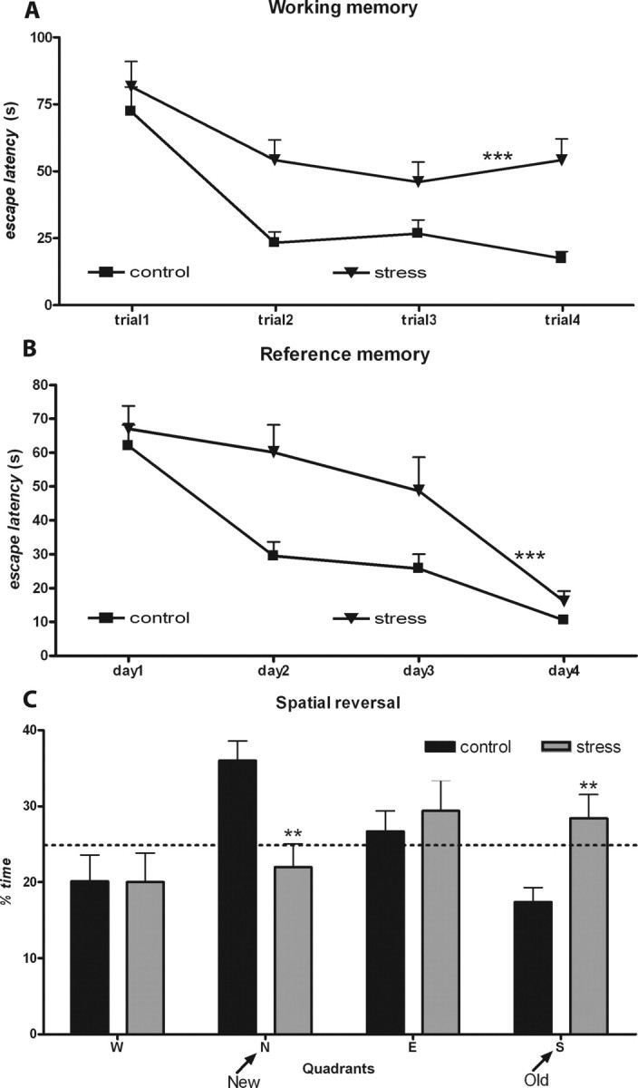
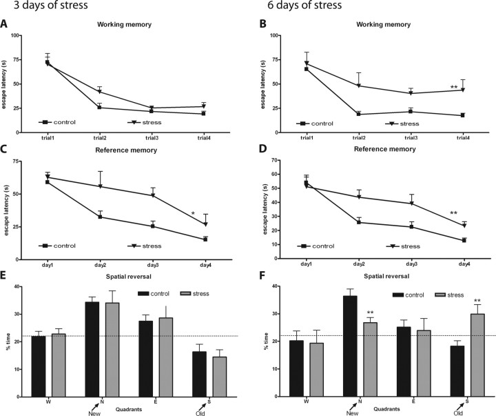
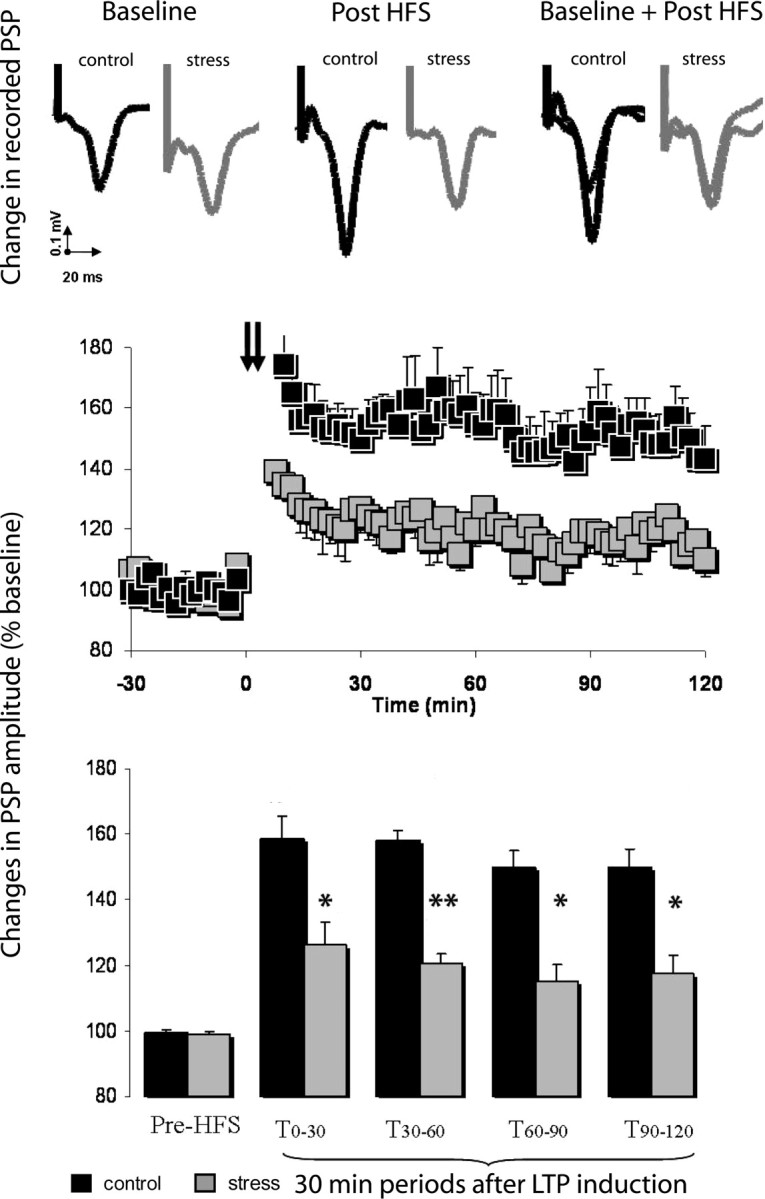
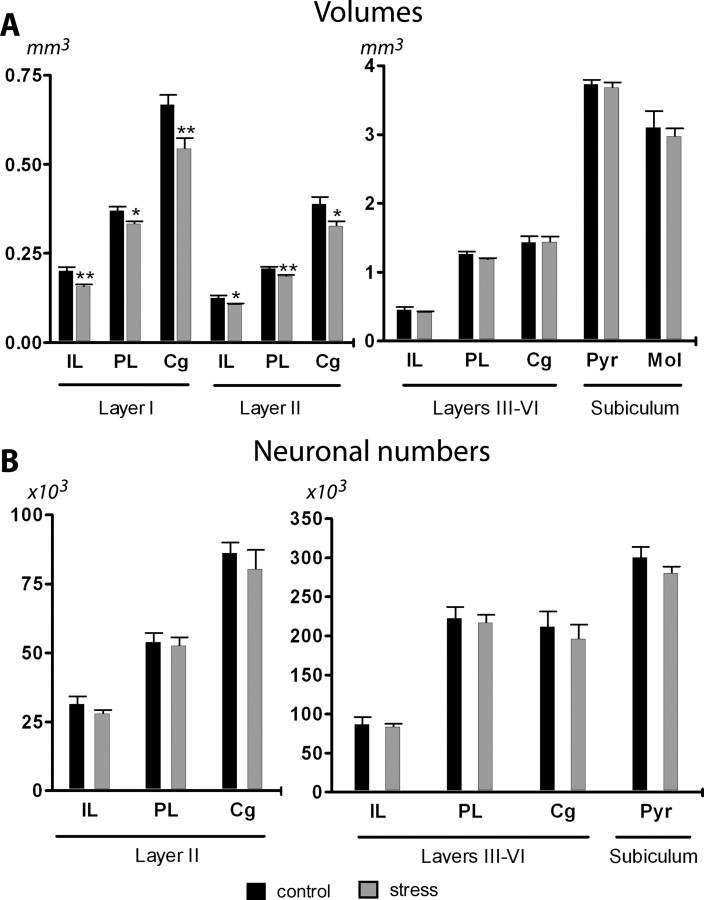
Research on the detrimental effects of stress in the brain has mainly focused on the hippocampus. Because prefrontal cortex (PFC) dysfunction characterizes many stress-related disorders, we here analyzed the impact of chronic stress in rats on the integrity of the hippocampal-PFC pathway, monitored by behavioral and electrophysiological function and morphological assessment. We show that chronic stress impairs synaptic plasticity by reducing LTP induction in the hippocampal-PFC connection; in addition, it induces selective atrophy within the PFC and severely disrupts working memory and behavioral flexibility, two functions that depend on PFC integrity. We also demonstrate that short periods of stress exposure induce spatial reference memory deficits before affecting PFC-dependent tasks, thus suggesting that the impairment of synaptic plasticity within the hippocampus-to-PFC connection is of relevance to the stress-induced PFC dysfunction. These findings evidence a fundamental role of the PFC in maladaptive responses to stress and identify this area as a target for intervention in stress-related disorders.


Figures

Figure 1.
Figure 1.
Behavioral impairments induced by chronic stress. A, B, Learning curves in the working (A) and reference memory task (B) of control (n = 10) and chronically stressed (n = 10) rats. The higher escape latencies of the stressed animals are easily appreciated. C, Results from the reverse task experiment. Average time spent on the four trials in each imaginary quadrant is given as a percentage of the total escape latency. Dotted line represents performance at the chance level (25%). Results of distance swam are not presented. **p < 0.01 and ***p < 0.001 compared with controls. Error bars represent SEM.
Figure 2.
Figure 2.
Behavioral tests after 3 and 6 d of unpredictable stress. A–D, Learning curves in the working (A, B) and reference memory task (C, D) of control (n = 10) and stressed (n = 10) rats. E, F, Results from the reverse task experiment. Average time spent on the four trials in each imaginary quadrant is given as a percentage of the total escape latency. Dotted line represents performance at the chance level (25%). Graphs on the left refer to animals stressed for 3 d, and those on the right refer to 6 d of stress. Results of distance swam are not presented. *p < 0.05 and **p < 0.01 compared with controls. Error bars represent SEM.
Figure 3.
Figure 3.
Effects of chronic stress on hippocampal-prefrontal cortex LTP. Top, Representative PSP recorded from the prefrontal cortex of chronic-stressed (n = 5) and control (n = 5) animals before and after hippocampal HFS. Middle, Chronically stressed rats displayed a deficit in HFS-induced LTP when compared with controls. Squares are mean ± SEM of the normalized PSP amplitude for 2 min periods. Hippocampal HFS is indicated by arrows. Bottom, LTP in chronically stressed rats and controls represented at different time periods. The first and following pairs of columns represent mean ± SEM of the average normalized PSP amplitude in consecutive 30 min periods before and after HFS, respectively. *p < 0.05 and **p < 0.01 compared with controls. Error bars represent SEM.
Figure 4.
Figure 4.
Effects of chronic stress on the volumes and number of neurons of the different mPFC areas and subiculum. A, Average volumes of layers I, II, and III–VI of the IL, PL, and Cg regions of the mPFC and the molecular (Mol) and pyramidal (Pyr) layers of the subiculum. B, Estimated number of neurons in layers II and III–VI of the IL, PL, and Cg regions and the Pyr layer of the subiculum. Note the different scales used and that with the exception of the numerical values for layers I and II, which should be read off against the left axis, all other values should be read off against the right one. Control animals, n = 5; stress rats, n = 5. *p < 0.05 and **p < 0.01 compared with CON. Error bars represent SEM.
Figure 5.
Figure 5.
Volumes and number of neurons of the different mPFC areas and subiculum after 3 and 6 d of unpredictable stress. A, B, Average volumes of layers I, II, and III–VI of the IL, PL, and Cg regions of the mPFC and the molecular (Mol) and pyramidal (Pyr) layers of the subiculum. C, D, Estimated number of neurons in layers II and III–VI of the IL, PL, and Cg regions and the Pyr layer of the subiculum. Graphs on the left refer to animals stressed for 3 d, and those on the right refer to 6 d of stress. Control animals, n = 5; stressed rats, n = 5. Error bars represent SEM.

Similar articles

Cited by

References

    1. Bagley J, Moghaddam B. Temporal dynamics of glutamate efflux in the prefrontal cortex and in the hippocampus following repeated stress: effects of pretreatment with saline or diazepam. Neuroscience. 1997;77:65–73. - PubMed
    1. Bayatti N, Behl C. The neuroprotective actions of corticotropin releasing hormone. Ageing Res Rev. 2005;4:258–270. - PubMed
    1. Brown SM, Henning S, Wellman CL. Mild, short-term stress alters dendritic morphology in rat medial prefrontal cortex. Cereb Cortex. 2005;15:1714–1722. - PubMed
    1. Cerqueira JJ, Pêgo JM, Taipa R, Bessa-Peixoto JM, Almeida OFX, Sousa N. Morphological correlates of corticosteroid-induced changes in prefrontal cortex-dependent behaviours. J Neurosci. 2005;25:7792–7800. - PMC - PubMed
    1. Cerqueira JJ, Taipa R, Uylings HBM, Almeida OFX, Sousa N. Specific configuration of dendritic degeneration in pyramidal neurons of the medial prefrontal cortex induced by differing corticosteroid regimens. Cereb Cortex. 2007 in press. - PubMed

Publication types