Comparative Study
. 2007 Mar 14;27(11):2846-57.
doi: 10.1523/JNEUROSCI.0116-07.2007.

NMDA receptor subunits have differential roles in mediating excitotoxic neuronal death both in vitro and in vivo

Affiliations
Comparative Study

NMDA receptor subunits have differential roles in mediating excitotoxic neuronal death both in vitro and in vivo

Yitao Liu et al. J Neurosci. .

Abstract

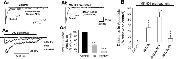
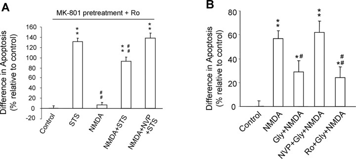
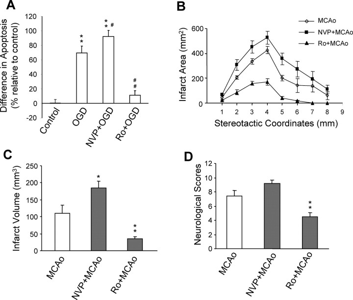
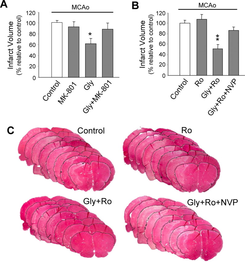
Well-documented experimental evidence from both in vitro and in vivo models of stroke strongly supports the critical involvement of NMDA receptor-mediated excitotoxicity in neuronal damage after stroke. Despite this, the results of clinical trials testing NMDA receptor antagonists as neuroprotectants after stroke and brain trauma have been discouraging. Here, we report that in mature cortical cultures, activation of either synaptic or extrasynaptic NR2B-containing NMDA receptors results in excitotoxicity, increasing neuronal apoptosis. In contrast, activation of either synaptic or extrasynaptic NR2A-containing NMDA receptors promotes neuronal survival and exerts a neuroprotective action against both NMDA receptor-mediated and non-NMDA receptor-mediated neuronal damage. A similar opposing action of NR2B and NR2A in mediating cell death and cell survival was also observed in an in vivo rat model of focal ischemic stroke. Moreover, we found that blocking NR2B-mediated cell death was effective in reducing infarct volume only when the receptor antagonist was given before the onset of stroke and not 4.5 h after stroke. In great contrast, activation of NR2A-mediated cell survival signaling with administration of either glycine alone or in the presence of NR2B antagonist significantly attenuated ischemic brain damage even when delivered 4.5 h after stroke onset. Together, the present work provides a molecular basis for the dual roles of NMDA receptors in promoting neuronal survival and mediating neuronal damage and suggests that selective enhancement of NR2A-containing NMDA receptor activation with glycine may constitute a promising therapy for stroke.


Figures

Figure 1.
Figure 1.
Functional NR2A- and NR2B-containing NMDA receptors are present in cultured neurons and are preferentially blocked by their respective antagonists. Whole-cell recording was performed at a holding membrane potential of −60 mV in an extracellular solution supplemented with 10 μm CNQX, 0.5 μm TTX, and 10 μm bicuculline. A, Example traces of whole-cell currents evoked by a brief perfusion of 50 μm NMDA (plus 10 μm glycine and 5 μm strychnine) from a multibarrel fast perfusion system in the absence or presence of specific NR2A (NVP-AAM077, 0.4 μm), NR2B antagonists (Ro 25–6981, 0.5 μm), or both. The percentage blockade of the NMDA-induced currents by sequential application of these two antagonists is summarized in the histogram in B. Preapplication of Ro 25–6981 did not alter the percentage blockade produced by NVP-AAM077 (p = 0.97; in the absence vs the presence of Ro 25–6981), whereas preapplication of NVP-AAM077 produced a small, albeit statistically insignificant, reduction in the percentage blockade of the currents produced by Ro 25–6981 (5.6%; p = 0.19).
Figure 2.
Figure 2.
NR2A- and NR2B-containing NMDA receptors exert opposing effects on NMDA-induced excitotoxic neuronal damage. Cortical neuronal cultures were treated without NMDA (control) or with NMDA (50 μm plus 10 μm glycine) for 20 min and examined for neuronal cell death after 20 h. A, NMDA-induced necrotic neuronal injury was mediated by NR1/NR2B- but not NR2A-containing NMDA receptors. Necrotic neuronal injury was quantitatively determined by measuring LDH release. NR2B preferential antagonist Ro 25–6981 (Ro; 0.5 μm), but not NR2A preferential antagonist NVP-AAM077 (NVP; 0.4 μm), prevented NMDA-induced necrosis. Data are presented as the difference in LDH levels as a percentage of control. **p < 0.01 compared with nontreated control; n = 18 tissue culture wells from three separate experiments for each group. B, C, NR2A-containing and NR1/NR2B receptors exert opposing effects on NMDA-induced neuronal apoptosis. Representative images of Hoechst-33342-stained neurons in B illustrate the differential effects of coapplication of NMDA with NR2A antagonist NVP-AAM077 (NVP; 0.4 μm) or NR2B antagonist Ro 25–6981 (Ro; 0.5 μm) on NMDA-induced neuronal apoptosis. NMDA stimulation produced neuronal damage such as chromatin condensation and/or fragmentation, which were aggravated in the presence of NVP-AAM077 but eliminated in the presence of Ro 25–6981. The cell death ELISA assay in C quantifies the differential effects of NVP-AAM077 and Ro 25–6981 on NMDA-induced neuronal apoptosis. Data are presented as the difference in apoptosis levels as a percentage of control. **p < 0.01 compared with nontreated control; #p < 0.05 and ##p < 0.01, compared with NMDA treatment alone; n = 18 tissue culture wells from three separate experiments for each group. D, NR2A-containing and NR1/NR2B receptors have opposing actions on cell survival (Akt activation) and death (caspase-3 activation) signaling pathways. Top, Cell lysates from cultured neurons treated as indicated were sequentially immunoblotted with antibodies specific to Akt phosphorylated on serine 473, the active form of the enzyme (P-Akt), and total Akt (Akt). Bottom, Cell lysates were sequentially immunoblotted with antibodies that specifically recognize cleaved caspase-3 (activated caspase-3; Casp3) and β-tubulin as a load control (Tubulin).
Figure 3.
Figure 3.
Genetic deletions of NR2A and NR2B subunits have differential effects on NMDA-induced neuronal apoptosis. Cultured mouse cortical neurons from NR2A−/− embryos or NR2B−/− embryos and WT littermate control mice were treated with NMDA (50 μm plus 10 μm glycine) for 20 min. Neurons were stained with Hoechst-33342 20 h after NMDA treatments and examined by microscopy. Apoptosis was quantified as the percentage of cells demonstrating cromatin condensation/fragmentation. NMDA-induced apoptosis was significantly reduced by genetic deletion of NR2B (B), but not NR2A (A), subunit (n = 4–6 tissue culture wells with >150 cells counted in each group). *p < 0.05 and **p < 0.01, compared with nontreated WT control; #p < 0.05, compared with NMDA-treated WT group. KO, Knock-out.
Figure 4.
Figure 4.
Activation of synaptic NR1/NR2B NMDA receptors produces a proapoptotic action, which is masked by a predominant synaptic NR2A-containing receptor-mediated cell survival-promoting effect. A, Functional synaptic NR2B-containing receptors are present in cortical neurons in culture. Spontaneous mEPSCs were recorded in whole-cell voltage-clamp mode at a holding membrane potential of −60 mV in the presence of TTX (0.5 μm) and bicuculline (10 μm) with 0 added Mg2+. Aa, Examples of mEPSC traces (averaged from 100 individual events) obtained in the absence (Control) and presence of Ro 25–6981 (Ro; 0.5 μm) or the broad spectrum NMDA receptor antagonist APV (APV; 50 μm). Ab, Total NMDA receptor-mediated component of mEPSCs was obtained by subtracting the averaged mEPSC recorded in the presence of APV from the averaged control mEPSC (Control-APV; shaded area). Ac, The NR1/NR2B receptor component was obtained by subtracting the averaged mEPSC recorded in the presence of Ro 25–6981 (Ro) from the averaged control mEPSC (Control-Ro; shaded area). Ad, Bar graph summarizes data obtained from five individual neurons. Charge transfer is equivalent to the area of the shaded regions. B, Enhanced activation of synaptic NR2A-containing and NR1/NR2B receptors exerts opposing actions on neuronal survival and death. Potentiation of synaptic NMDA receptor activation was achieved by increasing the presynaptic release of glutamate by incubating cultured neurons with bicuculline (Bic; 50 μm) for 4 h in the absence or presence of NR2-containing receptor-specific antagonists. Blockade of NR2A-containing (Bic+NVP), but not NR1/NR2B (Bic+Ro), NMDA receptors increased neuronal apoptosis. The NR2A blockade-induced apoptosis was prevented by an additional blockade of NR1/NR2B receptors (Bic+NVP+Ro). C, Spontaneously activated synaptic NR2A- and NR2B-containing receptors also have opposing roles in promoting neuronal survival and death. Incubation of neurons with NVP-AAM077 (NVP), but not Ro 25–6981 (Ro), for an extended duration (48 h) in the absence of bicuculline stimulation was sufficient to produce an increase in apoptosis. The NVP-AAM077-induced apoptosis was prevented by addition of Ro 25–6981 (NVP+Ro). Thus, both synaptic NR2A-containing and NR1/NR2B subpopulations of NMDA receptors are spontaneously activated by presynaptically released glutamate, exerting counteracting effects on cell survival and death, but synaptic NR2A-containing receptor activation is predominant and required for maintaining normal neuronal survival. **p < 0.01 compared with control; n = 16 (B) and 10–12 (C) for each group from three separate experiments.
Figure 5.
Figure 5.
Activation of extrasynaptic NR2A-containing NMDA receptors promotes cell survival, protecting against extrasynaptic NR1/NR2B receptor-mediated and non-NMDA receptor-dependent neuronal death. A, Functional NR2A-containing NMDA receptors are present at extrasynaptic sites. Whole-cell recordings were performed at a holding membrane potential of −60 mV. Aa, Averaged traces of mEPSCs showing an APV-sensitive (50 μm) NMDA receptor-mediated component (Control-APV; shaded area). Ab, Averaged traces of mEPSCs showing the blockade of synaptic NMDA receptors by the open channel blocker MK-801 (10 μm plus 50 μm bicuculline; 10 min), as demonstrated by the elimination of the NMDA receptor-mediated component of the mEPSCs (Control-APV; shaded area). Ac, Example traces of whole-cell currents evoked by NMDA (200 μm) after the blockade of synaptic NMDA receptors with MK-801 in the absence (A; control) or presence (B; 0.5 μm) of Ro 25–6981 or Ro 25–6981 plus NVP-AAM077 (C; 0.4 μm). NMDA receptor-mediated currents were evoked by fast application of NMDA within 10 min of washing out MK-801 and bicuculline. Currents remaining after the blockade of extrasynaptic NR1/NR2B receptors were virtually abolished by the addition of NVP-AAM077, suggesting the presence of functional extrasynaptic NR2A-containing NMDA receptors in these neurons. Ad, Histogram summarizes data from five individual neurons. B, Activation of extrasynaptic NR2A-containing NMDA receptors protects against neuronal death mediated by extrasynaptic NR1/NR2B NMDA receptors. Excitotoxic neuronal death was induced in cortical neurons by bath application of NMDA (50 μm; 20 min) after the blockade of synaptic NMDA receptors with MK-801 plus bicuculline, and cell death was assayed 20 h later. NMDA elicited neuronal apoptosis, which was exacerbated when the NR1/NR2B component was selectively stimulated (NVP+NMDA) but eradicated when the NR2A-containing component was specifically activated (Ro+NMDA). **p < 0.01 compared with control. #p < 0.05, ##p < 0.01 compared with NMDA treatment. n = 11–12 from two separate experiments for each group.
Figure 6.
Figure 6.
Selective activation of NR2A-containing NMDA receptors protects neurons from NMDA receptor-mediated or non-NMDA receptor-mediated neuronal apoptosis. A, Activation of extrasynaptic NR2A-containing NMDA receptors can counteract NMDA receptor-independent apoptosis. Bath application of STS (100 nm; 1 h) after blockade of synaptic NMDA receptors by pretreatment with MK-801 plus bicuculline and of extrasynaptic NR1/NR2B receptors by Ro 25–6981 (Control) induced a significant increase in neuronal apoptosis (STS). Brief application of NMDA (200 μm; 5 min) did not produce neuronal apoptosis on its own (NMDA) but significantly reduced the STS-induced neuronal apoptosis (NMDA+STS), and the NMDA-induced neuroprotective action was abrogated by coapplication of NVP-AAM077 (0.4 μm; NVP+NMDA+STS). **p < 0.01 compared with Ro 25–6981 treatment (Control). ##p < 0.01 compared with STS treatment. n = 8–12 for each group from three separate experiments. B, Pretreatment of neuronal cultures with glycine (300 μm plus 10 μm strychnine) for 10 min significantly reduced the neuronal apoptosis produced by NMDA applied thereafter (Gly+NMDA). This neuroprotective effect was abolished by coapplication of NR2A antagonist NVP-AAM077 (0.4 μm) with glycine (NVP+Gly+NMDA) but not by coapplication of NR2B-specific antagonist Ro 25–6981 (0.5 μm) with glycine (Ro+Gly+NMDA), indicating that the neuroprotective effect of glycine is primarily mediated through enhancing the activation of NR2A-containing NMDA receptors. *p < 0.05, **p < 0.01 compared with control. #p < 0.05, ##p < 0.01 compared with NMDA. n = 17–18 for each group from three separate experiments.
Figure 7.
Figure 7.
Pretreatments with NR2A- and NR2B-specific antagonists promote neuronal survival and death, respectively, in both in vitro and in vivo models of ischemia. A, NR2A-containing and NR1/NR2B receptors exert opposing effects in ischemic neuronal injuries in vitro. Cortical cultures were challenged with a 1 h OGD, and apoptosis was assayed 23 h after the challenge. OGD resulted in a significant increase in neuronal apoptosis compared with nonchallenged controls (Control), and the OGD-induced apoptosis was respectively potentiated by the NR2A preferential antagonist NVP-AAM077 (NVP+OGD; 0.4 μm) and inhibited by the NR2B antagonist Ro 25–6981 (Ro+OGD; 0.5 μm) when bath applied 30 min before, and during, the OGD challenge. **p < 0.01 compared with control. #p < 0.05, ##p < 0.01 compared with OGD. n = 17–18 for each group from three separate experiments. B, C, NR2A- and NR2B-containing receptors exert opposing effects in ischemic neuronal injuries in vivo. Adult rats were subjected to a 1 h focal cerebral ischemia produced by MCAo, and cerebral infarction was assessed 24 h after MCAo onset. Intravenous infusion 30 min before MCAo onset of NVP-AAM077 (NVP+MCAo; 2.4 mg/kg; n = 5) and Ro 25–6981 (Ro+MCAo; 6 mg/kg; n = 6) increased and decreased, respectively, both infarct area (B) and total infarct volume (C). *p < 0.05, **p < 0.01 compared with MCAo. D, Neurological scores assessed 24 h after stroke onset in the same groups of animals shown in B and C indicate that blockade of the NR2A-containing NMDA receptors resulted in a trend toward worsening neurological function, whereas blockade of NR1/NR2B NMDA receptors markedly improved neurological behavior. **p < 0.01 compared with MCAo.
Figure 8.
Figure 8.
Postischemic potentiation of NR2A-containing NMDA receptors through administration of glycine reduces ischemic brain damage in an in vivo focal ischemic stroke model. Adult rats received either drug or saline treatment 3 h after a 1.5 h MCAo challenge (4.5 h after MCAo onset). A, General blockade of NMDA receptors with nonsubunit-specific antagonist MK-801 (1 mg/kg; n = 8) after stroke was no longer neuroprotective, whereas a poststroke treatment with NMDA receptor coagonist glycine (Gly; 800 mg/kg; n = 8) significantly reduced total infarct volume. The glycine effect was fully blocked by coapplication of MK-801 (Gly+MK-801; n = 7), indicating mediation by NMDA receptors. *p < 0.05 compared with Control (MCAo alone). B, Postischemic treatment with NR2B-selective antagonist Ro 25–6981 (Ro; 6 mg/kg; n = 10) was ineffective, whereas the glycine effect persisted in the presence of Ro 25–6981 (6 mg/kg) (Gly+Ro; n = 9). The addition of NR2A antagonist NVP-AAM077 (2.4 mg/kg; Gly+Ro+NVP; n = 10) abolished the neuroprotection offered by glycine, indicating glycine acts via selective potentiation of NR2A-containing NMDA receptors. **p < 0.01 compared with control (MCAo alone). C, Representative rat brain sections stained with H&E from each treatment group in B. Pale staining indicates infarct.

Similar articles

Cited by

References

    1. Aarts M, Liu Y, Liu L, Besshoh S, Arundine M, Gurd JW, Wang YT, Salter MW, Tymianski M. Treatment of ischemic brain damage by perturbing NMDA receptor-PSD-95 protein interactions. Science. 2002;298:846–850. - PubMed
    1. Aarts M, Iihara K, Wei WL, Xiong ZG, Arundine M, Cerwinski W, MacDonald JF, Tymianski M. A key role for TRPM7 channels in anoxic neuronal death. Cell. 2003;115:863–877. - PubMed
    1. Albensi BC, Igoechi C, Janigro D, Ilkanich E. Why do many NMDA antagonists fail, while others are safe and effective at blocking excitotoxicity associated with dementia and acute injury? Am J Alzheimers Dis Other Demen. 2004;19:269–274. - PMC - PubMed
    1. Arundine M, Tymianski M. Molecular mechanisms of glutamate-dependent neurodegeneration in ischemia and traumatic brain injury. Cell Mol Life Sci. 2004;61:657–668. - PMC - PubMed
    1. Bederson JB, Pitts LH, Tsuji M, Nishimura MC, Davis RL, Bartkowski H. Rat middle cerebral artery occlusion: evaluation of the model and development of a neurologic examination. Stroke. 1986;17:472–476. - PubMed

Publication types

MeSH terms

LinkOut - more resources