. 2007 Jun;18(6):2081-9.
doi: 10.1091/mbc.e06-08-0695. Epub 2007 Mar 14.

Dynactin is required for coordinated bidirectional motility, but not for dynein membrane attachment

Affiliations

Dynactin is required for coordinated bidirectional motility, but not for dynein membrane attachment

Marjan Haghnia et al. Mol Biol Cell. 2007 Jun.

Abstract

Transport of cellular and neuronal vesicles, organelles, and other particles along microtubules requires the molecular motor protein dynein (Mallik and Gross, 2004). Critical to dynein function is dynactin, a multiprotein complex commonly thought to be required for dynein attachment to membrane compartments (Karki and Holzbaur, 1999). Recent work also has found that mutations in dynactin can cause the human motor neuron disease amyotrophic lateral sclerosis (Puls et al., 2003). Thus, it is essential to understand the in vivo function of dynactin. To test directly and rigorously the hypothesis that dynactin is required to attach dynein to membranes, we used both a Drosophila mutant and RNA interference to generate organisms and cells lacking the critical dynactin subunit, actin-related protein 1. Contrary to expectation, we found that apparently normal amounts of dynein associate with membrane compartments in the absence of a fully assembled dynactin complex. In addition, anterograde and retrograde organelle movement in dynactin deficient axons was completely disrupted, resulting in substantial changes in vesicle kinematic properties. Although effects on retrograde transport are predicted by the proposed function of dynactin as a regulator of dynein processivity, the additional effects we observed on anterograde transport also suggest potential roles for dynactin in mediating kinesin-driven transport and in coordinating the activity of opposing motors (King and Schroer, 2000).


Figures

Figure 1.
Figure 1.
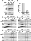
Axonal transport and the dynactin complex are disrupted in arp1 mutant larvae. (A and B) In contrast to wild-type larvae arp11 mutant animals exhibit the “tail-flip” phenotype common to axonal transport mutants in Drosophila. (C and D) Immunofluorescence with antibodies recognizing the synaptic cysteine string protein revealed abundant large accumulations within the segmental nerves of the arp11 mutant, whereas wild-type animals exhibited typical background staining. (E) Larval brain extracts from wild-type larvae and homozygous arp11 larvae were analyzed by Western blotting. The blots were probed with antibodies to different subunits of dynein and dynactin complexes. Tubulin antibody was used as a loading control. The p150Glued and p50 subunits are obviously reduced in the arp1 mutant larvae. (F) The dynactin complex is disrupted in the arp11 mutant. Third instar brains were homogenized, and a high-speed supernatant was sedimented on a 5–20% sucrose gradients. In wild type, sedimentation of the dynactin subunits peaks at ∼17S-, whereas in the arp11 mutant, both Arp1 and p50 sediment at ∼4S–8S. Asterisk (*) points to dynamitin in the Arp1 blot (the same blot has been reprobed). The peak of Arp1 reactivity detected at ∼4S in addition to the expected peak at 17S is due to cross reactivity of our Arp1 antibody with actin (see Materials and Methods and Figure 2).
Figure 2.
Figure 2.
The dynactin complex, but not dynein, is disrupted in the arp1 dsRNA-treated cells. (A) HSS and HSP were prepared from arp1 dsRNA and GFP dsRNA-treated cells. Western blot analysis shows that loss of arp1 leads to reduction in the levels of both p150Glued and p50, whereas KHC and DHC remain unchanged. Note that in the HSS sample, tubulin and actin shown as loading controls display a lower level in the GFP control compared with the Arp1-treated sample, which explains the lower levels of DHC and KHC in that sample. Although the knockdown of Arp1 protein is evident in the HSP where no actin is detected, in the HSS, the Arp1 antibody cross-reacts with actin (see Materials and Methods). (B) Quantitation of transcript amounts was done with RT-qPCR. Arbitrary values of transcripts levels were obtained from duplicate data points and changes in transcript levels for the arp1 RNAi-treated samples were compared with the GFP RNAi-treated samples and normalized to GAPDH. Knockdown of arp1 transcript is shown as percentage (mean of 3 experiments ± SEM). (C–F) HSS and HSP from arp1 dsRNA- and GFP dsRNA-treated cells were sedimented on a 5–20% sucrose gradients. In GFP dsRNA-treated cells, sedimentation of the dynactin subunits Arp1, p150Glued, and p50 are found in a broad peak at ∼17, whereas in the arp1 dsRNA-treated cells, Arp1 levels are reduced and both p150Glued and p50 are found at ∼8S. Note that in the arp1 dsRNA-treated samples, the levels of p50 and p150Glued are reduced, and Western blots for p50 and p150Glued required longer exposure times in D and F (see brackets and A). The Arp1 band detected at ∼8S in both GFP and arp1 dsRNA HSS corresponds to the actin cross-reactivity. This band is not observed in the HSP fraction where no actin can be detected. Dynein and kinesin sedimentation properties are not affected by reduction of Arp1, peaking at ∼17S and ∼8S, respectively. In C and D, arrowhead points to the 400-kDa dynein heavy chain.
Figure 3.
Figure 3.
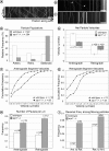
Vesicle movement is reduced in the arp11 mutant. (A) Kymograph from control movie displaying bidirectional movement in a dense field of APP–YFP particles. Bar, 10 μm. Movies and additional kymographs may be found in Supplemental Material and in Supplemental Figure 3. (B and C) Kymographs from arp11 larvae displaying altered patterns of movement. Kymograph in (B) shows a reduced number of APP–YFP particles, which are primarily stationary, with a few anterograde particles moving at velocities comparable with those in wild-type larvae (arrows). Kymograph in C shows primarily stationary particles, with some particles moving at a reduced velocity. Bar, 10 μm. Movies and additional kymographs may be found in Supplemental Material and in Supplemental Figure 3. (D) Populations of anterograde, retrograde, and stationary particles in arp11 mutant (gray) and wild-type (white) axons. (n = 284 particles analyzed from 17 arp11 mutant movies; n = 140 particles from 3 wild-type movies). (E) Net anterograde and retrograde velocities for moving particles in the arp11 mutant (gray) and wild-type (white) axons (mean ± SEM). (F) Cumulative frequency (defined as the summed percentage of particles with velocities less than or equal to velocity value on the x-axis) distribution of anterograde segment velocities of particles in arp11 mutant (circle) and wild-type (triangle) axons. (G) Cumulative frequency distribution of retrograde segment velocities of particles in arp11 mutant (circle) and wild-type (triangle) axons. (H) Pause frequency of arp11 mutant (gray) and wild-type (white) particles per micrometer of movement (mean ± SEM). Analysis excludes particles that do not pause. (I) Distribution of particle reversal frequency for arp11 mutant (gray) and wild-type (white) axons in anterograde and retrograde directions, expressed as frequency per total number of moving cargoes. Numbers above bars indicate sample size.
Figure 4.
Figure 4.
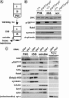
Dynein association with membranes is not affected by disruption of the dynactin complex. (A) Schematic representation of the flotation experiment. (B) Subcellular fractionation of third instar membranes on sucrose step gradients indicates that dynein is present in both soluble fraction and membrane fractions (35/8 interface) and that the amount of membrane-associated dynein is not altered in the arp11 mutant larvae. Rab8 and syntaxin are used as loading controls and syntaxin is used as a membrane control. (C) Similar results were obtained when dynein association with membranes is analyzed in arp1 dsRNA-treated S2 cells. Golgi membranes (d120) are enriched in the 35/8 interface, whereas mitochondria (cyt c) are enriched in the heavy membrane fraction. Importantly, as seen in A, levels of membrane-associated dynein are equivalent in both control and arp1 RNAi-treated cells. Note that actin is present in all fractions and is thus masking the Arp1 reduction detected with the Arp1 antibody in the arp1 dsRNA-treated cells (see Figure 2 and Materials and Methods).

Similar articles

Cited by

References

    1. Almenar-Queralt A., Goldstein L. S. Linkers, packages and pathways: new concepts in axonal transport. Curr. Opin. Neurobiol. 2001;11:550–557. - PubMed
    1. Bowman A. B., Kamal A., Ritchings B. W., Philp A. V., McGrail M., Gindhart J. G., Goldstein L. S. Kinesin-dependent axonal transport is mediated by the Sunday driver (SYD) protein. Cell. 2000;103:583–594. - PubMed
    1. Bowman A. B., Patel-King R. S., Benashski S. E., McCaffery J. M., Goldstein L. S., King S. M. Drosophila roadblock and Chlamydomonas LC 7, a conserved family of dynein-associated proteins involved in axonal transport, flagellar motility, and mitosis. J. Cell Biol. 1999;146:165–180. - PMC - PubMed
    1. Clark S. W., Meyer D. I. ACT 3, a putative centractin homologue in S. cerevisiae is required for proper orientation of the mitotic spindle. J. Cell Biol. 1994;127:129–138. - PMC - PubMed
    1. Deacon S. W., Serpinskaya A. S., Vaughan P. S., Lopez Fanarraga M., Vernos I., Vaughan K. T., Gelfand V. I. Dynactin is required for bidirectional organelle transport. J. Cell Biol. 2003;160:297–301. - PMC - PubMed

Publication types

MeSH terms