Comparative Study
. 2007 Mar 15;6(6):750-7.
doi: 10.4161/cc.6.6.3986. Epub 2007 Mar 7.

Differential gene expression of p27Kip1 and Rb knockout pituitary tumors associated with altered growth and angiogenesis

Affiliations
Comparative Study

Differential gene expression of p27Kip1 and Rb knockout pituitary tumors associated with altered growth and angiogenesis

Wei-Ming Chien et al. Cell Cycle. .

Abstract

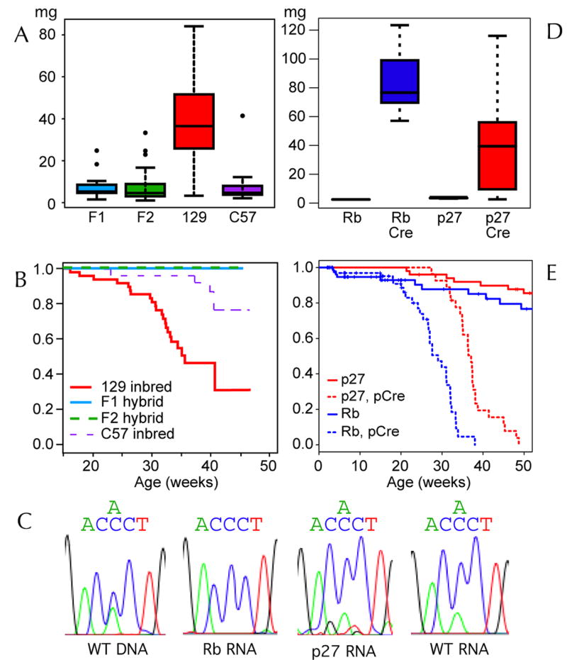
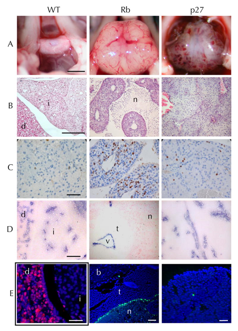
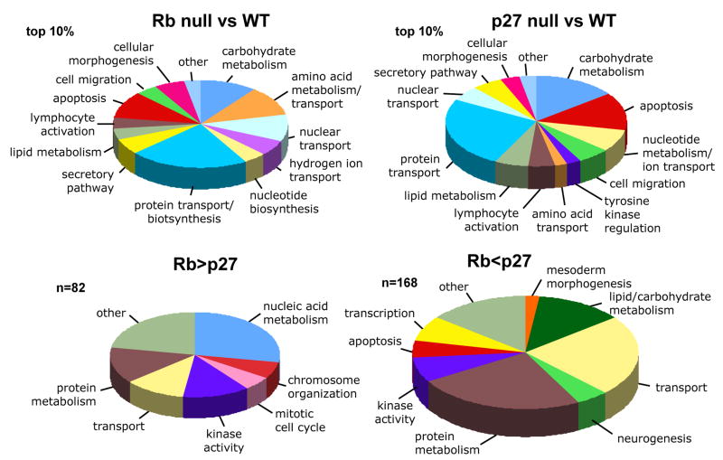
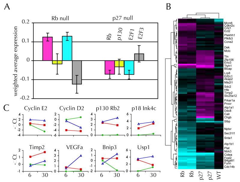
Mice lacking the p27Kip1 Cdk inhibitor, like mice lacking Rb, develop pituitary tumors involving pars intermedia melanotrophs, yet p27(Kip1) tumors are genetically distinct from Rb derived tumors as they exhibit haploid insufficiency. We compared tumors from mice with p27( Kip1) constitutive and tissue specific null mutations to tumors arising in tissue specific Rb knockout mice with the aim of determining whether they are distinguished by quantitative or qualitative differences. The rate of p27Kip1 knockout tumor development was strongly influenced by strain background due to polygenic strain modifiers in the C57BL/6J versus 129S4 strains but, unlike a prior report of Rb mutants, this impacted tumor incidence but not the tumor spectrum. p27Kip1 tumors were oligoclonal or polyclonal based on studies of X-chromosomal inactivation of Dock11. In contrast, Rb null tissue developed monoclonal neoplasms even in the absence of a requirement for Rb mutant clonal selection. Rb null tumors exhibited a higher proliferation rate and developed ischemic necrosis associated with an aberrant vasculature. p27Kip1 null tumors maintained normal vascular density, through a tumor cell dependent mechanism, but were more often hemorrhagic. Gene expression profiles distinguished p27Kip1 from Rb null tumors including significant differences in expression of Rb and E2F signature genes. Rb null tumors expressed higher levels of VEGF which, in other systems, is associated with dilated vessels, ineffective perfusion and tissue hypoxia. Mouse models lacking p27Kip1 and Rb may help us better understand the pathophysiology of MEN syndromes, retinoblastoma and other cancers that disrupt these important cell cycle inhibitors.


Figures

Figure 1
Figure 1. Pituitary tumor growth and mortality
Box and whisker plots demonstrating pituitary tumor mass (A) from p27−/− mice graphed as a function of genetic background. F1 and F2 hybrids are compared to 129S4 and C57BL/6J inbred animals. Mean values (center bar) are shown within the middle quartiles (box), the range (whiskers) and outliers (points). The fraction of surviving animals is plotted (B) as a function of strain background. Sequence plots (C) of a PCR amplified Dock11 SNP are shown using genomic DNA from F1 hybrid tissue (F1DNA), and RNA from normal pars intermedia (WT RNA), an Rb null tumor (Rb RNA) and a p27 null tumor (p27 RNA) as the starting material. Pituitary tumor mass in mice with tissue specific mutations (D) of Rb (Rb/Cre) and p27Kip1 (p27/Cre) is graphed with control animals which lack the Cre transgene. The survival (E) of Rb and p27Kip1 pituitary tissue specific animals is also plotted.
Figure 2
Figure 2. Histologic comparison of p27Kip1 vs Rb null pars intermedia tumors
Appearance of the pituitary in situ (A) on the floor of the cranium in a wildtype mouse (WT) comparison to a large RbF19/F19;POMC-Cre+ tumor (Rb) p27L+/L+;POMC-Cre+ pituitary tumor (p27) with extensive hemorrhage. Hematoxylin and eosin (B) stained normal pituitary showing the pars distalis (d), pars intermedia (i). An Rb null tumor with cords of viable tumor and ischemic necrosis (n), and a hemorrhagic p27 null pars intermedia tumor. Ki67 immunoperoxidase staining (C) indicates proliferating cells. TUNEL staining of Rb null tissue with normal brain (b) adjacent to tumor (t) and lined by a region of bright TUNEL positivity on the edge of an area of ischemic necrosis (n). MECCA32 immunostaining (D) highlights the endothelial cells and reveals the capillary density of normal pituitary compared Rb null tumor with a central vessel (v) surrounded circumferentially by viable tumor (t) and peripheral necrosis (n). Anti-p27 immunofluorescence (E left) of a p27L+/L+;POMC-Cre+ pituitary at 6 weeks demonstrates nearly complete loss of p27 expression (red) from the pars intermedia (i) compared to the adjacent pars distalis (d). TUNEL staining (E mid, right) is shown for a p27 null and Rb null tumor (green) showing brain (b), tumor (t) and necrotic (n) areas (blue, DAPI). Scale bars (A 2mm, B 0.5mm, C-E 0.1mm)
Figure 3
Figure 3. Gene ontologies of p27Kip1 and Rb null pituitary tumors
Pie charts summarizing functional annotation clustering results (based on GO biological process or molecular function level 5 annotations) of the top 10% of all genes ranked by differential expression of (A) p27−/− tumors vs normal wildtype pars intermedia, or (B) Rb null tumor vs. normal. The size of each segment represents the numbers of genes in each group and functional groups are ordered clockwise (from the top) by their statistical significance score. In contrast the genes (C) which are significantly increased in Rb null vs p27−/− tumors (n=82) are given in comparison to (D) the GO classification (BP or MF level 4–5) of genes increased in p27−/− tumors relative to Rb null tumors (n=168). For brevity only one or two descripitive terms for each functional cluster is listed.
Figure 4
Figure 4. Rb and p27Kip1 null pars intermedia tumor expression profiles
Average microarray expression levels of the subset of genes (A) previously described as either Rb, p130/p107, E2F1, or E2F3 signature genes are given for Rb null and p27Kip1 null tumors. Change in expression compared to wildtype for each gene were multiplied by the statistical weights (provided by Black EP et al. 2003, 2005) so the results are positive if they correlate with the reported signature regardless of whether the gene’s expression is increased or decreased. A heat map (B) showing differential expression of 100 genes with the most significant differences in expression in p27−/− vs. Rb null tumor tissue. Average values of replicate arrays are shown from 2 Rb and 2 p27Kip1 null tumors in comparison to arrays with negative control wildtype (WT) tissue. Locations of 41 selected genes are given after heirarchial clustering. QPCR results (C) are shown for 8 genes showing differential expression in Rb null (open triangle), p27Kip1 null (red square) and wildtype (green circle) pars intermedia tissue at 6 weeks of age and in tumors or 30 week old wildtype tissue. Expression values are given in terms of -∆Ct (change in PCR cycle number) relative to wildtype tissue at 6 weeks of age.

Similar articles

Cited by

References

    1. Sherr CJ, McCormick F. The RB and p53 pathways in cancer. Cancer Cell. 2002;2:103–12. - PubMed
    1. Williams BO, Schmitt EM, Remington L, Bronson RT, Albert DM, Weinberg RA, Jacks T. Extensive contribution of Rb-deficient cells to adult chimeric mice with limited histopathological consequences. Embo J. 1994;13:4251–9. - PMC - PubMed
    1. Hu N, Gutsmann A, Herbert DC, Bradley A, Lee WH, Lee EY. Heterozygous Rb-1 delta 20/+mice are predisposed to tumors of the pituitary gland with a nearly complete penetrance. Oncogene. 1994;9:1021–7. - PubMed
    1. Kiyokawa H, Kineman RD, Manova-Todorova KO, Soares VC, Hoffman ES, Ono M, Khanam D, Hayday AC, Frohman LA, Koff A. Enhanced growth of mice lacking the cyclin-dependent kinase inhibitor function of p27(Kip1) Cell. 1996;85:721–32. - PubMed
    1. Nakayama K, Ishida N, Shirane M, Inomata A, Inoue T, Shishido N, Horii I, Loh DY. Mice lacking p27(Kip1) display increased body size, multiple organ hyperplasia, retinal dysplasia, and pituitary tumors. Cell. 1996;85:707–20. - PubMed

Publication types

MeSH terms

Substances