. 2007 Apr 11;129(14):4386-92.
doi: 10.1021/ja068739h. Epub 2007 Mar 16.

Structure of an unprecedented G-quadruplex scaffold in the human c-kit promoter

Affiliations

Structure of an unprecedented G-quadruplex scaffold in the human c-kit promoter

Anh Tuân Phan et al. J Am Chem Soc. .

Abstract

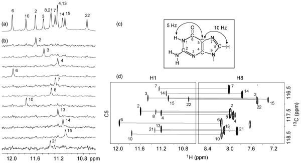
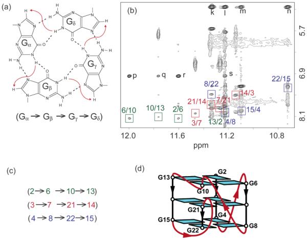
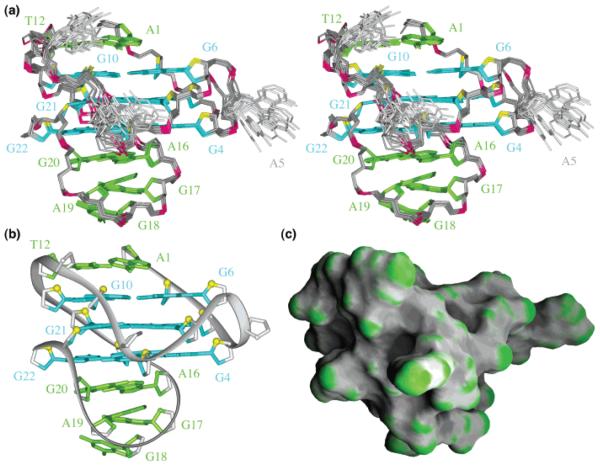
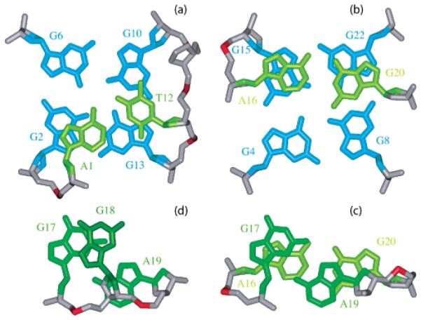
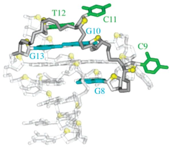
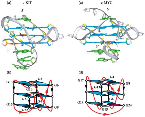
The c-kit oncogene is an important target in the treatment of gastrointestinal tumors. A potential approach to inhibition of the expression of this gene involves selective stabilization of G-quadruplex structures that may be induced to form in the c-kit promoter region. Here we report on the structure of an unprecedented intramolecular G-quadruplex formed by a G-rich sequence in the c-kit promoter in K+ solution. The structure represents a new folding topology with several unique features. Most strikingly, an isolated guanine is involved in G-tetrad core formation, despite the presence of four three-guanine tracts. There are four loops: two single-residue double-chain-reversal loops, a two-residue loop, and a five-residue stem-loop, which contain base-pairing alignments. This unique structural scaffold provides a highly specific platform for the future design of ligands specifically targeted to the promoter DNA of c-kit.


Figures

Figure 1
Figure 1
Spectra and assignments of c-kit87up. (a) The 600 MHz imino proton spectrum of the c-kit87up sequence in K+ solution at 25 °C. Unambiguous assignments are indicated. (b) Imino protons were assigned in 15N-filtered spectra of samples, 2% 15N-labeled at the indicated positions. (c) Long-range J couplings in a guanine used for imino-H8 correlation. (d) H8 proton assignments of c-kit87up by through-bond correlations between imino and H8 protons via 13C5 at natural abundance, using long-range J couplings shown in (c).
Figure 2
Figure 2
G-tetrad alignments and folding topology of c-kit87up. (a) Schematic of a G-tetrad with characteristic guanine imino-H8 connectivity patterns indicated by arrows. (b) NOESY spectrum (mixing time, 200 ms). The characteristic guanine imino-H8 cross-peaks for G-tetrads are framed and labeled with the imino proton assignment in the first position and that of the H8 proton in the second position. Peaks k, l, m, and n show close distances between G20(NH2) and the imino protons of guanines from the bottom tetrad; peaks p, q, r, and s show close distances between A1(H2) and the imino protons of guanines from the top tetrad. (c) Summary of characteristic guanine imino-H8 patterns observed for c-kit87up. (d) Topology of c-kit87up based on the G-tetrad alignments in (c).
Figure 3
Figure 3
Solution structure of the c-kit87up quadruplex in K+ solution. (a) Stereoview of 11 superpositioned refined structures. Guanine bases in the G-tetrad core are colored cyan; other bases, green; the backbone, gray; phosphorus atoms, red; O4′atoms, yellow. (b) Ribbon and (c) surface views of a representative refined structure.
Figure 4
Figure 4
Base pairs in loops. Stacking (a) between the Watson-Crick A1•T12 pair and the G2•G6•G10•G13 tetrad, (b) between the Watson-Crick A16•G20 pair and the G4•G8•G22•G15 tetrad, (c) between the Watson-Crick A16•G20 pair and the sheared G17•A19 pair, and (d) between the sheared G17•A19 pair and the single-base G18.
Figure 5
Figure 5
Highlight of the C9-G10-C11-T12 fragment within the c-kit87up quaduplex.
Figure 6
Figure 6
The 600 MHz imino proton spectra of the modified c-kit87up sequences in K+ solution at 25 °C.
Figure 7
Figure 7
Comparison between G-quadruplex structures observed in (a, b) c-kit (this work) and (c, d) c-myc promoters. In (a) and (c): color coded as in Figure 3, except that snapback residues are colored orange; the blue arrows indicate break points within interrupted G-tetrad cores. In (b) and (d): loops are colored red; anti and syn guanines are colored cyan and magenta, respectively.

References

    1. Yarden Y, Kuang WJ, Yang-Feng T, Coussens L, Munemitsu S, Dull TJ, Chen E, Schlessinger J, Francke U, Ullrich A. EMBO J. 1987;6:3341–3351. - PMC - PubMed
    1. Roskoski R., Jr. Biochem. Biophys. Res. Commun. 2005;337:1–13. - PubMed
    1. Taniguchi M, Nishida T, Hirota S, Isozaki K, Ito T, Nomura T, Matsuda H, Kitamura Y. Cancer Res. 1999;59:4297–4300. - PubMed
    1. Kitamura Y, Hirota S, Nishida T. Mutation Res. 2001;477:165–171. - PubMed
    1. Tian Q, Frierson HF, Jr., Krystal GW, Moskaluk CA. Amer. J. Pathol. 1999;154:1643–1647. - PMC - PubMed

Publication types