

Freshly isolated Valpha24+ CD4+ invariant natural killer T cells activated by alpha-galactosylceramide-pulsed B cells promote both IgG and IgE production
- PMID: 17362268
- PMCID: PMC1941929
- DOI: 10.1111/j.1365-2249.2007.03364.x
Freshly isolated Valpha24+ CD4+ invariant natural killer T cells activated by alpha-galactosylceramide-pulsed B cells promote both IgG and IgE production
Abstract
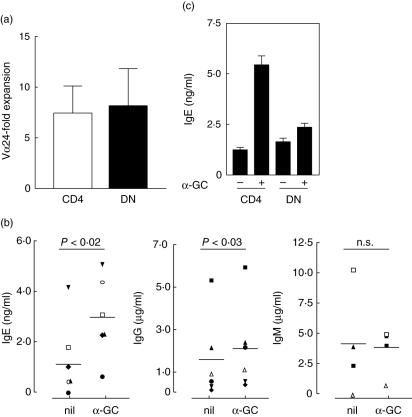
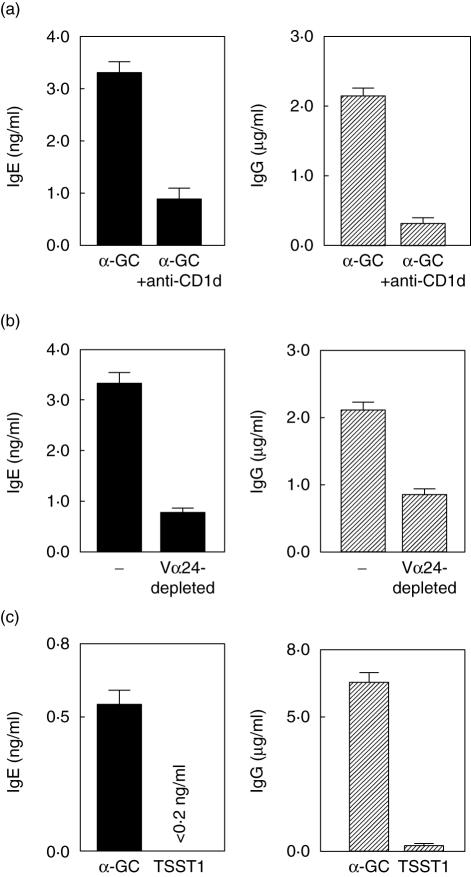
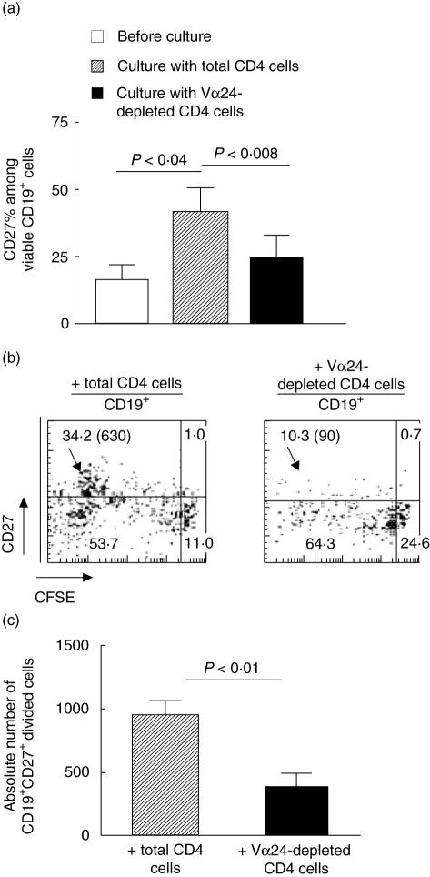
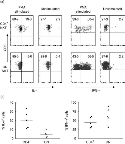
CD1d-restricted invariant natural killer T (iNK T) cells activated by their experimental ligand alpha-galactosylceramide (alpha-GC) can produce both T helper 1 (Th1) and Th2 cytokines and display regulatory functions. Recent studies identified CD4(+) and CD4(-) CD8(-) double-negative (DN) iNK T cells as the two major components of the human population and suggest that they display a Th2 and a Th1 profile, respectively. We compared the Th2-promoting activity of freshly isolated human CD4(+) and DN iNK T cells in terms of their capacity to induce Ig production by autologous B cells. Secretion of IgG and IgE but not IgM was enhanced by the CD4(+) T cell subset (including iNK T cells) but not by its DN counterpart. iNK T cells were directly responsible for this pro-Th2 effect, as demonstrated by the requirement for both alpha-GC stimulation and CD1d presentation, as well as by its disappearance upon iNK T cell depletion. Interaction with iNK T cells led to progressive accumulation of isotype-switched and activated B cells. Myeloid dendritic cells (DC) completely block the induction of Ig production in co-culture. This dominant inhibitory effect of myeloid DC was concomitant with a specific loss of interleukin (IL)-4 production by CD4(+) iNK T but not by conventional T cells. These data support the conclusion that, conversely to the interferon (IFN)-gamma-producing DN human iNK T cell population, interleukin (IL)-4-producing CD4(+) iNK T cells can activate and help B cells to produce both IgG and IgE through a CD1d-dependent mechanism, in keeping with a functional Th1/Th2 dichotomy between these subsets.
Figures

Similar articles
-
Human dendritic cells transfected with allergen-DNA stimulate specific immunoglobulin G4 but not specific immunoglobulin E production of autologous B cells from atopic individuals in vitro.Immunology. 2007 Oct;122(2):239-46. doi: 10.1111/j.1365-2567.2007.02633.x. Immunology. 2007. PMID: 17848164 Free PMC article.
-
IL-2 and a contact-mediated signal provided by TCR alpha beta + or TCR gamma delta + CD4+ T cells induce polyclonal Ig production by committed human B cells. Enhancement by IL-5, specific inhibition of IgA synthesis by IL-4.J Immunol. 1992 Mar 15;148(6):1674-84. J Immunol. 1992. PMID: 1347306
-
Nonredundant roles for CD1d-restricted natural killer T cells and conventional CD4+ T cells in the induction of immunoglobulin E antibodies in response to interleukin 18 treatment of mice.J Exp Med. 2003 Apr 21;197(8):997-1005. doi: 10.1084/jem.20021701. Epub 2003 Apr 14. J Exp Med. 2003. PMID: 12695491 Free PMC article.
-
Role of alpha-galactosylceramide-activated Valpha14 natural killer T cells in the regulation of allergic diseases.Allergol Int. 2007 Mar;56(1):1-6. doi: 10.2332/allergolint.R-06-136. Epub 2007 Jan 29. Allergol Int. 2007. PMID: 17259803 Review.
-
Invariant natural killer T cell-based immunotherapy for cancer.Immunotherapy. 2009 Jan;1(1):73-82. doi: 10.2217/1750743X.1.1.73. Immunotherapy. 2009. PMID: 20635975 Review.
Cited by
-
Encapsulation of MERS antigen into α-GalCer-bearing-liposomes elicits stronger effector and memory immune responses in immunocompetent and leukopenic mice.J King Saud Univ Sci. 2022 Jul;34(5):102124. doi: 10.1016/j.jksus.2022.102124. Epub 2022 May 27. J King Saud Univ Sci. 2022. PMID: 35663348 Free PMC article.
-
Invariant natural killer T cells in lupus patients promote IgG and IgG autoantibody production.Eur J Immunol. 2015 Feb;45(2):612-23. doi: 10.1002/eji.201444760. Epub 2014 Nov 27. Eur J Immunol. 2015. PMID: 25352488 Free PMC article.
-
Can invariant Natural Killer T cells drive B cell fate? a look at the humoral response.Front Immunol. 2025 Feb 18;16:1505883. doi: 10.3389/fimmu.2025.1505883. eCollection 2025. Front Immunol. 2025. PMID: 40040714 Free PMC article. Review.
-
Human invariant NKT cell subsets differentially promote differentiation, antibody production, and T cell stimulation by B cells in vitro.J Immunol. 2013 Aug 15;191(4):1666-76. doi: 10.4049/jimmunol.1202223. Epub 2013 Jul 12. J Immunol. 2013. PMID: 23851681 Free PMC article.
-
Activation and Regulation of B Cell Responses by Invariant Natural Killer T Cells.Front Immunol. 2018 Jun 18;9:1360. doi: 10.3389/fimmu.2018.01360. eCollection 2018. Front Immunol. 2018. PMID: 29967611 Free PMC article. Review.
References
-
- Godfrey DI, Hammond KJL, Poulton LD, Smyth MJ, Baxter AG. NKT cells: facts, functions and fallacies. Immunol Today. 2000;21:573–83. - PubMed
-
- Kronenberg M, Gapin L. The unconventional lifestyle of NKT cells. Nat Rev Immunol. 2002;2:557–68. - PubMed
-
- MacDonald HR. Development and selection of NKT cells. Curr Opin Immunol. 2002;14:250–4. - PubMed
Publication types
MeSH terms
Substances
LinkOut - more resources
Full Text Sources
Research Materials
Miscellaneous
