

Expression of S100B during embryonic development of the mouse cerebellum
- PMID: 17362503
- PMCID: PMC1832187
- DOI: 10.1186/1471-213X-7-17
Expression of S100B during embryonic development of the mouse cerebellum
Abstract
Background: In the cerebellum of newborn S100B-EGFP mice, we had previously noted the presence of a large population of S100B-expressing cells, which we assumed to be immature Bergmann glial cells. In the present study, we have drawn on this observation to establish the precise spatio-temporal pattern of S100B gene expression in the embryonic cerebellum.
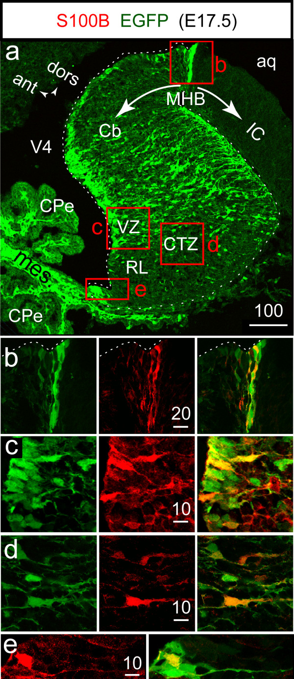
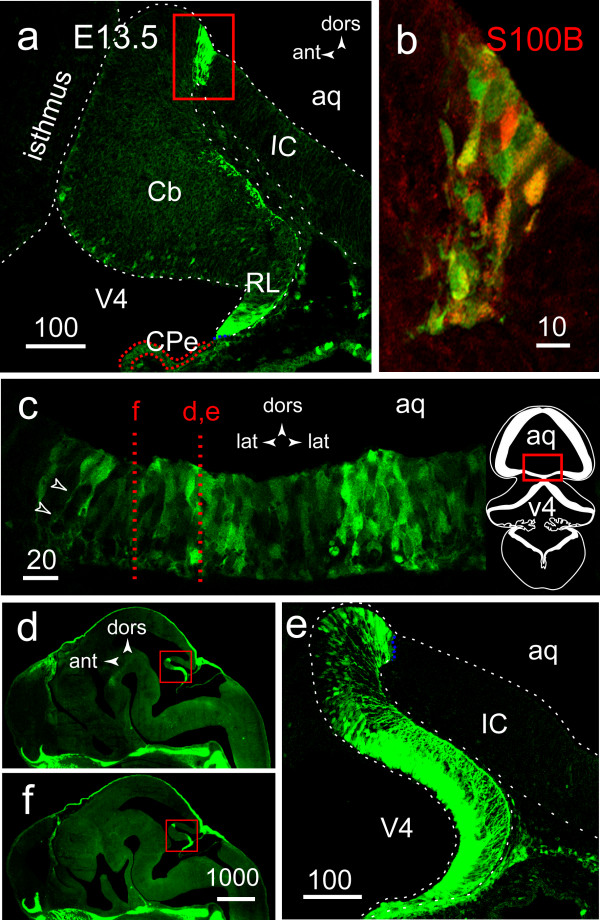
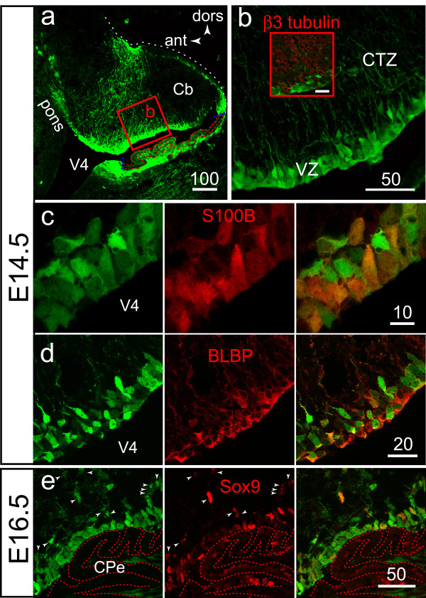
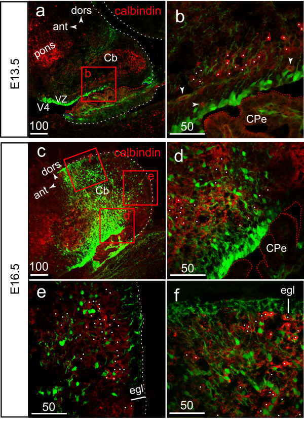
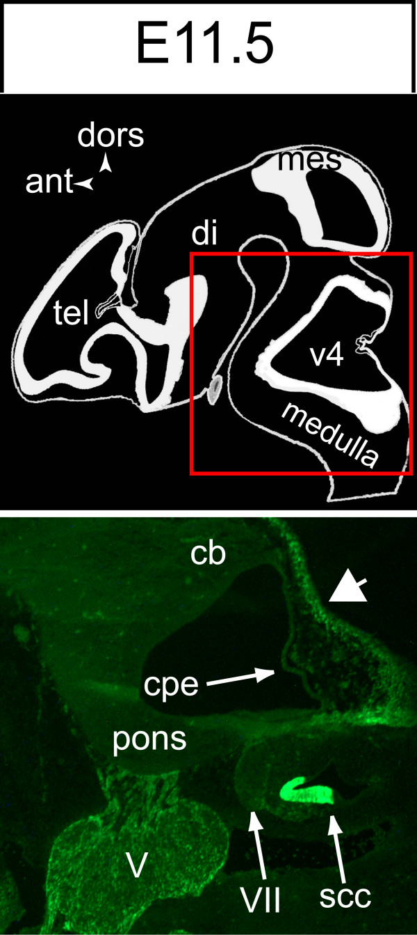
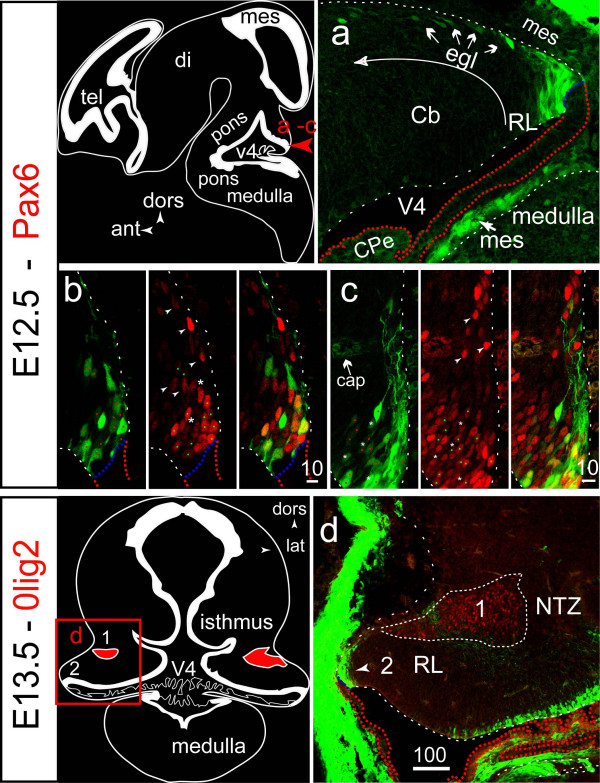
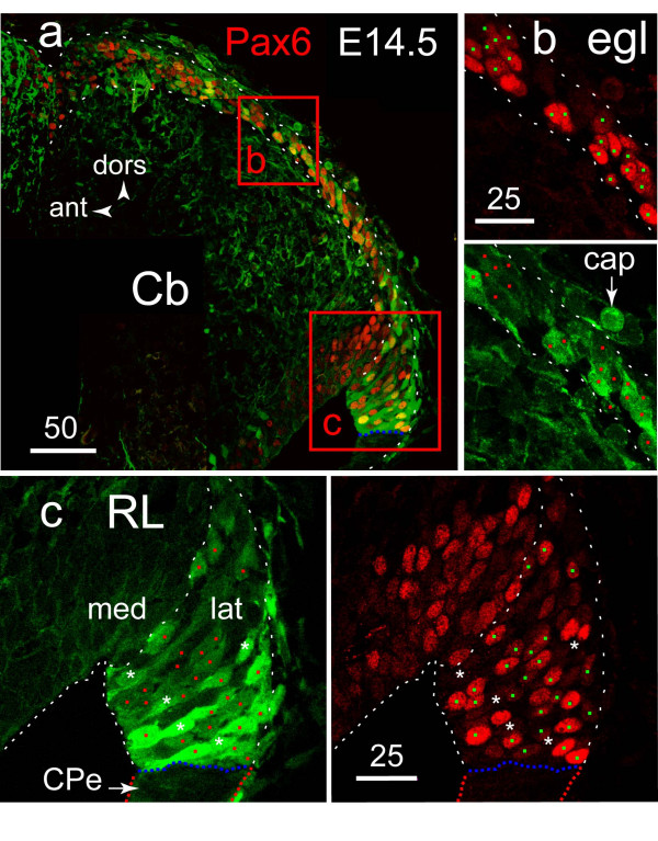
Results: From E12.5 until E17.5, S100B was expressed in the primary radial glial scaffold involved in Purkinje progenitor exit from the ventricular zone and in the Sox9+ glial progenitors derived from it. During the same period coinciding with the primary phase of granule neuron precursor genesis, transient EGFP expression tagged the Pax6+ forerunners of granule precursors born in the cerebellar rhombic lip.
Conclusion: This study provides the first characterization of S100B-expressing cell types of the embryonic mouse cerebellum in a high-resolution map. The transient activation of the S100B gene distinguishes granule neuron precursors from all other types of precursors so far identified in the rhombic lip, whereas its activation in radial glial precursors is a feature of Bergmann cell gliogenesis.
Figures
Similar articles
-
Visualization of S100B-positive neurons and glia in the central nervous system of EGFP transgenic mice.J Comp Neurol. 2003 Mar 17;457(4):404-19. doi: 10.1002/cne.10552. J Comp Neurol. 2003. PMID: 12561079
-
Enhanced calcium transients in glial cells in neonatal cerebellar cultures derived from S100B null mice.Exp Cell Res. 2000 Jun 15;257(2):281-9. doi: 10.1006/excr.2000.4902. Exp Cell Res. 2000. PMID: 10837142
-
Close homologue of adhesion molecule L1 promotes survival of Purkinje and granule cells and granule cell migration during murine cerebellar development.J Comp Neurol. 2009 Apr 10;513(5):496-510. doi: 10.1002/cne.21981. J Comp Neurol. 2009. PMID: 19226508
-
Development and developmental disorders of the human cerebellum.J Neurol. 2003 Sep;250(9):1025-36. doi: 10.1007/s00415-003-0199-9. J Neurol. 2003. PMID: 14504962 Review.
-
[Development expression of voltage-gated potassium channels in mouse cerebellar granule cells].Tanpakushitsu Kakusan Koso. 2000 Feb;45(3 Suppl):248-54. Tanpakushitsu Kakusan Koso. 2000. PMID: 10707626 Review. Japanese. No abstract available.
Cited by
-
Restoring microglial and astroglial homeostasis using DNA immunization in a Down Syndrome mouse model.Brain Behav Immun. 2019 Jan;75:163-180. doi: 10.1016/j.bbi.2018.10.004. Epub 2018 Oct 25. Brain Behav Immun. 2019. PMID: 30389461 Free PMC article.
-
Targeting S100B in Cerebral Ischemia and in Alzheimer's Disease.Cardiovasc Psychiatry Neurol. 2010;2010:687067. doi: 10.1155/2010/687067. Epub 2010 Sep 2. Cardiovasc Psychiatry Neurol. 2010. PMID: 20862385 Free PMC article.
-
Glial-neuronal interactions--implications for plasticity and drug addiction.AAPS J. 2009 Mar;11(1):123-32. doi: 10.1208/s12248-009-9085-4. Epub 2009 Feb 24. AAPS J. 2009. PMID: 19238557 Free PMC article. Review.
-
Differential temporal expression of S100β in developing rat brain.Front Cell Neurosci. 2015 Mar 18;9:87. doi: 10.3389/fncel.2015.00087. eCollection 2015. Front Cell Neurosci. 2015. Retraction in: Front Cell Neurosci. 2023 Oct 05;17:1289247. doi: 10.3389/fncel.2023.1289247. PMID: 25852479 Free PMC article. Retracted.
-
Purkinje Neurons with Loss of STIM1 Exhibit Age-Dependent Changes in Gene Expression and Synaptic Components.J Neurosci. 2021 Apr 28;41(17):3777-3798. doi: 10.1523/JNEUROSCI.2401-20.2021. Epub 2021 Mar 18. J Neurosci. 2021. PMID: 33737457 Free PMC article.
References
-
- Joyner AL, Zervas M. Genetic inducible fate mapping in mouse: Establishing genetic lineages and defining genetic neuroanatomy in the nervous system. Dev Dyn. 2006 - PubMed
-
- Yuasa S. Bergmann glial development in the mouse cerebellum as revealed by tenascin expression. Anat Embryol (Berl) 1996;194:223–234. - PubMed
MeSH terms
Substances
LinkOut - more resources
Full Text Sources
Molecular Biology Databases
Research Materials
Miscellaneous
