

Culture of murine aortic explants in 3-dimensional extracellular matrix: a novel, miniaturized assay of angiogenesis in vitro
- PMID: 17363012
- PMCID: PMC1952682
- DOI: 10.1016/j.mvr.2007.02.002
Culture of murine aortic explants in 3-dimensional extracellular matrix: a novel, miniaturized assay of angiogenesis in vitro
Abstract
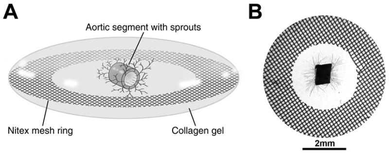
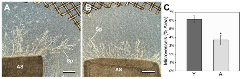
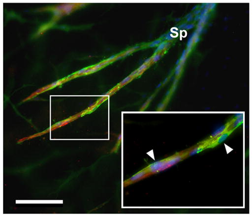
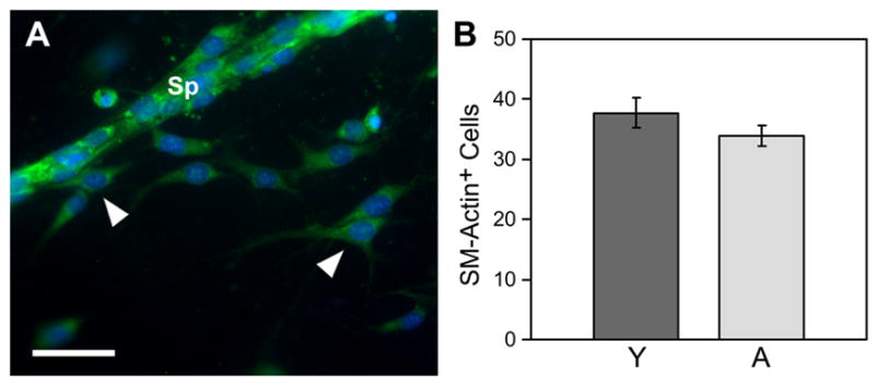
Assays of angiogenesis in vitro are critical to the study of vascular morphogenesis and to the evaluation of therapeutic compounds that promote or inhibit vascular growth. Culture of explanted aortic segments from rats or mice in a 3-dimensional extracellular matrix (ECM) is one of the most effective ways to generate capillary-like endothelial sprouts in vitro. We have modified the classic aortic explant model by placing the aortic segments from mice within small (5.6 mm diameter, 30 microl volume) lenticular hydrogels of type I collagen supported at the edge by nylon mesh rings. This method of culture, referred to as the "miniature ring-supported gel" (MRSG) assay, optimizes handling, cytological staining, and conventional imaging of the specimen and permits use of minimal volumes of reagents in a 96-well tissue culture format. We have used the MRSG assay to quantify the impaired angiogenic response of aged mice relative to young mice and to show that aged mice have significantly decreased sprout formation, but have similar levels of invasion of vascular smooth muscle cells into the supportive ECM. The MRSG assay, which combines low volume, physically robust gels in conjunction with mouse aortic segments, may prove to be a highly useful tool in studies of the process and control of vascular growth.
Figures
Similar articles
-
Miniaturized assays of angiogenesis in vitro.Methods Mol Biol. 2012;843:87-98. doi: 10.1007/978-1-61779-523-7_9. Methods Mol Biol. 2012. PMID: 22222524 Free PMC article.
-
Direct comparison of angiogenesis in natural and synthetic biomaterials reveals that matrix porosity regulates endothelial cell invasion speed and sprout diameter.Acta Biomater. 2021 Nov;135:260-273. doi: 10.1016/j.actbio.2021.08.038. Epub 2021 Aug 29. Acta Biomater. 2021. PMID: 34469789 Free PMC article.
-
Angiogenesis In Vitro Utilizing Murine Vascular Explants in Miniaturized 3-Dimensional Collagen Gels.Open Circ Vasc J. 2011 May 9;4:12-17. doi: 10.2174/1877382601104010012. Open Circ Vasc J. 2011. PMID: 24701258 Free PMC article.
-
A novel, quantitative model for study of endothelial cell migration and sprout formation within three-dimensional collagen matrices.Microvasc Res. 1999 Mar;57(2):118-33. doi: 10.1006/mvre.1998.2122. Microvasc Res. 1999. PMID: 10049660
-
Paracrine regulation of angiogenesis by different cell types in the aorta ring model.Int J Dev Biol. 2011;55(4-5):447-53. doi: 10.1387/ijdb.103222rn. Int J Dev Biol. 2011. PMID: 21858770 Review.
Cited by
-
Endothelial progenitor cells: from senescence to rejuvenation.Semin Nephrol. 2014 Jul;34(4):365-73. doi: 10.1016/j.semnephrol.2014.06.003. Epub 2014 Jun 13. Semin Nephrol. 2014. PMID: 25217265 Free PMC article. Review.
-
Defective angiogenesis, endothelial migration, proliferation, and MAPK signaling in Rap1b-deficient mice.Blood. 2008 Mar 1;111(5):2647-56. doi: 10.1182/blood-2007-08-109710. Epub 2007 Nov 9. Blood. 2008. PMID: 17993608 Free PMC article.
-
Manipulating the microvasculature and its microenvironment.Crit Rev Biomed Eng. 2013;41(2):91-123. doi: 10.1615/critrevbiomedeng.2013008077. Crit Rev Biomed Eng. 2013. PMID: 24580565 Free PMC article. Review.
-
Sirtuin 1 ablation in endothelial cells is associated with impaired angiogenesis and diastolic dysfunction.Am J Physiol Heart Circ Physiol. 2014 Dec 15;307(12):H1691-704. doi: 10.1152/ajpheart.00281.2014. Epub 2014 Sep 19. Am J Physiol Heart Circ Physiol. 2014. PMID: 25239805 Free PMC article.
-
Endothelium-specific deletion of Nox4 delays retinal vascular development and mitigates pathological angiogenesis.Angiogenesis. 2021 May;24(2):363-377. doi: 10.1007/s10456-020-09757-3. Epub 2020 Nov 17. Angiogenesis. 2021. PMID: 33201372 Free PMC article.
References
-
- Abramoff MD. Image Processing with ImageJ. Biophotonics International. 2004;11(7):36–42.
-
- Agah A, Kyriakides TR, Letrondo N, Bjorkblom B, Bornstein P. Thrombospondin 2 levels are increased in aged mice: consequences for cutaneous wound healing and angiogenesis. Matrix Biol. 2004;22(7):539–47. - PubMed
-
- Arthur WT, Vernon RB, Sage EH, Reed MJ. Growth factors reverse the impaired sprouting of microvessels from aged mice. Microvasc Res. 1998;55(3):260–70. - PubMed
-
- Barandier C, Montani JP, Yang Z. Mature adipocytes and perivascular adipose tissue stimulate vascular smooth muscle cell proliferation: effects of aging and obesity. Am J Physiol Heart Circ Physiol. 2005;289(5):H1807–13. - PubMed
-
- Davis GE, Camarillo CW. An alpha 2 beta 1 integrin-dependent pinocytic mechanism involving intracellular vacuole formation and coalescence regulates capillary lumen and tube formation in three-dimensional collagen matrix. Exp Cell Res. 1996;224(1):39–51. - PubMed
Publication types
MeSH terms
Substances
Grants and funding
LinkOut - more resources
Full Text Sources
Other Literature Sources
Medical
