. 2007 Jun 1;581(Pt 2):479-93.
doi: 10.1113/jphysiol.2006.123414. Epub 2007 Mar 15.

Modulation of hERG potassium currents in HEK-293 cells by protein kinase C. Evidence for direct phosphorylation of pore forming subunits

Affiliations

Modulation of hERG potassium currents in HEK-293 cells by protein kinase C. Evidence for direct phosphorylation of pore forming subunits

S L Cockerill et al. J Physiol. .

Abstract

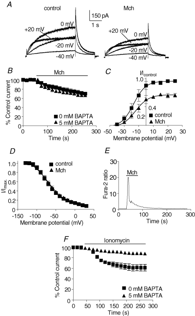
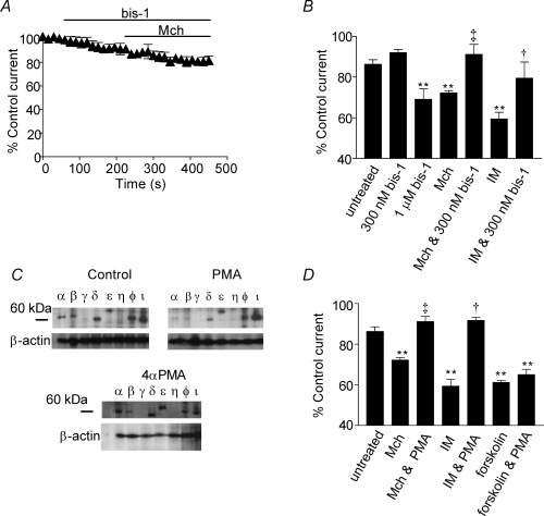
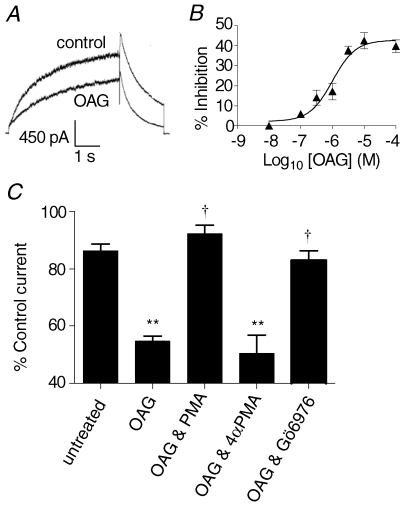
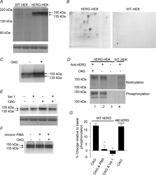
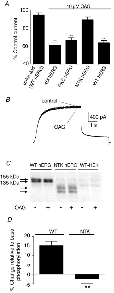
The human ether-a-go-go related gene (hERG) potassium channel is expressed in a variety of tissues including the heart, neurons and some cancer cells. hERG channels are modulated by several intracellular signalling pathways and these provide important mechanisms for regulating cellular excitability. In this study, we investigated muscarinic modulation of hERG currents and direct phosphorylation of channel subunits expressed in HEK-293 cells at physiologically relevant temperatures by protein kinase C (PKC). Activation of G(alpha q/11)-coupled M(3)-muscarinic receptors with methacholine, reduced current amplitudes at all potentials with minor effects on the voltage dependence of activation and inactivation. The response to methacholine was insensitive to intracellular BAPTA, but was attenuated by either acute inhibition of PKC with 300 nm bisindolylmaleimide-1 (bis-1) or chronic down-regulation of PKC isoforms by 24 h pretreatment of cells with phorbol 12-myristate 13-acetate (PMA). Stimulation of PKC with 1-oleoyl 2-acetylglycerol (OAG), an analogue of diacylglycerol (DAG), mimicked the actions of muscarinic receptor stimulation. Direct phosphorylation of hERG was measured by [(32)P]orthophosphate labelling of immunoprecipitated protein with an anti-hERG antibody. Basal phosphorylation was high in unstimulated cells and further increased by OAG. The OAG dependent increase was abolished by bis-1 and down-regulation of PKC, but basal levels of phosphorylation were unchanged. Deletion of the amino-terminus of hERG prevented both the modulation of channel activity and the increase of phosphorylation by OAG. Our results are consistent with calcium and/or DAG sensitive isotypes of PKC modulating hERG currents through a mechanism that involves direct phosphorylation of sites on the amino terminus of hERG.


Figures

Figure 1
Figure 1. Suppression of hERG currents by M3 muscarinic receptor stimulation
hERG channels were transiently expressed in HEK-293 cells stably expressing M3 muscarinic receptors. A, representative currents elicited with 5 s depolarizations to the indicated test potentials before (control) and during application of a maximal concentration (1 mm) of the agonist methacholine (Mch). Tail currents were obtained with repolarization to −50 mV. B, time course of response to methacholine in cells dialysed for 10 min via the patch pipette with intracellular solutions containing 0 or 5 mm BAPTA. Cells were repetitively depolarized for 5 s to 0 mV and peak tail currents measured upon repolarization to −50 mV. Values were normalized to peak tail currents before methacholine application and mean values plotted as a function of time. C, activation curves before and during application of methacholine. The V0.5,act before and after muscarinic receptor stimulation was −17 ± 2 mV and −14 ± 2 mV, respectively (n = 5). Slope factors were 8 ± 2 mV and 9 ± 2 mV before and during methacholine application, respectively. D, muscarinic receptor stimulation had no significant effects on voltage-dependent inactivation. E, representative intracellular Ca2+ response to methacholine assessed by fura-2 imaging of a single cell. The time course of the Ca2+ response is notably different from the hERG current response in B and shows the characteristic initial transient rise in Ca2+ followed by the more slowly declining component of the response. F, comparison of time-course effects of ionomycin in cells dialysed for 10 min with 0 or 5 mm BAPTA. Values were normalized to peak tail currents before ionomycin application and mean values plotted as a function of time.
Figure 2
Figure 2. hERG current modulation by methcholine and ionomycin can be attenuated by inhibition or down-regulation of PKC
A, mean hERG current in response to methacholine (Mch) in HEK-M3 cells following inhibition of PKC with 300 nm bis-1. The protocol and tail current analysis are the same as described in Fig. 1B. Bis-1 was applied for 3 min before co-application with methacholine. B, hERG current modulation is attenuated by 300 nm bis-1. Mean percentage values for tail current remaining in control conditions after 5 min (untreated), and upon application of the following: 300 nm bis-1 for 3 min; 1 μm bis-1 for 3 min; methacholine (Mch) for 2 min; 3 min perfusion with bis-1 followed by co-application of bis-1 with Mch for 2 min; 5 μm ionomycin (IM) for 2 min; 3 min perfusion with bis-1 followed by co-application of IM for 2 min. C, representative Western blots showing PKC isotype expression in hERG-HEK cells treated with 0.1% DMSO (control), 1 μm PMA, or 1 μm 4αPMA for 24 h. Control and PMA blots were performed in pairs to enable direct comparisons to be made. 4αPMA blots were performed separately. Similar results were observed in 4 other experiments. The lower panels show the blots after being stripped and re-probed for β-actin to show protein loading across all lanes. D, down-regulation of PKC isotypes abolishes hERG current modulation by Mch and IM. hERG current modulation in untreated cells and cells incubated in 1 μm PMA for 24 h (PMA). Tail current amplitudes were measured after > 2 min application of Mch, IM (5 μm) or forskolin (40 μm) and expressed as a percentage of current before test compound application. Experiments with Mch activation of muscarinic receptors were performed in HEK-M3 cells transiently expressing hERG and all other experiments were performed on hERG-HEK cells. **Significantly different from untreated (P < 0.01); ‡significantly different from Mch-only response (P < 0.01); †significantly different from IM-only response (P < 0.05). Note, for clarity not all significant differences are presented.
Figure 3
Figure 3. Activation of PKC mimics modulation of hERG currents by M3-muscarinic receptor stimulation
A, representative currents at a test potential of 0 mV from hERG-HEK cells in control conditions and during application of 10 μm OAG. B, OAG-dependent inhibition of hERG tail current was concentration dependent. The mean results were fitted with a Hill function with a pIC50 of 5.9 ± 0.1 (∼1 μm). C, summary of mean results for OAG effects on hERG tail current amplitudes. hERG current suppression by 10 μm OAG could be significantly reduced by 100 nm Gö6976 (P < 0.01 compared to OAG, n = 5), a selective antagonist of α and β1 PKC isotyes, and by 24 h incubation with 1 μm PMA (OAG & PMA), but not 4αPMA (OAG & 4αPMA). **Significantly different from untreated (P < 0.01); †significantly different from OAG (P < 0.01). Not all comparisons are shown.
Figure 4
Figure 4. Phosphorylation of hERG is increased by activation of PKC
Phospho-proteins were immunoprecipitated from WT-HEK cells and hERG-HEK cells using anti-hERG antibody. A, upper panel, representative autoradiograph showing phospho-proteins were only pulled down from hERG-expressing cells. Samples were run in duplicate to demonstrate reproducibility of results. The molecular masses of the two bands correspond with the immature (135 kDa) and mature (155 kDa) forms of hERG. Both hERG proteins are clearly phosphorylated in untreated cells. The lower panel shows Coomassie blue staining of the gel before drying and demonstrates there is equal antibody loading and immunoprecipitation across lanes. B, representative phospho-peptide maps following tryptic digest and separation by 2D electrophoresis of phospho-proteins immunoprecipitated from untreated hERG-HEK cells and WT-HEK cells. The hERG-HEK cell plate was exposed for 10 days and the WT HEK cell plate for longer (14 days). Phospho-peptides can only be detected from hERG expressing cells. C, representative autoradiograph showing the increase of phosphorylation in cells treated with 10 μm OAG for 5 min. D, quantification of biotinylation and phosphorylation of immunoprecipitated proteins. Protein samples transferred to nitrocellulose were first probed with streptavidin (upper panel), and subsequently exposed to autoradiography film for phospho-protein detection (lower panel). The streptavidin labelling was equivalent in OAG treated cells (lane 1) and untreated cells (lane 2), indicating equal amounts of immunoprecipitated hERG in each lane. Comparison of hERG phosphorylation levels in lanes 1 and 2 indicate there was a 17% increase over basal levels with OAG stimulation. No phospho-proteins or streptavidin labelled proteins were detected if anti-hERG serum was not added to the lysates (lane 3). Faint streptavidin-labelled bands were detected in some WT-HEK lysates (lane 4), but the proteins were not phosphorylated and were present in only small quantities. Similar results were seen in two other experiments. E, bis-1 inhibits OAG-dependent increase of hERG subunit phosphorylation. Bis-1 at 3 μm did not alter basal phosphorylation, but blocked the increase of phosphorylation by OAG. Lower panels, Coomassie blue staining of gel indicating equal immunoprecipitation across lanes. F, incubation of cells with 1 μm PMA for 24 h to down-regulate PKC isotypes also prevented the increase in phosphorylation by OAG. There was also no change in phosphorylation in untreated versus PMA-treated cells. G, mean change of phosphorylation for WT hERG and 4M-hERG (a functional channel lacking PKA phosphorylation). The percentage change of phosphorylation relative to basal levels was measured in untreated cells. Chronic PMA preincubation and bis-1 treatment abolished the increase of WT hERG phosphorylation in response to OAG. In cells transiently expressing 4M-hERG, the phosphorylation response to OAG was the same as WT hERG. Measurements for each experimental treatment have been repeated a minimum of 3 times. *Significantly different from WT hERG response to OAG alone (P < 0.05).
Figure 5
Figure 5. PKC dependent modulation of hERG channel function and phosphorylation is abolishd in N-truncated hERG
A, mean current amplitudes following application of 10 μm OAG for 5 min in cells transiently tranfected with WT, 4 m, ΔPKC or N-truncated (NTK) hERG channels. Tail current amplitudes in the presence of OAG were calculated as a percentage of control current. B, representative current traces before (control) and during application of 10 μm OAG from HEK cells transiently transfected with NTK-hERG. C, phosphorylation of transiently expressed WT and NTK hERG channels immunoprecipitated with pan-hERG1 antibody. hERG phosphorylation bands are indicated by arrows. Phosphoproteins at the molecular masses of WT or NTK hERG were absent in untransfected (WT HEK) cells. D, mean percentage change of phosphorylation in response to OAG of WT hERG and NTK hERG phosphoproteins (n = 3). **Significantly different from OAG response of WT hERG (P < 0.001).

Similar articles

Cited by

References

    1. Arcangeli A. Expression and role of hERG channels in cancer cells. In: Chadwick DJ, Goode J, editors. The hERG Cardiac Potassium Channel: Structure, Function and Long QT Syndrome. Chichester, UK: John Wiley & Sons Ltd; 2005. pp. 225–232. Novartis Foundation Symposium 266.
    1. Barros F, Gomez-Varela D, Viloria CG, Palomero T, Giraldez T, De La Pena P. Modulation of human erg K+ channel gating by activation of a G protein-coupled receptor and protein kinase C. J Physiol. 1998;511:333–346. - PMC - PubMed
    1. Bauer CK, Wulfsen I, Schafer R, Glassmeier G, Wimmers S, Flitsch J, Ludecke DK, Schwarz JR. HERG K+ currents in human prolactin-secreting adenoma cells. Pflugers Arch. 2003;445:589–600. - PubMed
    1. Boyle WJ, Van Der Geer P, Hunter T. Phosphopeptide mapping and phosphoamino acid analysis by two-dimensional separation on thin-layer cellulose plates. Methods Enzymol. 1991;201:110–149. - PubMed
    1. Budd D, Challiss R, Young K, Tobin A. Cross talk between M3-muscarinic and β2-adrenergic receptors at the level of receptor phosphorylationand desensitization. Mol Pharmacol. 1999;56:813–823. - PubMed

Publication types

MeSH terms