. 2007 Jul 1;110(1):142-50.
doi: 10.1182/blood-2006-02-002931. Epub 2007 Mar 15.

CD13/APN regulates endothelial invasion and filopodia formation

Affiliations

CD13/APN regulates endothelial invasion and filopodia formation

Nenad Petrovic et al. Blood. .

Abstract

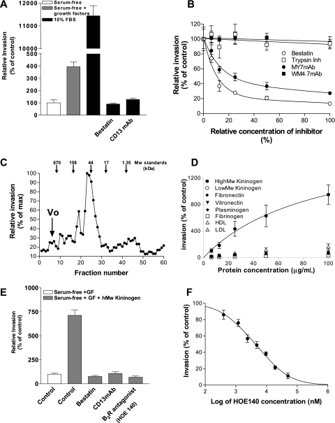
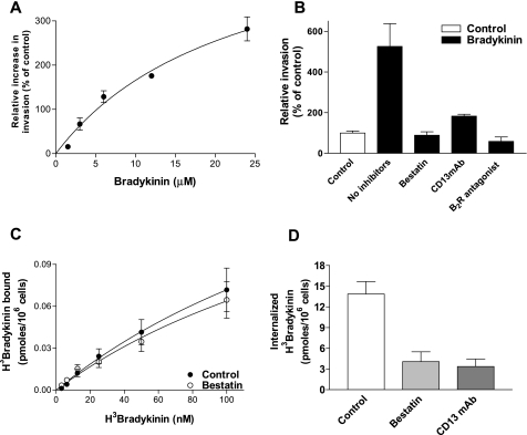
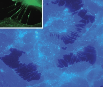
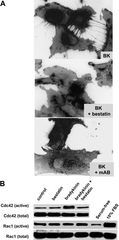
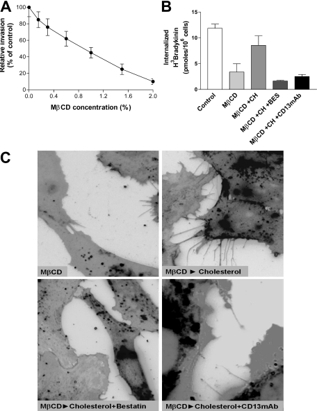
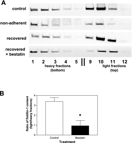
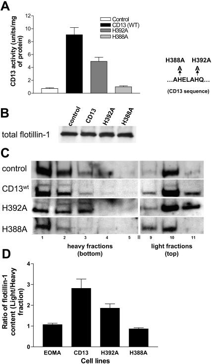
CD13/aminopeptidase N is a transmembrane peptidase that is induced in the vasculature of solid tumors and is a potent angiogenic regulator. Here, we demonstrate that CD13 controls endothelial cell invasion in response to the serum peptide bradykinin by facilitating signal transduction at the level of the plasma membrane. Inhibition of CD13 abrogates bradykinin B(2) receptor internalization, leading to the attenuation of downstream events such as bradykinin-induced activation of Cdc42 and filopodia formation, and thus affects endothelial cell motility. Investigation into mechanisms underlying this block led us to focus on B(2)R internalization via membrane-dependent mechanisms. Membrane disruption by depletion of cholesterol or trypsinization halts B(2)R internalization, invasion, and filopodia formation, which can be recovered with addition of cholesterol. However, this functional recovery is severely impaired in the presence of CD13 antagonists, and the distribution of membrane proteins is disordered in treated cells, suggesting a role for CD13 in plasma membrane protein organization. Finally, exogenous expression of wild-type but not mutant CD13 further alters protein distribution, suggesting peptidase activity is required for CD13's regulatory activity. Therefore, CD13 functions as a novel modulator of signal transduction and cell motility via its influence on specific plasma membrane organization, thus regulating angiogenesis.


Figures

Figure 1
Figure 1
Serum proteins modulate CD13 dependent endothelial invasion in vitro. (A) Primary endothelial cells (HUVECs) were plated in the top chamber of Matrigel-coated transwell plates in the presence (▩) or absence (□) of added growth factors or 10% FBS (■) and the number of cells invading through the Matrigel barrier assessed after 24 hours. The CD13 functional antagonists bestatin (100 μg/mL) and MY7 antibody (1:100 dilution; CD13 mAb) were added to medium containing 10% FBS. (B) Invasion was assessed in the presence of increasing doses of the CD13 antagonists or irrelevant inhibitors or noninhibitory CD13 antibodies. The highest concentration of each agent used is set at 100%. For bestatin (○) and soybean trypsin inhibitor (□), 100% equals 100 μg/mL, while for antibodies MY7 (●; neutralizing mAb) and WM4.7 (■; noninhibitory mAb) 100% equals 1:40 dilution. (C) FBS was fractionated by gel filtration, and fractions were assayed for their ability to induce endothelial invasion. (D) Invasion was assessed in the presence of increasing concentrations (“Materials and methods”) of the plasma components fibronectin (♦), vitronectin (▾), plasminogen (▴), fibrinogen (□), high-molecular-weight kininogen (HK; ●), and low-molecular-weight kininogen (○), LDL (*), and HDL (▵). (E) HK (100 μg/mL; ▩)–induced invasion was assessed in the presence of the CD13 inhibitors bestatin or MY7 antibody (CD13 mAb, 1:100 dilution) or the bradykinin B2 receptor antagonist HOE140 (10 μM). (F) Effect of increasing concentrations of B2 receptor antagonist HOE140 on invasion induced by 10% FBS. Relative invasion in treated cells is expressed as percentage of controls. Data are shown as means plus or minus a standard deviation (SD), n = 3.
Figure 2
Figure 2
CD13 regulates bradykinin-induced invasion and internalization, but not its binding to endothelial cells. (A) Effects of increasing concentrations of bradykinin (in serum-free medium containing growth factor supplement) on endothelial invasion. (B) Sensitivity of bradykinin-induced invasion (12.5 μM; ■) to CD13 inhibition by bestatin (100 μg/mL) or MY7 antibody (CD13 mAb, 1:100 dilution) or inhibition of bradykinin signaling by the B2R antagonist HOE140 (10 μM). (C) Binding of 3H-bradykinin to HUVECs at 0°C in the presence or absence of bestatin. (D) Effects of CD13 inhibition by bestatin (100 μg/mL) or MY7 antibody (CD13 mAb, 1:100 dilution) on 3H-bradykinin (50 nM) internalization at 37°C. Data are shown as mean (± SD, n = 3).
Figure 3
Figure 3
Bradykinin induces filopodia formation in endothelial cells. HUVECs were plated in serum-free medium containing growth factor supplement followed by the addition of bradykinin (15 minutes; 200 nM). Cells were fixed and stained with filipin; images were captured with a Zeiss Axioplan 2 (Thornwood, NJ) fluorescence microscope with 63×/1.4 NA oil objective, a Zeiss Axiocam HRC digital camera with plug-in v. 2.0 acquisition software, and processed with Photoshop 1 (Adobe, San Jose, CA). Inset shows immunofluorescence analysis of the filopodia marker fascin.
Figure 4
Figure 4
Suppression of CD13 activity inhibits bradykinin-induced filopodia formation. (A) HUVECs were trypsinized and replated with bradykinin in the presence or absence of bestatin (100 μg/mL) or MY7 antibody (CD13 mAb, 1:100 dilution) and stained with filipin as in Figure 3. (B) Bestatin (100 μg/mL) inhibits bradykinin signal transduction, resulting in inhibition of Cdc42 but not Rac activation in HUVECs.
Figure 5
Figure 5
Cholesterol depletion suppresses bradykinin-induced filopodia formation. (A) HUVEC invasion in the presence of increasing concentrations of the cholesterol depleting agent M-β-CD (0.15%-2%, 0.5 hours). (B) Effects on internalization of 3H-bradykinin in HUVECs depleted of cholesterol with M-β-CD (MβCD; 2%, 0.5 hours) or depleted with M-β-CD then replenished with cholesterol (CH; 20 μg/mL, 3 hours) in the presence of the CD13 antagonists bestatin (100 μg/mL) or MY7 antibody (CD13 mAb; 1:100 dilution). (C) Effects on bradykinin-induced filopodia formation in HUVECs depleted of cholesterol with M-β-CD or depleted with M-β-CD and replenished with cholesterol in the presence of bestatin (100 μg/mL) or MY7 antibody (CD13 mAb; 1:100 dilution) Data are shown as means (± SD, n = 3).
Figure 6
Figure 6
CD13 is necessary for the efficient restoration of membrane organization. (A) HUVECs were analyzed for distribution of the marker protein flotillin-1 under conditions that inhibit filopodia formation. Control HUVECs (control) indicate normal flotillin-1 distribution. Sonicated cell lysates containing equivalent total protein levels from trypsinized HUVECs were plated in noncoated plastic dishes and rocked in serum-containing medium (nonadherent), plated in serum-containing medium in tissue culture–coated dishes in the absence (recovered), or the presence of bestatin (recovered + bestatin; 100 μg/mL) were analyzed by sucrose gradient separation and fractions assayed for flotillin-1 by Western blot analysis. Heavy fractions (fractions 1-5) represent higher-density protein complexes, whereas light fractions (fractions 9-12) contain proteins complexed with lipids, such as lipid rafts/caveolae. Results are representative of 3 separate experiments. (B) The relative distribution of flotillin-1 in light versus heavy sucrose gradient fractions in cells “recovered” in the presence of bestatin versus control vehicle was calculated using densitometric quantification of Western blots. The overall distribution of flotillin-1 in the gradient fractions was significantly different between the 2 conditions (P < .02). Data are shown as means (± SD, n = 3).
Figure 7
Figure 7
Enzymatic activity of CD13 is needed for optimal organization of membrane proteins. (A) CD13 enzyme activity was determined in vector-transfected EOMA cells (control) and in cells transfected with expression plasmids containing wild-type human CD13 (CD13 [WT]), CD13 point mutants His388Ala (H388A) or His392Ala (H392A). (B) Total levels of flotillin-1 protein are equivalent in cell lysates from all 4 EOMA lines by Western blot analysis. (C) Triton-solubilized cell lysates from control, wild-type, or mutant-transfected cells were analyzed by sucrose gradient separation and fractions assayed for flotillin-1 by Western blot analysis. High-density fractions (fractions 1-5) represent soluble proteins and light fractions (fractions 9-12) contain proteins complexed with lipids. (D) Ratios of flotillin-1 detected in light and heavy sucrose gradient fractions were calculated using densitometric quantification of Western blots of the 4 EOMA lines. Data are shown as means (± SD, n = 3).

Similar articles

Cited by

References

    1. Blaukat A. Structure and signalling pathways of kinin receptors. Andrologia. 2003;35:17–23. - PubMed
    1. Prado GN, Taylor L, Zhou X, Ricupero D, Mierke DF, Polgar P. Mechanisms regulating the expression, self-maintenance, and signaling-function of the bradykinin B2 and B1 receptors. J Cell Physiol. 2002;193:275–286. - PubMed
    1. Hayashi I, Amano H, Yoshida S, et al. Suppressed angiogenesis in kininogen-deficiencies. Lab Invest. 2002;82:871–880. - PubMed
    1. Colman RW, Pixley RA, Sainz IM, et al. Inhibition of angiogenesis by antibody blocking the action of proangiogenic high-molecular-weight kininogen. J Thromb Haemost. 2003;1:164–170. - PubMed
    1. Song JS, Sainz IM, Cosenza SC, et al. Inhibition of tumor angiogenesis in vivo by a monoclonal antibody targeted to domain 5 of high molecular weight kininogen. Blood. 2004;104:2065–2072. - PubMed

Publication types