. 2007 Jun 1;175(11):1139-50.
doi: 10.1164/rccm.200610-1426OC. Epub 2007 Mar 15.

Control of virus reactivation arrests pulmonary herpesvirus-induced fibrosis in IFN-gamma receptor-deficient mice

Affiliations

Control of virus reactivation arrests pulmonary herpesvirus-induced fibrosis in IFN-gamma receptor-deficient mice

Ana L Mora et al. Am J Respir Crit Care Med. .

Abstract

Rationale: Idiopathic pulmonary fibrosis (IPF) is a chronic progressive fibrotic lung disorder of unknown cause. Several studies suggest an association between Epstein-Barr virus pulmonary infection and the development of IPF.

Objectives: To determine whether reduction of gamma-herpesvirus reactivation from latency would alter progressive lung fibrogenesis in an animal model of virus-induced pulmonary fibrosis.

Methods: IFN-gamma receptor-deficient (IFN-gammaR(-/-)) mice infected intranasally with murine gamma-herpesvirus 68 (MHV68) develop lung fibrosis that progresses for up to at least 180 days after initial infection. Viral replication during the chronic phase of infection was controlled by two methods: the administration of cidofovir, an antiviral drug effective at clearing lytic but not latent virus, and by using a mutant gamma-herpesvirus defective in virus reactivation from latency.

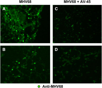
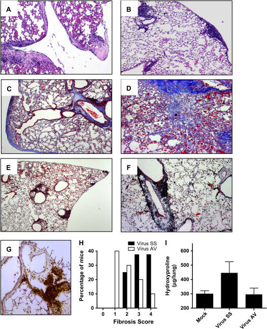
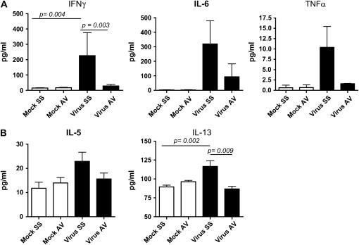
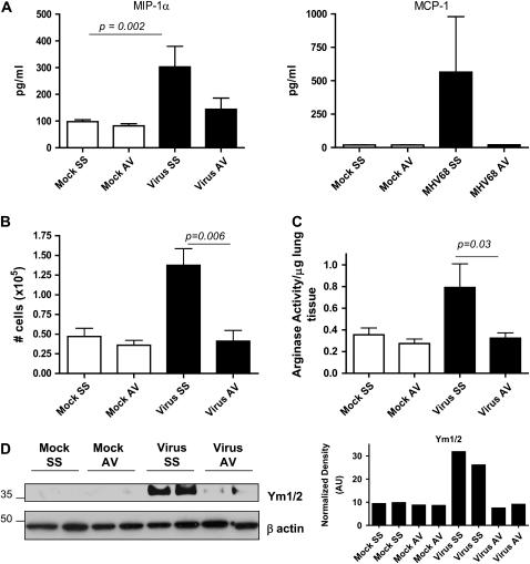
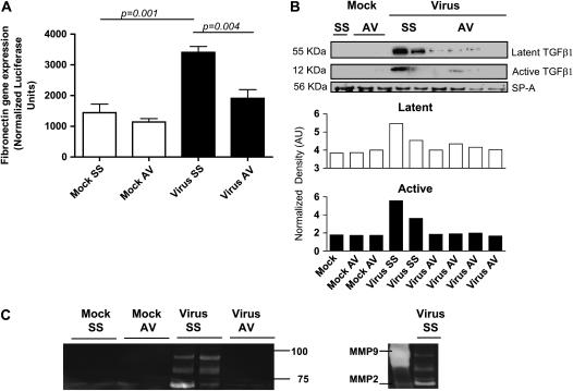
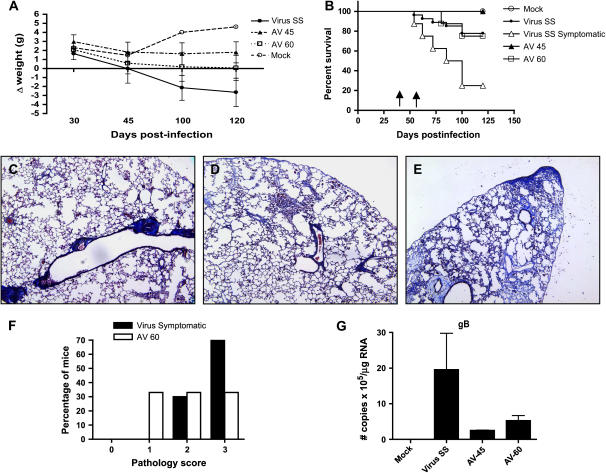
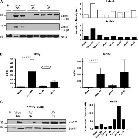
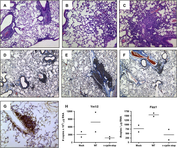
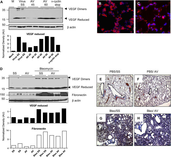
Measurements and main results: Ten percent of the asymptomatic MHV68-infected animals that received antiviral treatment beginning on Day 45 postinfection had severe pulmonary fibrosis compared with 40% of the control saline-treated animals. Absence of severe fibrosis was also observed in IFN-gammaR(-/-) mice infected with the defective reactivation mutant MHV68 v-cyclin stop. Decreased fibrosis was associated with lower levels of transforming growth factor-beta, vascular endothelial growth factor, and markers of macrophage alternative activation. When antiviral treatment was administered on Day 60 in symptomatic animals, survival improved from 20 to 80% compared with untreated symptomatic animals, but lung fibrosis persisted in 60% of the mice.

Conclusions: MHV68-induced fibrosis is a result of viral lytic replication during chronic lung herpesvirus infection in mice. We speculate that antiviral therapy might help to control lung fibrosis in humans with IPF and associated herpesvirus infection.


Figures

<b>Figure 1.</b>
Figure 1.
Viral antigen clearance after cidofovir treatment in murine γ-herpesvirus 68 (MHV68)-infected IFN-γ receptor–deficient (IFN-γR−/−) mice. (AD) Immunostained frozen lung sections from MHV68-infected IFN-γR−/− mice, taken on Day 120 postinfection: (A and B) Mice that received saline solution beginning on Day 45; (C and D) mice that received antiviral treatment beginning on Day 45 (AV-45). Each panel represents a different animal. Original magnification: (AD) ×10.
<b>Figure 2.</b>
Figure 2.
Prevention of lung fibrosis and persistent lymphocytic infiltrates in virus-infected mice treated with cidofovir. (A) Hematoxylin and eosin (H&E) staining of MHV68-infected lung on Day 120. Notice the thickening of the pleura and alveolar walls. (B) H&E-stained section from MHV68-infected mouse receiving antiviral treatment from Day 45 of infection. Lymphocytic infiltrates are observed in the subpleural, perivascular, and peribronchial areas, but there is no thickening of the pleura or alveolar walls. (C and D) Masson trichrome staining of lung section from MHV68-infected mice on Day 120; mice were treated with saline solution. Collagen deposition is demonstrated by blue staining. (E and F) Masson trichrome staining of lung section from MHV68-infected lung receiving antiviral treatment. Notice the absence of fibrosis. Each panel represents a different animal. Original magnification: (AC and E), ×10; (D and F) ×20. (G) Immunohistochemical analysis of lung from antiviral-treated mice, using anti-B220–specific antibody. (H) Semiquantitative morphometric analysis of lung histopathology in virus-infected mice with (Virus AV) or without (Virus SS) antiviral therapy; analyzed on Day 120 of infection. Infected mice showed higher pathology scores corresponding to thickening of the interalveolar septa and thickening of the pleura. In contrast, mice receiving antiviral treatment had lymphocytic infiltrates. (n = 18 for Virus SS and n = 12 for Virus AV). AV = antiviral agent; SS = saline solution. (I) Hydroxyproline determination in lung lysates from mock and infected mice shows that the antiviral treatment decreases collagen content compared with virus-infected lungs of saline-treated animals.
<b>Figure 3.</b>
Figure 3.
Decreased levels of cytokines after treatment in MHV68-infected IFN-γR−/− mice. (A) IFN-γ, IL-6, and tumor necrosis factor (TNF)-α levels were measured in bronchoalveolar lavage (BAL) fluid from mock and MHV68-infected IFN-γR−/− mice after treatment with saline solution (SS) or the antiviral agent (AV), which was begun on Day 45 postinfection. Levels of cytokines were determined in a multiplex bead immunoassay on Day 120. (B) IL-5 and IL-13 levels were measured in BAL fluid 120 days postinfection in mock and MHV68-infected IFN-γR−/− mice treated with saline solution or antiviral agent. Shown are means and SEM (n = 4–10 mice in each group).
<b>Figure 4.</b>
Figure 4.
Antiviral treatment decreases recruitment and activation via the alternative pathway of macrophages in MHV68-infected IFN-γR−/− mice. (A) Macrophage inflammatory protein (MIP)-1α and monocyte chemotactic protein (MCP)-1 levels were measured in bronchoalveolar lavage (BAL) fluid from mock and MHV68-infected IFN-γR−/− mice on Day 120 after chronic treatment with saline solution (SS) or antiviral agent (AV) begun on Day 45. Levels of cytokines were determined in a multiplex bead immunoassay. Shown are means and SEM (n = 4–10 per group). (B) BAL was performed in mock and virus-infected animals after 4 months of saline solution or antiviral therapy (n = 4–8 mice in each group). Cell counts show a significant diminution in the numbers of cells after antiviral therapy. (C) Measurement of arginase I activity in lung samples of mock and infected animals on Day 120 after treatment with saline solution or antiviral begun on Day 45. Arginase activity significantly diminished after antiviral treatment. Shown are means and SEM (n = 3–7 per group). (D) Lung homogenate from mock and virus-infected mice treated with saline solution or antiviral were subjected to Western blot analysis for Ym1/2 proteins. Antiviral treatment in infected mice was associated with lower levels of Ym proteins in the lungs. Blots were stripped and reprobed with an anti–β-actin antibody to normalize expression of Ym1/2.
<b>Figure 5.</b>
Figure 5.
Control of virus replication diminished fibrogenesis in MHV68 IFN-γR−/− infected mice. (A) NIH3T3 cells stably transfected with a fibronectin reporter were cultured for 24 hours in the presence of bronchoalveolar lavage (BAL) fluid from mock and virus-infected animals treated with saline solution (SS) or antiviral (AV). Afterward, the cells were harvested and fibronectin gene transcription was measured by luminescence (n = 4 per group). MHV68 infection stimulates gene transcription of a reporter under control of the fibronectin promoter. AV treatment significantly diminished fibronectin transcription. (B) Western blot analysis for the latent and active forms of transforming growth factor (TGF)-β in BAL fluid samples collected on Day 120. The blot was stripped and reprobed with an anti-surfactant A (SP-A) antibody to normalize expression of latent (open columns) and active (solid columns) TGF-β. Decreased levels of active TGF-β were found in infected mice treated with the antiviral agent. (C) Gelatin zymography of BAL fluid samples from mock and MHV68-infected animals at the indicated times points after infection. High gelatinolytic activity was observed in samples from infected mice compared with mock animals and infected animals treated with antiviral agent. Purified matrix metalloproteinase (MMP)–2 and MMP-9 were used to identify zymography bands in the samples from virus-infected animals treated with SS.
<b>Figure 6.</b>
Figure 6.
Antiviral treatment in symptomatic mice improved clinical disease and survival. (A) Body weight was tracked for mock (open circles) and MHV68-infected mice treated with saline solution (Virus SS; solid circles) or antiviral from Day 45 (AV-45; solid triangles) or from Day 60 (AV-60; open squares). Data are presented as the difference in body weight from Day 0 of infection. More severe illness was observed in SS-treated mice. A beneficial effect was observed with the antiviral treatment. Number of mice: mock (n = 10), SS (n = 9), AV-45 (n = 5), AV-60 (n = 6). Data are representative of three different experiments. (B) Survival is plotted versus time postinfection for mock (open circles), MHV68-infected mice treated with saline solution (Virus SS; solid circles), MHV68-infected mice treated with antiviral begun on Day 45 (AV-45; solid triangles), and symptomatic MHV68-infected mice treated with antiviral (AV-60; open squares) or saline solution begun on Day 60 (virus symptomatic; open triangles). Number of mice: mock (n = 20), SS (n = 26), AV-45 (n = 19), AV-60 (n = 8), virus symptomatic (n = 8). Data represent three pooled different experiments. The Kaplan-Meier survival curves were significantly different as follows: mock versus SS (p = 0.0087), SS versus AV-45 (p = 0.03), virus symptomatic versus AV-60 (p = 0.03). Arrows indicate the time points when AV treatment was begun on Day 45 and Day 60. (CE) Masson trichrome staining of lung sections from MHV68-infected mice receiving antiviral treatment from Day 60 postinfection. Each panel represents a different animal. Notice the perivascular fibrosis in (C), mild interstitial fibrosis in (D), and the severe interstitial and subpleural deposition of collagen in (E). Original magnification: ×10. (F) Semiquantitative morphometric analysis of lung histopathology in symptomatic virus-infected mice with or without antiviral therapy analyzed on Day 120 of infection. Virus-infected symptomatic mice showed a high proportion of multiple fibrotic foci with thickness of the pleura (score, 4). A reduction in the severity of the fibrosis was found in symptomatic mice receiving antiviral treatment. Number of mice: virus symptomatic (n = 8); AV-60 (n = 8). (G) Real-time reverse transcription-polymerase chain reaction (RT-PCR) quantifying viral transcripts for the late lytic gene gB in the lungs of mock and virus-infected mice treated with SS or AV (n = 3). Negative amplification was obtained in mock-infected mice. A lower number of copies was observed in MHV68-infected mice treated with cidofovir.
<b>Figure 7.</b>
Figure 7.
Antiviral treatment in symptomatic mice fails to control alternative activation of macrophages. (A) Western blot analysis for the latent and active forms of TGF-β in bronchoalveolar lavage (BAL) fluid samples collected on Day 120. Blot was stripped and reprobed with an anti-surfactant A (SP-A) antibody to normalize expression of latent (open columns) and active (solid columns) TGF-β. Decreased levels of active TGF-β were found in symptomatic infected mice treated with the antiviral agent. (B) IFN-γ and monocyte chemotactic protein (MCP)–1 levels were measured in BAL fluid from mock and MHV68-infected IFN-γR−/− mice after treatment with saline solution (SS) or antiviral, which was begun on Day 45 or 60 postinfection (AV-45 and AV-60, respectively). Levels of cytokines were determined in a multiplex bead immunoassay on Day 120. Shown are means and SEM. Number of mice: mock (n = 5); AV-45 (n = 5); virus SS (n = 6); AV-60 (n = 6). (C) Lung homogenate from mock and virus-infected mice treated with saline solution or antiviral were subjected to Western blot analysis for Ym1/2 proteins. Antiviral begun on Day 60 postinfection failed to lower levels of Ym proteins in the lungs. Blot was stripped and reprobed with an anti–β-actin antibody to normalize expression of Ym1/2.
<b>Figure 8.</b>
Figure 8.
Infection with the reactivation-deficient v-cyclin stop MHV68 failed to produce lung fibrosis and alternative activation of macrophages. (AC) Hematoxylin-and-eosin staining of v-cyclin stop MHV68–infected lung on Day 20. v-Cyclin stop MHV68 has an acute replication similar to that of wild-type virus. Notice the lymphocytic infiltrates around blood vessels and airways, and the accumulation of alveolar macrophages and fibroblasts. (DF) Masson trichrome staining of lung sections from v-cyclin stop MHV68–infected mice on Day 150. Collagen deposition is demonstrated by blue staining. Notice the absence of interstitial fibrosis. Each panel represents a different animal. Original magnification: (A and DF) ×10; (B and C) ×20. (G) Immunohistochemical analysis of v-cyclin stop virus–infected lung, using an anti-B220 antibody. (H and I) Quantitative reverse transcription–polymerase chain reaction was used to determine the levels of Ym and Fizz1, respectively, in lungs of mock, wild-type (WT)–infected, and v-cyclin stop MHV68–infected IFN-γR−/− mice on Day 120 postinfection. Data are normalized against β-actin.
<b>Figure 9.</b>
Figure 9.
Cidofovir treatment in a bleomycin fibrosis model is ineffective in controlling vascular endothelial growth factor (VEGF) expression and fibrosis. (A) Western blot analysis, using an anti-VEGF antibody in lung homogenates collected on Day 120–150. High levels of VEGF were found in wild-type MHV68–infected IFN-γR−/− mice receiving saline solution (Virus SS) and symptomatic infected mice treated with the antiviral agent from Day 60 of infection (AV-60). Low VEGF levels were obtained in infected mice treated with antiviral from Day 45 of infection (AV-45) and also in mice infected with the v-cyclin stop mutant MHV68. The blot was stripped and reprobed with an anti–β-actin antibody to normalize expression of reduced VEGF. (B) VEGF expression was detected in hyperplastic alveolar epithelial cells and alveolar macrophages by immunofluorescence analysis of lung of MHV68-infected mice on Day 120 (red). Slides were counterstained with 4′,6-diamidino-2-phenyindole, which stains nuclei blue. (C) Frozen section from a mouse treated with antiviral from Day 45 and stained with anti-VEGF antibody (red) shows decreased VEGF expression. (D) VEGF and fibronectin expression were determined in lung lysates from mock and bleomycin-treated mice receiving saline solution (SS) or antiviral (AV). Comparable VEGF and fibronectin up-regulation was observed after bleomycin treatment in mice with or without antiviral treatment. The blot was stripped and reprobed with an anti–β-actin antibody to normalize expression of reduced VEGF and fibronectin. (EH) Masson trichrome staining of lungs of IFN-γR−/− mice on Day 21 after intratracheal inoculation of phosphate-buffered saline or bleomycin and after receiving subcutaneously cidofovir (antiviral, AV) or saline solution (SS) every 3 days. Collagen deposition is denoted in blue.

Comment in

Similar articles

Cited by

References

    1. American Thoracic Society; European Respiratory Society.Idiopathic pulmonary fibrosis: diagnosis and treatment [International Consensus Statement]. Am J Respir Crit Care Med 2000;161:646–664. - PubMed
    1. Selman M, Pardo A. The epithelial/fibroblastic pathway in the pathogenesis of idiopathic pulmonary fibrosis. Am J Respir Cell Mol Biol 2003;29:S93–S97. - PubMed
    1. Perez A, Rogers RM, Dauber JH. The prognosis of idiopathic pulmonary fibrosis. Am J Respir Cell Mol Biol 2003;29:S19–S26. - PubMed
    1. Stewart JP, Egan JJ, Ross AJ, Kelly BG, Lok SS, Hasleton PS, Woodcock AA. The detection of Epstein-Barr virus DNA in lung tissue from patients with idiopathic pulmonary fibrosis. Am J Respir Crit Care Med 1999;159:1336–1341. - PubMed
    1. Tang YW, Johnson JE, Browning PJ, Cruz-Gervis RA, Davis A, Graham BS, Brigham KL, Oates JA Jr, Loyd JE, Stecenko AA. Herpesvirus DNA is consistently detected in lungs of patients with idiopathic pulmonary fibrosis. J Clin Microbiol 2003;41:2633–2640. - PMC - PubMed

Publication types

MeSH terms