. 2007 Apr 4;26(7):1853-64.
doi: 10.1038/sj.emboj.7601642. Epub 2007 Mar 15.

The human Nup107-160 nuclear pore subcomplex contributes to proper kinetochore functions

Affiliations

The human Nup107-160 nuclear pore subcomplex contributes to proper kinetochore functions

Michela Zuccolo et al. EMBO J. .

Abstract

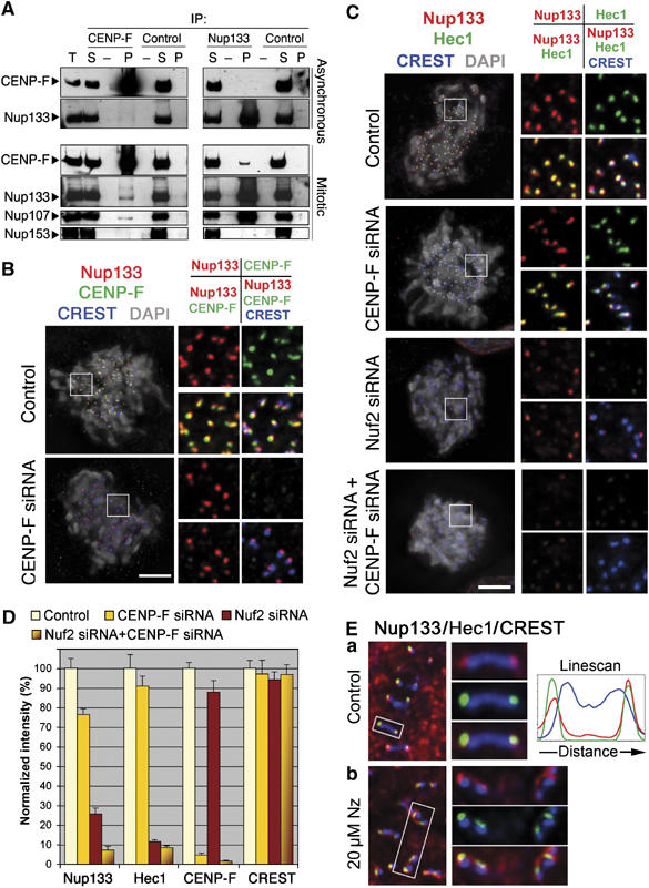
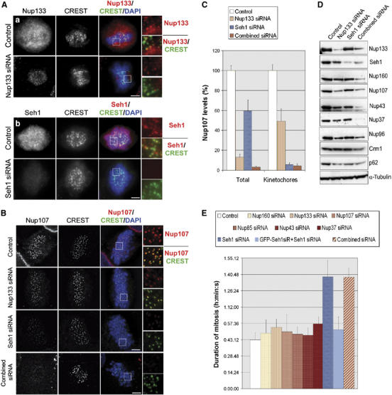
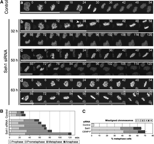
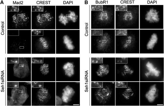
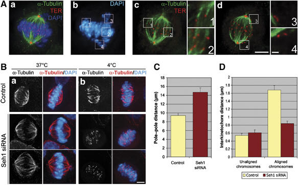
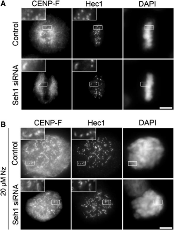
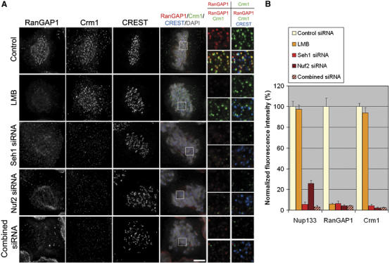
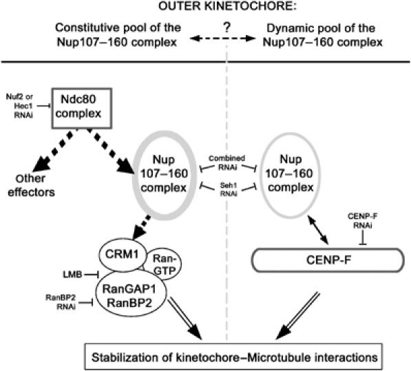
We previously demonstrated that a fraction of the human Nup107-160 nuclear pore subcomplex is recruited to kinetochores at the onset of mitosis. However, the molecular determinants for its kinetochore targeting and the functional significance of this localization were not investigated. Here, we show that the Nup107-160 complex interacts with CENP-F, but that CENP-F only moderately contributes to its targeting to kinetochores. In addition, we show that the recruitment of the Nup107-160 complex to kinetochores mainly depends on the Ndc80 complex. We further demonstrate that efficient depletion of the Nup107-160 complex from kinetochores, achieved either by combining siRNAs targeting several of its subunits excluding Seh1, or by depleting Seh1 alone, induces a mitotic delay. Further analysis of Seh1-depleted cells revealed impaired chromosome congression, reduced kinetochore tension and kinetochore-microtubule attachment defects. Finally, we show that the presence of the Nup107-160 complex at kinetochores is required for the recruitment of Crm1 and RanGAP1-RanBP2 to these structures. Together, our data thus provide the first molecular clues underlying the function of the human Nup107-160 complex at kinetochores.


Figures

Figure 1
Figure 1
Nup107–160 complex targeting at kinetochores depends on CENP-F and the Ndc80 complex. (A) Immunoprecipitation of asynchronous or colchicine-arrested (mitotic) HeLa cell extracts using either anti-CENP-F or anti-myc (control) mouse IgG, or affinity-purified anti-hNup133 or anti-GST (control) rabbit IgG. Equivalent amounts of total extracts (T) and immune supernatants (S) and a 10-fold equivalent of the immune pellets (P) were analyzed by immunoblot using anti-CENP-F, anti-hNup133, anti-hNup107 and as control the mAb414 antibody that recognizes Nup153, a nucleoporin that does not belong to the Nup107–160 complex. Because of the lower amounts of CENP-F in asynchronous as compared with mitotic extracts, CENP-F immunoblots were exposed to yield comparable input signals. (B) HeLa cells treated for 3 days with control or CENP-F siRNA duplexes were pre-extracted, fixed and stained with anti-hNup133 (red), anti-CENP-F (green), CREST serum (blue) and DAPI (gray). (C) Cells treated for 3 days with control, CENP-F, Nuf2 or a combination of CENP-F and Nuf2 siRNA duplexes were processed for immunofluorescence as above using anti-hNup133 (red), anti-Hec1 (green), CREST serum (blue) and DAPI (gray). Maximum projection of deconvolved Z-stack images of all the four staining (left) or various combinations of overlay images (insets, right) is presented. Scale bar, 5 μm. (D) Quantification of the fluorescence intensity at kinetochores was performed as described in Materials and methods. (E) Single Z-plane of deconvolved immunofluorescence images from (a) control or (b) nocodazole-treated (Nz, 20 μM, 3 h) cells stained for Nup133 (red), Hec1 (green) or CREST antigens (blue). Right: enlargements of the marked areas stained with CREST and either Nup133 (top), Hec1 (center) or Nup133+Hec1 (bottom). Line scan through a single kinetochore pair, performed using the ‘Linescan' function accessed using the Metamorph software system, is also shown.
Figure 2
Figure 2
Seh1 RNAi, which prevents the Nup107–160 complex from localizing at kinetochores, or efficient depletion of the Nup107–160 complex, induces a mitotic delay. (A, B) HeLa cells transfected with the indicated siRNA duplexes were pre-extracted before fixation and stained with (Aa) anti-Nup133, (Ab) anti-Seh1 or (B) anti-Nup107 antibodies (red), CREST serum (green) and DAPI (blue). Wide-field microscopy images (A) or maximum projections of deconvolved Z-stacks (B) of representative prometaphase cells and three-fold enlargements of the marked area are shown. Scale bar, 5 μm. (C) Quantification of Nup107 fluorescence intensity in mitotic HeLa cells. Total levels were measured on wide-field images from non-extracted cells (n⩾20) and levels at kinetochores were measured on pre-extracted cells as in Figure 1D. (D) Whole-cell extracts from control, Nup133, Seh1 or ‘combined siRNA'-treated HeLa cells were prepared 3 days after transfection and analyzed by Western blot using the indicated antibodies. α-Tubulin is shown as loading control. (E) HeLa cells treated with the indicated siRNA duplexes were recorded by time-lapse videomicroscopy from 24 to 96 h post siRNA transfection, acquiring images every 5 or 10 min. Duration of mitosis was calculated as the time spent between the first frame where cells rounded up and the first frame where two distinct daughter cells could be observed. Average values of at least 50 cells for each condition are presented. For the complementation experiment (GFP-Seh1siR+Seh1 siRNA), a HeLa cell line stably expressing an siRNA-resistant form of GFP-Seh1 was used (see Supplementary Figure 4B).
Figure 3
Figure 3
Seh1-depleted cells display extended prometaphase and metaphase. (A) HeLa cells transiently expressing histone H2B-GFP were treated with control (a) or Seh1 (b–d) siRNAs. At 32 h (b), 50 h (c) or 63 h (d) after siRNA transfection, randomly chosen prophase cells were imaged every 90 s for several hours with a spinning disk confocal microscope. Maximum intensity projections of 14 Z-stack planes for each time point are presented. Arrowheads indicate misaligned chromosomes. Light gray bars indicate frames from prophase to metaphase, whereas dark gray bars outline time spent from metaphase to anaphase. (B) Mitotic progression in two control and six representative Seh1-depleted cells selected at random. (C) Quantification of misaligned chromosomes (defined as in Meraldi and Sorger (2005), as chromosomes located outside a rectangular area encompassing the central 30% of the spindle) in control, Seh1- or CENP-F siRNA-treated cells. The number of cells displaying 1–2 (light gray), 3–4 (dark gray) or >4 (black) misaligned chromosomes is expressed as percentage of total metaphase cells analyzed (at least 100 per condition).
Figure 4
Figure 4
Seh1 depletion activates the spindle assembly checkpoint. Immunofluorescence of metaphase or prometaphase HeLa cells upon 72 h treatment with control or Seh1 siRNA stained for Mad2 (A) or BubR1 (B), CREST and DAPI. Three-fold magnifications of the marked areas are presented. Scale bar, 5 μm.
Figure 5
Figure 5
Seh1 depletion impairs chromosome congression and reduces tension between sister kinetochores. (A) Seh1-depleted cells were fixed and then stained with anti-α-tubulin (green), the TER serum that labels both the centromeres and centrosomes (red), and DAPI (blue). A maximum intensity projection of the deconvolved Z-stack from a representative mitotic cell is presented in (a) and (b). Single planes of the same cell illustrating kinetochore–microtubule attachment on chromosomes of the metaphase plate (c) and lack of microtubules on misaligned chromosomes (d). Scale bar, 5 μm. Higher magnifications of the marked areas are presented (scale bar, 1 μm). (B) Immunofluorescence analysis of control or Seh1-depleted cells placed either at 37°C (a) or at 4°C (b) for 10 min before fixation and stained with anti-α-tubulin antibody (red) and DAPI (blue). Scale bar, 5 μm. (C) Pole–pole distance was calculated as the distance between the two centrosomes labelled with the TER serum as in (A). (D) Interkinetochore distance between aligned or unaligned sister kinetochores situated in the same focal plane were calculated on deconvolved images. Values for unaligned chromosomes in control cells was obtained after treatment with 20 μM nocodazole for 3 h. At least 50 kinetochore pairs in eight different cells were measured.
Figure 6
Figure 6
Seh1 depletion induces MT-dependent stripping of CENP-F. Cells transfected with control or Seh1 siRNAs were either fixed and stained with anti-Hec1 and anti-CENP-F antibodies and DAPI (A), or treated for 3 h with 20 μM nocodazole before fixation (B). Three-fold enlargements of the marked areas are presented. Scale bar, 5 μm.
Figure 7
Figure 7
Mislocalization of the Nup107–160 complex from kinetochores impairs the kinetochore targeting of Crm1 and RanGAP1. (A) Cells were treated for 20 min with 10 ng/ml leptomycin B or for 72 h with control, Seh1, Nuf2 or the ‘combined siRNA' duplexes and subsequently pre-extracted, fixed and probed with anti-RanGAP1 (red) and anti-Crm1 (green) antibodies, CREST serum (blue) and DAPI. Single planes from deconvolved Z-stack images (scale bar, 5 μm) and three-fold enlargements of the boxed areas are presented. (B) Remaining levels of Nup133, RanGAP1 and Crm1 at kinetochores in control cells, upon treatment with Seh1, Nuf2 or the combined RNAi, or after LMB treatment, were quantified based on deconvolved Z-stack images (see Materials and methods).
Figure 8
Figure 8
Model summarizing the protein network involved in the targeting and function of the Nup107–160 complex at kinetochores. Dotted arrows indicate direct or indirect interactions. As outlined in Discussion, Seh1 is comprised within the Nup107–160 complex in this model.

Similar articles

Cited by

References

    1. Arnaoutov A, Azuma Y, Ribbeck K, Joseph J, Boyarchuk Y, Karpova T, McNally J, Dasso M (2005) Crm1 is a mitotic effector of Ran-GTP in somatic cells. Nat Cell Biol 7: 626–632 - PubMed
    1. Arnaoutov A, Dasso M (2003) The Ran GTPase regulates kinetochore function. Dev Cell 5: 99–111 - PubMed
    1. Arnaoutov A, Dasso M (2005) Ran-GTP regulates kinetochore attachment in somatic cells. Cell Cycle 4: 1161–1165 - PubMed
    1. Babu JR, Jeganathan KB, Baker DJ, Wu X, Kang-Decker N, Van Deursen JM (2003) Rae1 is an essential mitotic checkpoint regulator that cooperates with Bub3 to prevent chromosome missegregation. J Cell Biol 160: 341–353 - PMC - PubMed
    1. Belgareh N, Rabut G, Bai SW, van Overbeek M, Beaudouin J, Daigle N, Zatsepina OV, Pasteau F, Labas V, Fromont-Racine M, Ellenberg J, Doye V (2001) An evolutionarily conserved NPC subcomplex, which redistributes in part to kinetochores in mammalian cells. J Cell Biol 154: 1147–1160 - PMC - PubMed

Publication types

MeSH terms