. 2007 Apr 4;26(7):1820-30.
doi: 10.1038/sj.emboj.7601628. Epub 2007 Mar 15.

An alternative branch of the nonsense-mediated decay pathway

Affiliations

An alternative branch of the nonsense-mediated decay pathway

Wai-Kin Chan et al. EMBO J. .

Abstract

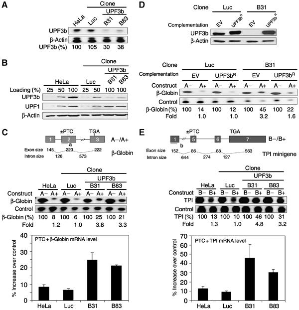
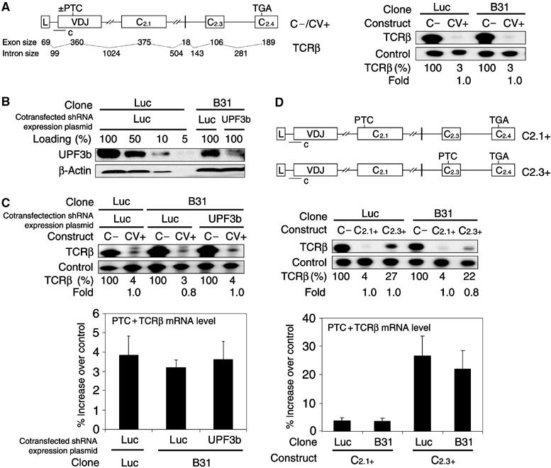
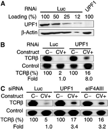
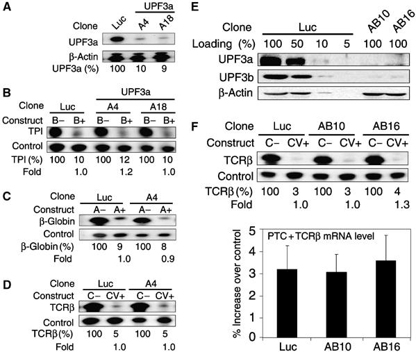
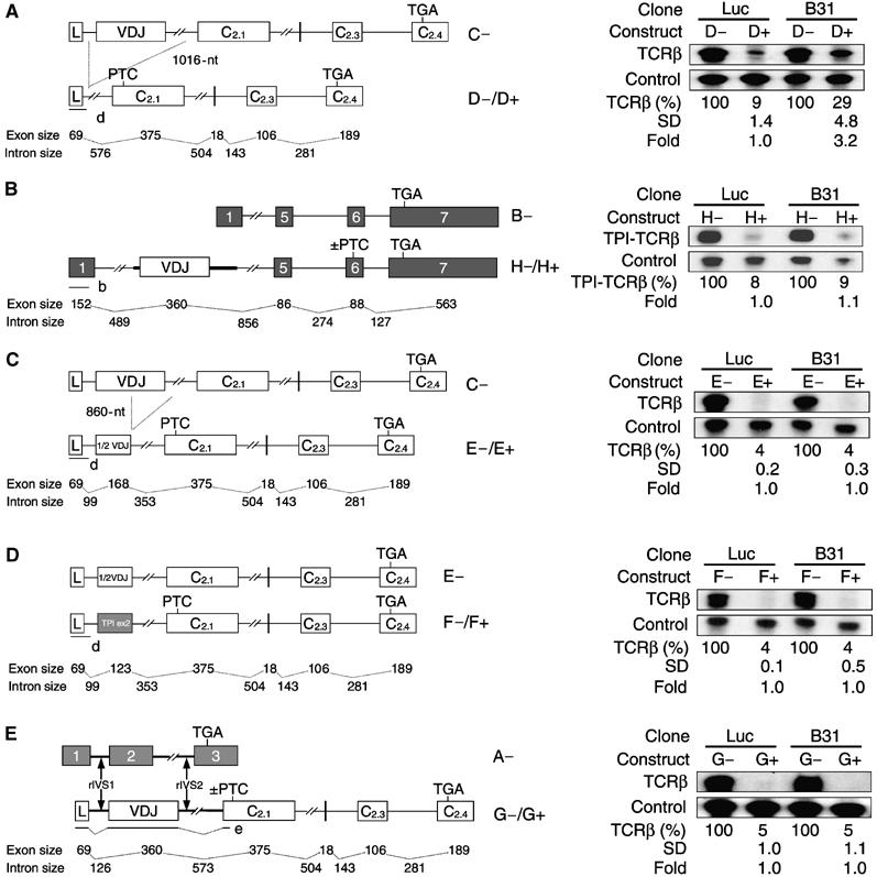
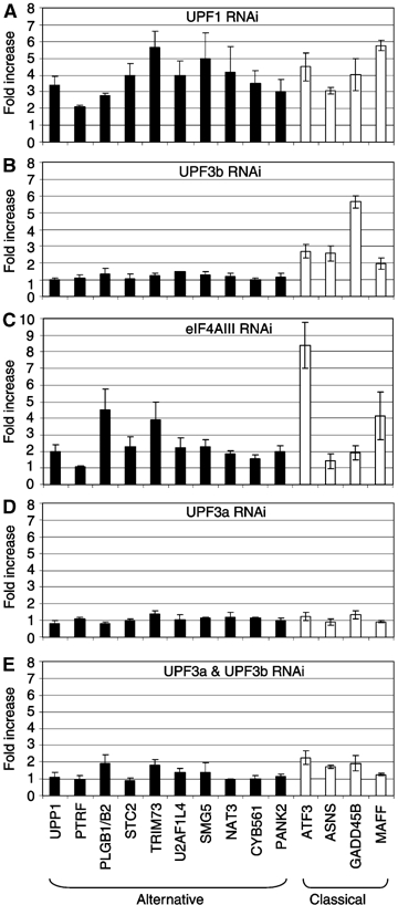
The T-cell receptor (TCR) locus undergoes programmed rearrangements that frequently generate premature termination codons (PTCs). The PTC-bearing transcripts derived from such nonproductively rearranged genes are dramatically downregulated by the nonsense-mediated decay (NMD) pathway. Here, we show that depletion of the NMD factor UPF3b does not impair TCRbeta NMD, thereby distinguishing it from classical NMD. Depletion of the related factor UPF3a, by itself or in combination with UPF3b, also has no effect on TCRbeta NMD. Mapping experiments revealed the identity of TCRbeta sequences that elicit a switch to UPF3b dependence. This regulation is not a peculiarity of TCRbeta, as we identified many wild-type genes, including one essential for NMD, that transcribe NMD-targeted mRNAs whose downregulation is little or not affected by UPF3a and UPF3b depletion. We propose that we have uncovered an alternative branch of the NMD pathway that not only degrades aberrant mRNAs but also regulates normal mRNAs, including one that participates in a negative feedback loop controlling the magnitude of NMD.


Figures

Figure 1
Figure 1
NMD is impaired in HeLa cells stably expressing UPF3b shRNA. (A) RNase protection analysis (using probe f, complementary to UPF3b) of total cellular RNA from HeLa cell clones stably transfected with a UPF3b shRNA expression plasmid (B31 and B83) or a luciferase shRNA expression plasmid (Luc). The Luc clone and untransfected HeLa cells served as negative controls. The numbers below the blot are UPF3b mRNA relative levels normalized to β-actin mRNA levels. (B) Western blot analysis of the cells in panel A. To assess the efficiency of UPF3b shRNA knockdown, serial dilutions of the negative control lysates were loaded. For loading controls, the membrane was reprobed with β-actin and UPF1 antibodies. (C) RNase protection analysis (using probe a) of total cellular RNA isolated from the cells in panel A transiently transfected with PTC+ and PTC− versions of the β-globin construct shown (A+ and A−, respectively). The internal control is a cotransfected TCRβ construct (C−). The numbers below the blot are relative mRNA levels (PTC− is set to 100) and the fold difference in PTC−/PTC+ ratio relative to Luc control cells. (D) Functional complementation of UPF3b depletion. Upper: Western blot analysis of UPF3b depletion and complementation. For complementation of UPF3b depletion, the Luc and B31 clones were transfected with an empty expression vector (EV) or an RNAi-resistant UPF3b expression vector (UPF3bR). Lower: RNase protection analysis of total cellular RNA isolated from Luc and B31 clones cotransfected with A− or A+ constructs (as described in panel C) and EV or UPF3bR. (E) RNase protection analysis (using probe b) of total RNA isolated from the cells in panel A transiently transfected with PTC+ and PTC− versions of the TPI construct shown (B+ and B−, respectively). The internal control was a cotransfected β-globin construct (J−). The values in panels C and E are the means of three independent transfection experiments. The results in panel D are representative of two independent experiments. Error bars indicate standard deviation.
Figure 2
Figure 2
TCRβ NMD is unperturbed by depletion of UPF3b. (A) RNase protection analysis (using probe c) of total cellular RNA isolated from the B31 and Luc cell clones transiently transfected with construct C− or CV+. (B) Western blot analysis (performed as in Figure 1B) of the UPF3b-depleted B31 cell clone transiently transfected with a UPF3b shRNA expression plasmid to further reduce the level of UPF3b. The Luc cell clone and Luc shRNA expression plasmid served as negative controls. (C) RNase protection analysis (using probe c) of total cellular RNA isolated from cells transiently transfected as in panel B, along with the indicated NMD reporter constructs. (D) RNase protection analysis (using probe c) of total cellular RNA isolated from the B31 and Luc cell clones transiently transfected (as in panel C) with the indicated constructs. The values in panel A are representatives of two independent experiments. The results in panels C and D are the means of these independent transfection experiments. Error bars indicate standard deviation.
Figure 3
Figure 3
TCRβ NMD in UPF3b-depleted cells requires UPF1 and eIF4AIII. (A) Western blot analysis of HeLa cells transiently transfected with siRNAs specific for UPF1 or luciferase (negative control). (B) RNase protection analysis (using probe c) of total RNA isolated from the cells in panel A transiently transfected with the TCRβ constructs C− and CV+ 48 h after siRNAs were transfected. (C) RNase protection analysis (using probe c) of total cellular RNA isolated from the B31 cell clone transiently transfected with constructs C− and CV+ 48 h after siRNAs were transfected. The results in panels B and C are representative of two independent experiments.
Figure 4
Figure 4
TCRβ NMD is unperturbed by depletion of both UPF3a and UPF3b. (A) RNase protection analysis (using probe g, complementary to UPF3a) of the UPF3a-deficient cell clones A4 and A18 (described in Supplementary Figure S5) transiently transfected with a UPF3a shRNA expression plasmid to further reduce the level of UPF3a. (BD) RNase protection analysis of total cellular RNA isolated from cells transiently transfected as in panel A, along with the indicated NMD reporter constructs. (E) Western blot analysis of the UPF3a- and UPF3b-deficient HeLa cell clones AB10 and AB16 (described in Supplementary Figure S6) transiently transfected with UPF3a and UPF3b shRNA expression plasmids to further reduce UPF3a and UPF3b levels. (F) RNase protection analysis of total cellular RNA isolated from cell clones transiently transfected as in panel E, along with the indicated NMD reporter constructs. The results in panels B–D are representative of two independent experiments. The values in panel F are the means of three independent transfection experiments. Error bars indicate standard deviation.
Figure 5
Figure 5
NMD of TCRβ is rendered UPF3b dependent by deletion of the VDJ exon and adjacent intron sequences. (A–E) RNase protection analysis of total cellular RNA isolated from the UPF3b-deficient B31 cell clone transiently transfected with the UPF3b shRNA expression plasmid to further reduce UPF3b levels (as described in Figure 2B). The Luc cell clone served as the negative control, as described in Figure 1A. All cells were also transiently transfected with the TCRβ constructs indicated in each panel. Construct D lacks the VDJ exon and part of the flanking introns. Construct E lacks the 3′ half of the VDJ exon and the 5′ portion of the downstream intron. Construct F has TPI exon 2 in place of the VDJ exon. Construct G has intron 1 (rIVS1) and intron 2 (rIVS2) from rabbit β-globin (indicated in bold) in place of TCRβ IVS1 and IVS2, respectively. Construct H is a TCRβ TPI-chimera construct that has the TCRβ VDJ exon and flanking intron sequences. Expression of each construct was quantified as in Figure 1C using the probes indicated in each panel. The values below each blot are the mean mRNA levels±standard deviation and fold change in PTC−/PTC+ ratio, determined as in Figure 1C from three independent transfection experiments, except that the blot in panel B is representative of two independent experiments.
Figure 6
Figure 6
Endogenous NMD targets of the classical and alternative branches of the NMD pathway. (AE) Real-time PCR analysis of the abundance of endogenous transcripts in cells depleted of UPF3b, UPF3a, both UPF3a and UPF3b, UPF1, or eIF4AIII (UPF1, UPF3a, UPF3b, and eIF4AIII levels were reduced to ∼15, ∼10, ∼10, and ∼20%, respectively; see Materials and methods for further details). The values shown are the average fold (n⩾3) increase relative to negative control Luc cells. Expression levels were normalized to that of the housekeeping transcript L19. Error bars indicate standard deviation.

Similar articles

Cited by

References

    1. Anders KR, Grimson A, Anderson P (2003) SMG-5, required for C. elegans nonsense-mediated mRNA decay, associates with SMG-2 and protein phosphatase 2A. EMBO J 22: 641–650 - PMC - PubMed
    1. Belgrader P, Cheng J, Maquat L (1993) Evidence to implicate translation by ribosomes in the mechanism by which nonsense codonsreduce the nuclear level of human triosephosphate isomerase mRNA. Proc Natl Acad Sci USA 90: 482–486 - PMC - PubMed
    1. Bruce SR, Wilkinson MF (2003) Nonsense mediated decay: a surveillance pathway that detects faulty TCR and BCR. In Recent Res Devel in Immunity, Pandalai SG (ed) Vol I, pp 1–12. Kerala, India: Research Signpost
    1. Buhler M, Paillusson A, Muhlemann O (2004) Efficient downregulation of immunoglobulin mu mRNA with premature translation-termination codons requires the 5′-half of the VDJ exon. Nucleic Acids Res 32: 3304–3315 - PMC - PubMed
    1. Buhler M, Steiner S, Mohn F, Paillusson A, Muhlemann O (2006) EJC-independent degradation of nonsense immunoglobulin-mu mRNA depends on 3′ UTR length. Nat Struct Mol Biol 13: 462–464 - PubMed

Publication types

MeSH terms

LinkOut - more resources