. 2007 Apr 4;26(7):1794-805.
doi: 10.1038/sj.emboj.7601622. Epub 2007 Mar 15.

Phosphorylation and ubiquitination of the IkappaB kinase complex by two distinct signaling pathways

Affiliations

Phosphorylation and ubiquitination of the IkappaB kinase complex by two distinct signaling pathways

Prashant B Shambharkar et al. EMBO J. .

Abstract

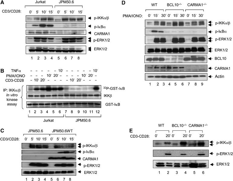
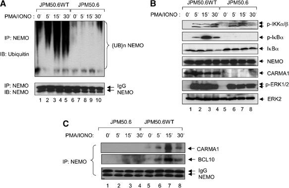
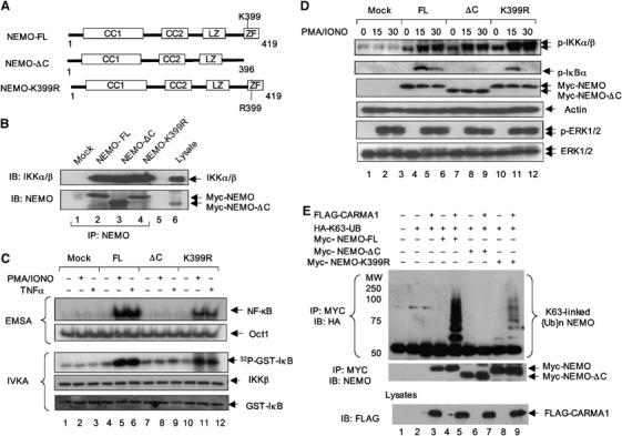
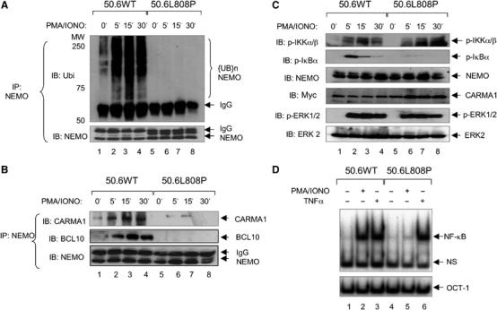
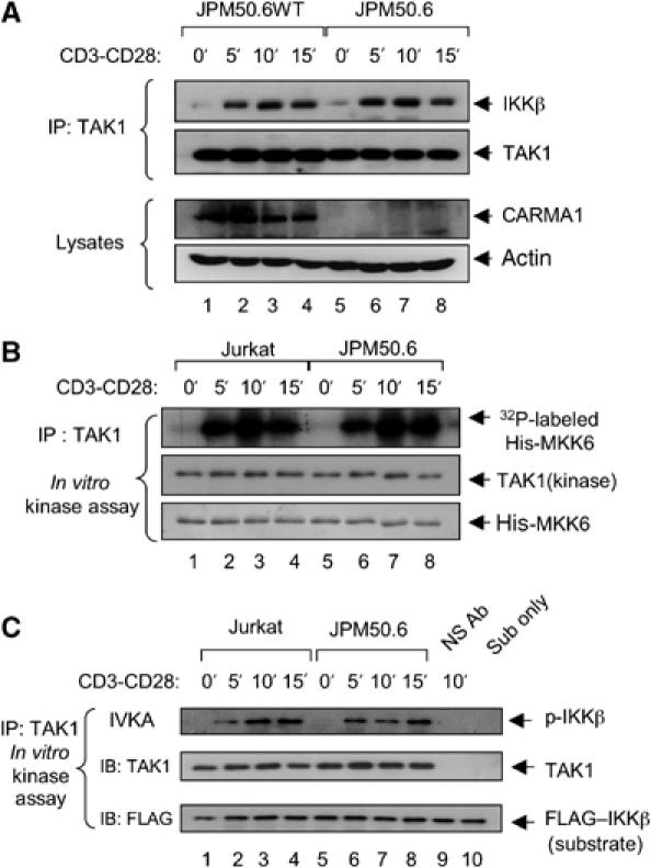
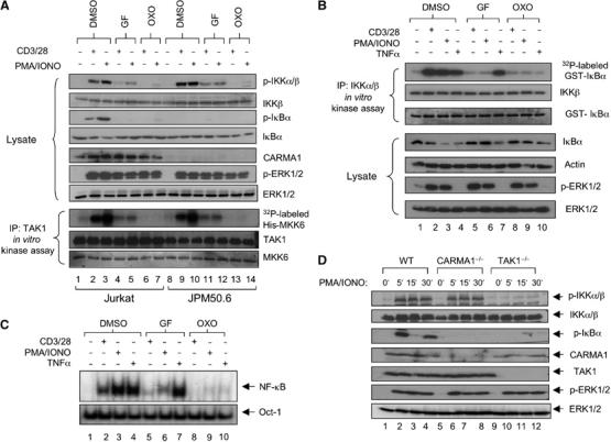
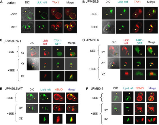
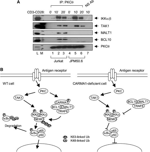
The IkappaB kinase (IKK) complex serves as the master regulator for the activation of NF-kappaB by various stimuli. It contains two catalytic subunits, IKKalpha and IKKbeta, and a regulatory subunit, IKKgamma/NEMO. The activation of IKK complex is dependent on the phosphorylation of IKKalpha/beta at its activation loop and the K63-linked ubiquitination of NEMO. However, the molecular mechanism by which these inducible modifications occur remains undefined. Here, we demonstrate that CARMA1, a key scaffold molecule, is essential to regulate NEMO ubiquitination upon T-cell receptor (TCR) stimulation. However, the phosphorylation of IKKalpha/beta activation loop is independent of CARMA1 or NEMO ubiquitination. Further, we provide evidence that TAK1 is activated and recruited to the synapses in a CARMA1-independent manner and mediate IKKalpha/beta phosphorylation. Thus, our study provides the biochemical and genetic evidence that phosphorylation of IKKalpha/beta and ubiquitination of NEMO are regulated by two distinct pathways upon TCR stimulation.


Figures

Figure 1
Figure 1
TCR-induced phosphorylation of IKKα/β is independent on CARMA1 or BCL10. (A) Jurkat or JPM50.6 cells (8 × 10⁶/sample) were stimulated with or without anti-CD3 and anti-CD28 (6 and 3 μg/ml, respectively) for various time points. Cell lysates were subjected to SDS–PAGE and analyzed by immunoblotting with indicated antibodies. (B) Jurkat or JPM50.6 cells (8 × 10⁶/sample) were stimulated with anti-CD3–CD28, PMA/ionomycin, or TNFα for various time points. The IKK complex was immunoprecipitated using anti-IKKα/β, and the immunoprecipitate was incubated with GST-IκBα(1–62) in the kinase reaction buffer for 30 min at 30°C. The reaction mixtures were subjected to SDS–PAGE and analyzed by autoradiography. (C) JPM50.6 cells or JPM50.6 cells reconstituted with wt CARMA1 (JPM50.6WT) were stimulated as indicated. Cell lysates were subjected to SDS–PAGE and analyzed by immunoblotting as described in (A). (D, E) Peripheral lymph nodes T cells from wt, CARMA1−/−, or Bcl10−/− mice were stimulated with PMA and ionomycin (D) or plate-bound anti-CD3–CD28 (E). Cell lysates were subjected to SDS–PAGE and analyzed by immunoblotting using indicated antibodies.
Figure 2
Figure 2
CARMA1 is required for the signal-induced ubiquitination of NEMO. (A, B) JPM50.6WT and JPM50.6 cells (2 × 10⁷/sample) were stimulated with or without PMA/ionomycin for various time points. The cell lysates were subjected to immunoprecipitation using anti-NEMO (A) or directly SDS–PAGE (B) and then analyzed by immunoblotting using indicated antibodies. (C) JPM50.6WT and JPM50.6 cells (3 × 10⁷/sample) were stimulated with or without PMA/ionomycin for various time points. NEMO was immunoprecipitated using anti-NEMO, and the resulting immunoprecipitates were subjected to SDS–PAGE and immunoblotting with indicated antibodies.
Figure 3
Figure 3
NEMO, but not its ubiquitination, is essential for IKKα/β phosphorylation. (A) Schematic representation of NEMO constructs. Domains: CC1, coiled coil 1; CC2, coiled coil 2; LZ, leucine zipper; ZF, zinc-finger. (B) NEMO-deficient Jurkat T cells (NEMO−/−) were reconstituted by transducing with lentiviral vectors encoding NEMO-FL, NEMO-ΔC, or NEMO-K399R. Mock-infected NEMO−/− cells were used as negative controls. NEMO was immunoprecipitated from the NEMO-reconstituted cells (mock, FL, ΔC, and K399R) (8 × 10⁶/sample) and the immunoprecipitates were probed for the constitutive association with IKKα/β. (C) The NEMO-reconstituted cells (mock, FL, ΔC, and K399R) (1 × 10⁷/sample) were stimulated with or without PMA/ionomycin or TNFα for 30 min. Nuclear extracts (10 μg) from these cells were incubated with 32P-labeled NF-κB or OCT-1 probes, followed by electrophoresis and autoradiography (upper panels). IKKα/β were immunoprecipitated from the cell lysates and subjected to in vitro kinase assay as described in Figure 1B (lower panels). (D) The NEMO-reconstituted cells (8 × 10⁶/sample) were stimulated with PMA/ionomycin for various time points. Cell lysates were subjected to SDS–PAGE and analyzed by immunoblotting using indicated antibodies. (E) NEMO constructs were coexpressed with or without CARMA1 in the presence of HA-K63-ubiquitin. NEMO constructs were immunoprecipitated and analyzed by immunoblotting.
Figure 4
Figure 4
Localization of CARMA1 in the lipid rafts is critical for NEMO ubiquitination. (A) JPM50.6 cells (2 × 10⁷) reconstituted with CARMA1 (50.6WT) or CARMA1(L808P) (50.6L808P) were stimulated with or without PMA/ionomycin for various time points. The immunoprecipitates were subjected to SDS–PAGE and analyzed by immunoblotting. (B) For detecting CARMA1-mediated association of BCL10 and NEMO in 50.6WT and 50.6L808P cells, co-immunoprecipitation was performed as described in Figure 2C. (C) The above lysates were also directly subjected to SDS–PAGE and analyzed by immunoblotting using indicated antibodies. (D) 50.6WT and 50.6L808P cells (1 × 10⁷/sample) were stimulated with or without PMA/ionomycin or TNFα for 30 min. Nuclear extracts (10 μg) from these cells were incubated with 32P-labeled NF-κB or OCT1 probe, followed by electrophoresis and autoradiography.
Figure 5
Figure 5
TAK1 is activated in a CARMA1-independent manner and associates with IKK upon TCR stimulation. Jurkat and JPM50.6 cells were stimulated with anti-CD3–CD28 for indicated time points. TAK1 was immunoprecipitated by using anti-TAK1 from these cells. The immunoprecipitates were subjected to immunoblotting using indicated antibodies (A) or to an in vitro kinase assay using either His-MKK6 (B) or IKKβ(K44A) (C) as substrates. In (C), lane 9 is a negative control using non-immune antibodies for stimulated Jurkat T cell lysates, whereas lane 10 is the untreated substrate incubated in kinase reaction buffer only. Kinase reaction mixtures were then subjected to SDS–PAGE and analyzed by autoradiography or immunoblotting using indicated antibodies.
Figure 6
Figure 6
PKC regulates TAK1, leading to IKKα/β phosphorylation and NF-κB activation in antigen receptor signaling pathway. (A) Jurkat and JPM50.6 (8 × 10⁶/sample) were preincubated with DMSO, PKC inhibitor, GF109203X (100 nM for 15 min) (GF), or TAK1 inhibitor, 5Z-7-oxozeaenol (2 μM for 30 min) (OXO), followed by stimulation with anti-CD3/CD28 antibodies or PMA/ionomycin. Cell lysates were prepared and subjected to SDS–PAGE followed by immunoblotting with indicated antibodies (upper panels). Part of these lysates was immunoprecipitated using anti-TAK1 and then subjected to in vitro kinase assay using MKK6 as substrates (lower panels). (B) The IKK complex was immunoprecipitated from Jurkat T cells following pretreatment with the above TAK1 or PKC inhibitors and stimulated with or without anti-CD3–CD28, PMA/ionomycin, or TNFα. The resulted immunoprecipitates were subjected to an in vitro kinase assay using GST-IκBα as substrates and detected by autoradiography. The resulted immunoprecipitates and cell lysates were also analyzed by immunoblotting using indicated antibodies. (C) By inhibiting TAK1 and PKC using the same conditions as in (A), nuclear extracts were prepared from Jurkat cells stimulated with or without anti-CD3–CD28, PMA/ionomycin, or TNFα, and subjected to EMSA using 32P-labeled NF-κB or Oct-1 probes. (D) wt, CARMA1−/−, and TAK1−/− chicken DT40 B cells were stimulated with PMA plus ionomycin for various time points. Total cell lysates were subjected to SDS–PAGE, followed by immunoblotting with indicated antibodies.
Figure 7
Figure 7
TAK1 is recruited to the immunological synapse in a CARMA1-independent manner. (A) Jurkat T cells or (B) JPM50.6 cells were labeled with FITC-conjugated cholera toxin B (FITC-CtxB) to localize the lipid rafts (green). The cells were incubated with SEE-untreated (upper panels) or SEE-treated (lower panels) Raji cells. The cells were stained with mouse anti-TAK1 and followed with ALEXA-conjugated goat anti-mouse antibodies for the localization of TAK1 (red). The formation of immunological synapse and the recruitment of TAK1 were visualized by confocal microscopy. (C) JPM50.6WT cells or (D) JPM50.6 cells were transfected with vectors encoding TAK1-GFP. Twenty-four hours after transfection, the transfected cells were first labeled with ALEXA-conjugated CTxB (red) and then incubated with SEE-untreated (top panels) or SEE-primed (middle and bottom panels) Raji cells. Images of Raji–T-cell conjugates and localization of the lipid rafts (red) are shown in DIC plus red and red-only channels. TAK1-GFP is shown in green. Yellow in the merge indicates the colocalization of TAK1-GFP with lipid rafts. The colocalization of TAK1-GFP and GM1-associated lipid rafts at the immunological synapse in SEE-primed cells is also shown in XZ focal axis images. (E) JPM50.6WT or (F) JPM50.6 cells were labeled with FITC-CtxB (green). The cells were incubated with SEE-untreated (upper panels) or SEE-primed (lower panels) Raji cells to allow the formation of Raji cell–T-cell conjugates. The cells were stained with Rabbit anti-NEMO antibodies and followed with ALEXA-conjugated goat anti-rabbit antibodies for the localization of NEMO (red). The formation of immunological synapse and the recruitment of NEMO were visualized using confocal microscopy. Images of Raji cell–T-cell conjugates and localization of the lipid rafts (green) are shown in DIC plus green, green-only, and red-only channels. Yellow in merge indicates the colocalization of NEMO with lipid rafts. The colocalization of NEMO and GM1-associated lipid rafts at the immunological synapse in SEE-treated cells is also shown in XZ focal axis images. All results are representative of three independent experiments. Asterisks indicate Raji cells.
Figure 8
Figure 8
The role of CARMA1 in antigen receptor signaling pathway. (A) Jurkat or JPM50.6 cells (4 × 10⁷/sample) were stimulated with or without anti-CD3/CD28 antibodies for various time points. PKCθ was immunoprecipitated using anti-PKCθ. The immunoprecipitates were subjected to SDS–PAGE and analyzed by immunoblotting using indicated antibodies. Lane ‘L' is the cell lysate from unstimulated Jurkat T cells and lane ‘7' is the immunoprecipitated sample of stimulated Jurkat T cells using non-immune antibodies. Lane ‘M' is a molecular weight marker. (B) The working model of antigen receptor-induced signaling events. In the wt cells, stimulation of antigen receptors leads to activation and initiation of receptor proximal signaling events that lead to activation of PKC. The activated PKC further activates TAK1. TAK1 associates and phosphorylates IKKα/β in a CARMA1-independent manner, whereas PKC phosphorylates CARMA1 which leads to the recruitment of the BCL10–MALT1–TRAF6 complex. The complex induces the ubiquitination of NEMO. The combined IKKα/β phosphorylation and NEMO ubiquitination activate the IKK complex, leading to the phosphorylation and degradation of IκBα, and subsequent NF-κB activation. In CARMA1-deficient cells, the BCL10–MALT1–TRAF6 complex is not activated and thus fails to induce the ubiquitination of NEMO. Without NEMO ubiquitination, the IKKα/β phosphorylation alone is insufficient to activate the IKK complex.

Similar articles

Cited by

References

    1. Baldwin AS Jr (1996) The NF-kappa B and I kappa B proteins: new discoveries and insights. Annu Rev Immunol 14: 649–683 - PubMed
    1. Bromley SK, Burack WR, Johnson KG, Somersalo K, Sims TN, Sumen C, Davis MM, Shaw AS, Allen PM, Dustin ML (2001) The immunological synapse. Annu Rev Immunol 19: 375–396 - PubMed
    1. Che T, You Y, Wang D, Tanner MJ, Dixit VM, Lin X (2004) MALT1/paracaspase is a signaling component downstream of CARMA1 and mediates T-cell receptor-induced NF-kappaB activation. J Biol Chem 279: 15870–15876 - PubMed
    1. Chen ZJ, Bhoj V, Seth RB (2006) Ubiquitin, TAK1 and IKK: is there a connection? Cell Death Differ 13: 687–692 - PubMed
    1. Egawa T, Albrecht B, Favier B, Sunshine MJ, Mirchandani K, O'Brien W, Thome M, Littman DR (2003) Requirement for CARMA1 in antigen receptor-induced NF-kappa B activation and lymphocyte proliferation. Curr Biol 13: 1252–1258 - PubMed

Publication types

MeSH terms