Review
. 2007 Apr;7(4):267-78.
doi: 10.1038/nri2054.

Scientific barriers to developing vaccines against avian influenza viruses

Affiliations
Review

Scientific barriers to developing vaccines against avian influenza viruses

Kanta Subbarao et al. Nat Rev Immunol. 2007 Apr.

Abstract

The increasing number of reports of direct transmission of avian influenza viruses to humans underscores the need for control strategies to prevent an influenza pandemic. Vaccination is the key strategy to prevent severe illness and death from pandemic influenza. Despite long-term experience with vaccines against human influenza viruses, researchers face several additional challenges in developing human vaccines against avian influenza viruses. In this Review, we discuss the features of avian influenza viruses, the gaps in our understanding of infections caused by these viruses in humans and of the immune response to them that distinguishes them from human influenza viruses, and the current status of vaccine development.


Conflict of interest statement

Kanta Subbarao and Tomy Joseph

Scientific barriers to developing vaccines against avian influenza viruses. Nature Reviews Immunology, published online 16 March 2007; doi: 10.1038/nri2054.

The laboratory of Dr Subbarao has a cooperative research and development agreement with MedImmune Vaccines to develop vaccines against potential pandemic strains of influenza.

Figures
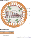
Figure 1
Figure 1. Schematic of an influenza A virus.
a | The influenza A virus particle has a lipid envelope that is derived from the host cell membrane. Three envelope proteins — haemagglutinin (HA), neuraminidase (NA) and an ion channel protein (matrix protein 2, M2) — are embedded in the lipid bilayer of the viral envelope. HA (rod shaped) and NA (mushroom shaped) are the main surface glycoproteins of influenza A viruses. The ratio of HA to NA molecules in the viral envelope usually ranges from 4:1 to 5:1. b | The HA glycoprotein is synthesized as an HA0 molecule that is post-translationally cleaved into HA1 and HA2 subunits; this cleavage is essential for virus infectivity. The HA glycoprotein is responsible for binding of the virus to sialic-acid residues on the host cell surface and for fusion of the viral envelope with the endosomal membrane during virus uncoating. The NA glycoprotein cleaves sialic-acid receptors from the cell membrane and thereby releases new virions from the cell surface. M2 functions as a pH-activated ion channel that enables acidification of the interior of the virion, leading to uncoating of the virion. Matrix protein 1 (M1), which is the most abundant protein in the virion, underlies the viral envelope and associates with the ribonucleoprotein (RNP) complex. Inside the M1 inner layer are eight single-stranded RNA molecules of negative sense that are encapsidated with nucleoprotein (NP) and associated with three RNA polymerase proteins — polymerase basic protein 1 (PB1), PB2 and polymerase acidic protein (PA) — to form the RNP complex. The PB1, PB2 and PA proteins are responsible for the transcription and replication of viral RNA. The virus also encodes a non-structural protein (NS) that is expressed in infected cells and a nuclear export protein (NEP). The location of NEP in the virion is not known.
Figure 2
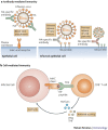
Figure 2. The adaptive immune response during infection with influenza virus.
Influenza viruses attach to the epithelial cell surface of host cells through binding of the viral haemagglutinin (HA) glycoprotein to cell-surface sialic-acid residues. The virion is internalized through endocytosis and fusion between host and viral membranes occurs in acidic vacuoles. Opening of the ion channel formed by matrix protein 2 (M2) triggers this fusion and the release of viral genes into the cytoplasm, through which they travel to the nucleus. Viral mRNAs are transported from the nucleus to the cytoplasm, where viral proteins are translated and progeny virions assemble and bud from the cell membrane. The release of progeny virions requires the action of the viral neuraminidase (NA) glycoprotein, which cleaves sialic-acid receptors from the host cell membrane. a | Antibodies specific for HA block virus attachment, thereby preventing infection of cells, or they can prevent fusion. Antibodies specific for NA bind virus to the cell, thereby preventing the release of virions. Antibodies specific for M2 bind virus to the cell and prevent the release of viral particles into the extracellular fluid. b | Cell-mediated immunity contributes to host resistance when CD8+T cells specific for viral proteins such as nucleoprotein (NP) or the RNA polymerase proteins polymerase basic protein 2 (PB2) and polymerase acidic protein (PA) recognize viral peptides presented by MHC class I molecules, resulting in the release of cytokines with antiviral activity — such as interferon-γ (IFNγ) and tumour-necrosis factor (TNF) — and perforins that mediate cytolysis of the infected cell. Lysis of the infected cell decreases the amount of virus released by the cell. The latter three mechanisms, NA-specific antibodies, M2-specific antibodies and CD8+ T cells, operate after a cell becomes infected. Only antibodies specific for HA can prevent infection; this is probably why they are the most effective mediators of immunity in vivo. TCR, T-cell receptor.
Figure 3
Figure 3. The eight-plasmid reverse-genetics system.
Generation of recombinant vaccines for pandemic influenza. a | Six plasmids encoding the internal proteins of the high-growth influenza A/Puerto Rico/8/34 (PR8) donor virus or the attenuated, cold-adapted (ca) H2N2 A/Ann Arbor/6/60 (AA) donor virus are co-transfected with two plasmids encoding the avian influenza virus haemagglutinin (HA; modified to remove virulence motifs, if necessary) and neuraminidase (NA) glycoproteins into qualified mammalian cells and the recombinant virus is then isolated. Recombinant viruses containing internal protein genes from the PR8 virus are used to prepare inactivated influenza virus vaccines. Recombinant viruses containing internal protein genes from the attenuated, cold-adapted AA virus are used to prepare live attenuated influenza virus vaccines. b | The generation of pandemic influenza vaccine viruses by classical reassortment. The reassortant viruses derive six internal protein genes from the vaccine donor virus and the HA and NA genes from the circulating avian influenza virus. The reassortant virus is selected using antisera specific for the HA and NA glycoproteins of the donor virus. M, matrix protein; NP, nucleoprotein; NS, non-structural protein; PA, polymerase acidic protein; PB, polymerase basic protein.

References

    1. Morens DM, Folkers GK, Fauci AS. The challenge of emerging and re-emerging infectious diseases. Nature. 2004;430:242–249. doi: 10.1038/nature02759. - DOI - PMC - PubMed
    1. Crawford PC, et al. Transmission of equine influenza virus to dogs. Science. 2005;310:482–485. doi: 10.1126/science.1117950. - DOI - PubMed
    1. Wright PF, Webster RG. Fields Virology. 2001. pp. 1533–1579.
    1. Fouchier RA, et al. Characterization of a novel influenza A virus hemagglutinin subtype (H16) obtained from black-headed gulls. J. Virol. 2005;79:2814–2822. doi: 10.1128/JVI.79.5.2814-2822.2005. - DOI - PMC - PubMed
    1. Treanor JJ, Tierney EL, Zebedee SL, Lamb RA, Murphy BR. Passively transferred monoclonal antibody to the M2 protein inhibits influenza A virus replication in mice. J. Virol. 1990;64:1375–1377. - PMC - PubMed

Publication types

Substances