

Gene expression profiling identifies FKBP39 as an inhibitor of autophagy in larval Drosophila fat body
- PMID: 17363962
- PMCID: PMC2084463
- DOI: 10.1038/sj.cdd.4402123
Gene expression profiling identifies FKBP39 as an inhibitor of autophagy in larval Drosophila fat body
Abstract
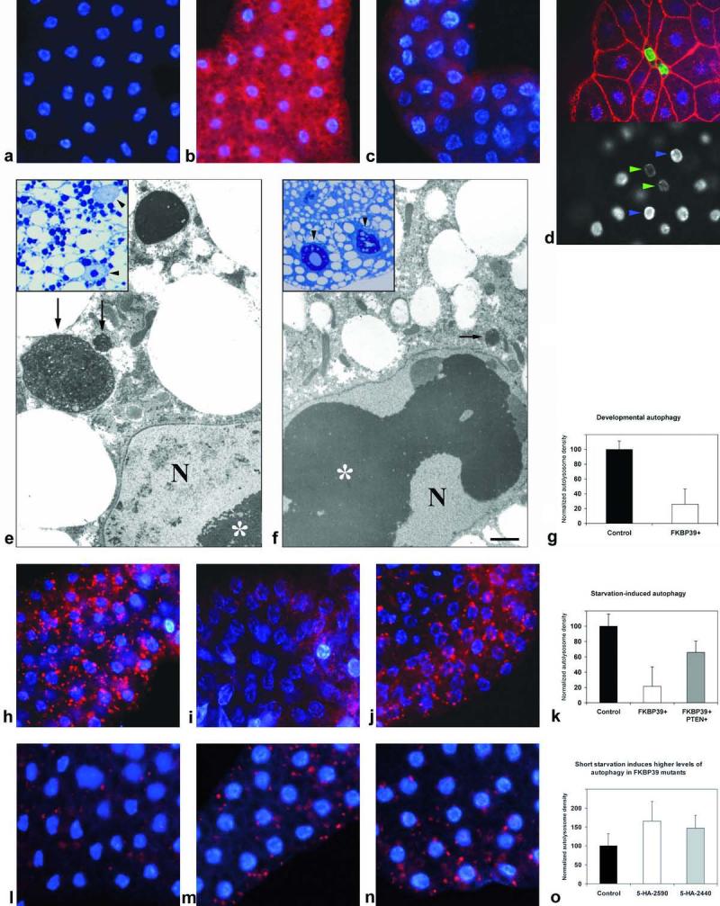
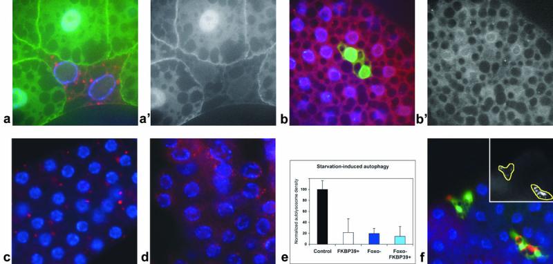
In Drosophila, the fat body undergoes a massive burst of autophagy at the end of larval development in preparation for the pupal transition. To identify genes involved in this process, we carried out a microarray analysis. We found that mRNA levels of the homologs of Atg8, the coat protein of early autophagic structures, and lysosomal hydrolases were upregulated, consistent with previous results. Genes encoding mitochondrial proteins and many chaperones were downregulated, including the inhibitor of eIF2alpha kinases and the peptidyl-prolyl cis-trans isomerase FK506-binding protein of 39 kDa (FKBP39). Genetic manipulation of FKBP39 expression had a significant effect on autophagy, potentially through modulation of the transcription factor Foxo. Accordingly, we found that Foxo mutants cannot properly undergo autophagy in response to starvation, and that overexpression of Foxo induces autophagy.
Figures
Similar articles
-
Loss of the starvation-induced gene Rack1 leads to glycogen deficiency and impaired autophagic responses in Drosophila.Autophagy. 2012 Jul 1;8(7):1124-35. doi: 10.4161/auto.20069. Epub 2012 May 7. Autophagy. 2012. PMID: 22562043 Free PMC article.
-
Balancing crosstalk between 20-hydroxyecdysone-induced autophagy and caspase activity in the fat body during Drosophila larval-prepupal transition.Insect Biochem Mol Biol. 2013 Nov;43(11):1068-78. doi: 10.1016/j.ibmb.2013.09.001. Epub 2013 Sep 12. Insect Biochem Mol Biol. 2013. PMID: 24036278
-
A dual function for Deep orange in programmed autophagy in the Drosophila melanogaster fat body.Exp Cell Res. 2006 Jul 1;312(11):2018-27. doi: 10.1016/j.yexcr.2006.03.002. Epub 2006 Apr 4. Exp Cell Res. 2006. PMID: 16600212
-
Molecular determinants of Drosophila immunophilin FKBP39 nuclear localization.Biol Chem. 2018 Apr 25;399(5):467-484. doi: 10.1515/hsz-2017-0251. Biol Chem. 2018. PMID: 29337690
-
Assays to monitor autophagy in Drosophila.Methods. 2014 Jun 15;68(1):134-9. doi: 10.1016/j.ymeth.2014.03.014. Epub 2014 Mar 22. Methods. 2014. PMID: 24667416 Free PMC article. Review.
Cited by
-
FKBP39 controls nutrient dependent Nprl3 expression and TORC1 activity in Drosophila.Cell Death Dis. 2021 Jun 2;12(6):571. doi: 10.1038/s41419-021-03860-z. Cell Death Dis. 2021. PMID: 34078879 Free PMC article.
-
XBP-1u suppresses autophagy by promoting the degradation of FoxO1 in cancer cells.Cell Res. 2013 Apr;23(4):491-507. doi: 10.1038/cr.2013.2. Epub 2013 Jan 1. Cell Res. 2013. PMID: 23277279 Free PMC article.
-
Relationship between growth arrest and autophagy in midgut programmed cell death in Drosophila.Cell Death Differ. 2012 Aug;19(8):1299-307. doi: 10.1038/cdd.2012.43. Epub 2012 May 4. Cell Death Differ. 2012. PMID: 22555456 Free PMC article.
-
MAPK/JNK signalling: a potential autophagy regulation pathway.Biosci Rep. 2015 Apr 22;35(3):e00199. doi: 10.1042/BSR20140141. Biosci Rep. 2015. PMID: 26182361 Free PMC article. Review.
-
Autophagy genes protect against Salmonella typhimurium infection and mediate insulin signaling-regulated pathogen resistance.Proc Natl Acad Sci U S A. 2009 Aug 25;106(34):14564-9. doi: 10.1073/pnas.0813319106. Epub 2009 Aug 10. Proc Natl Acad Sci U S A. 2009. PMID: 19667176 Free PMC article.
References
-
- Klionsky DJ, Cregg JM, Dunn WA, Jr., Emr SD, Sakai Y, Sandoval IV, Sibirny A, Subramani S, Thumm M, Veenhuis M, Ohsumi Y. A unified nomenclature for yeast autophagy-related genes. Dev Cell. 2003;5(4):539–45. - PubMed
-
- Levine B, Klionsky DJ. Development by self-digestion: molecular mechanisms and biological functions of autophagy. Dev Cell. 2004;6(4):463–77. - PubMed
-
- Mizushima N. The pleiotropic role of autophagy: from protein metabolism to bactericide. Cell Death Differ. 2005;12(Suppl 2):1535–41. - PubMed
-
- Sass M, Kovacs J. The effect of ecdysone on the fat body cells of the penultimate larvae of Mamestra brassicae. Cell Tissue Res. 1977;180(3):403–9. - PubMed
Publication types
MeSH terms
Substances
Grants and funding
LinkOut - more resources
Full Text Sources
Molecular Biology Databases
