

doi: 10.1016/j.cplett.2006.10.067.
Orientation Determination of Membrane-Disruptive Proteins Using Powder Samples and Rotational Diffusion: A Simple Solid-State NMR Approach
Affiliations
- PMID: 17364006
- PMCID: PMC1826912
- DOI: 10.1016/j.cplett.2006.10.067
Item in Clipboard
Orientation Determination of Membrane-Disruptive Proteins Using Powder Samples and Rotational Diffusion: A Simple Solid-State NMR Approach
Chem Phys Lett.
.
Abstract
The orientation of membrane proteins undergoing fast uniaxial rotation around the bilayer normal can be determined without macroscopic alignment. We show that the motionally averaged powder spectra exhibit their 0° frequency, [Formula: see text], at the same position as the peak of an aligned sample with the alignment axis parallel to the magnetic field. This equivalence is exploited to determine the orientation of a β-sheet antimicrobial peptide not amenable to macroscopic alignment, using (13)CO and (15)N chemical shifts from powder spectra. This powder sample approach permits orientation determination of naturally membrane-disruptive proteins in diverse environments and under magic-angle spinning.
Figures
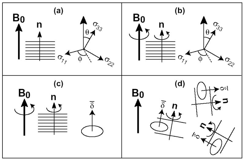
Schematics showing the equivalence between the 0°-aligned spectra and unoriented spectra. (a) Rigid 0°-aligned sample. (b, c) Mobile aligned sample. In (c), the motionally averaged tensor has the unique axis along the bilayer normal. (d) Mobile unoriented sample.
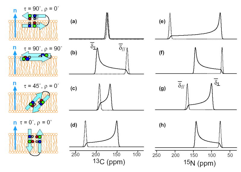
Calculated 13CO (a-d) and 15N (e-h) powder spectra (solid lines) and 0°-aligned spectra (dotted lines) of a uniaxially mobile β-sheet peptide for various orientations. (a, e) τ = 90°, ρ = 0°. (b, f) τ = 90°, ρ = 90°. (c, g) τ = 45°, ρ = 0°. (d, h) τ = 0°, ρ = 0°. Note the identity between the frequency of the 0°-aligned spectra and the
position of the powder spectra.
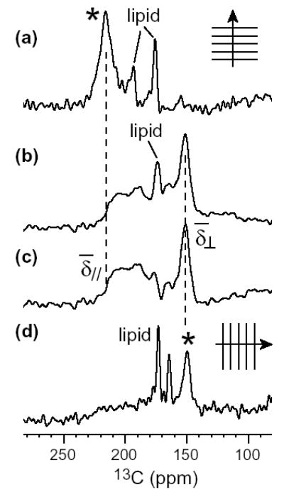
13CO spectra of Val16-labeled PG-1 in DLPC membrane. (a) 0°-aligned spectrum from ref. (7). (b) Powder spectrum obtained with CP. (c) Difference spectrum after subtracting the lipid background signal, showing only the peptide signal. (d) Spectrum of a 90°-aligned sample from ref. (7). The peptide signals in (a) and (d) are indicated by an asterisk.
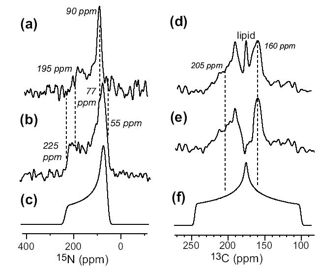
15N (a, b) and 13CO (d, e) static powder spectra of 15N-Phe4 and 13CO-Val6 labeled TP-I in DLPC membrane (1:15 molar ratio). 15N spectrum at 303 K (a) and 243 K (b) differ in the CSA. (c) Simulated rigid-limit 15N powder pattern. (d) 13CO spectrum of the peptide and the lipids at 303 K. (e) 13CO spectrum of the peptide after subtracting the lipid background signal. (f) Simulated rigid-limit 13CO spectrum.
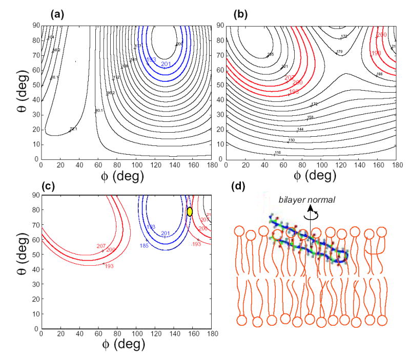
TP-I orientation from 15N and 13CO chemical shifts. (a) Phe4 15N chemical shift as a function of (θ, φ) of the bilayer normal in the molecule-fixed PDB system. (b) Val6 13CO chemical shift as a function of (θ, φ). The measured chemical shifts with the associated uncertainty are colored. (c) The 13CO and 15N chemical shifts overlap at (θ, φ) = (78°, 155°). (d) TP-I orientation with the bilayer normal in the vertical direction. The peptide and DLPC bilayers are drawn to scale.
References
Grants and funding
LinkOut - more resources
Full Text Sources
Molecular Biology Databases