. 2007 Apr;117(4):997-1007.
doi: 10.1172/JCI30379. Epub 2007 Mar 15.

Artificial lymph nodes induce potent secondary immune responses in naive and immunodeficient mice

Affiliations

Artificial lymph nodes induce potent secondary immune responses in naive and immunodeficient mice

Noriaki Okamoto et al. J Clin Invest. 2007 Apr.

Abstract

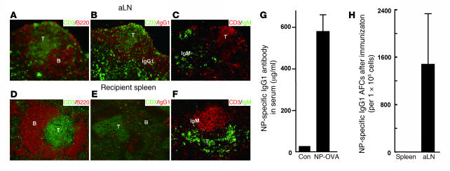
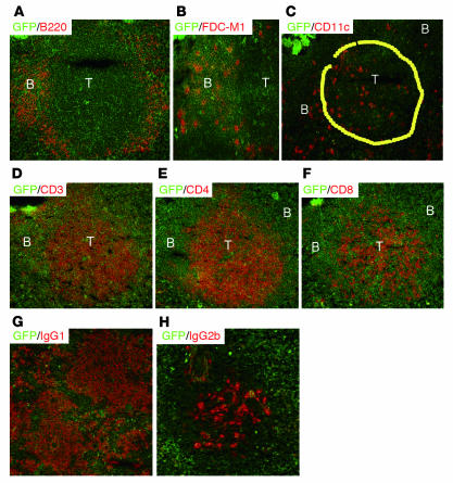
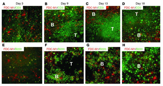
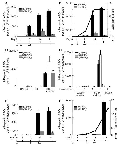
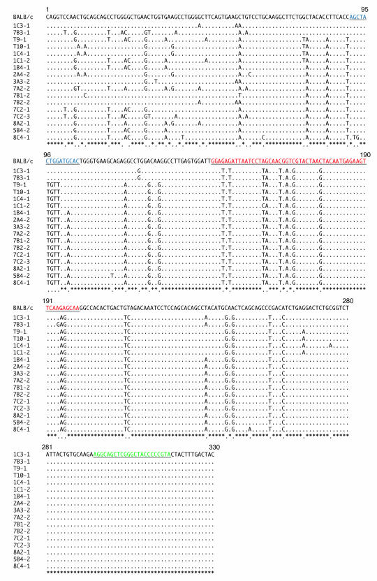
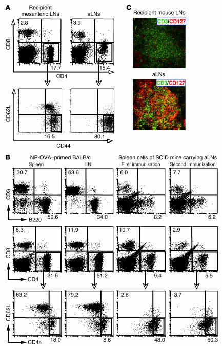
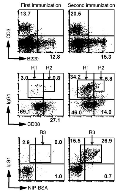
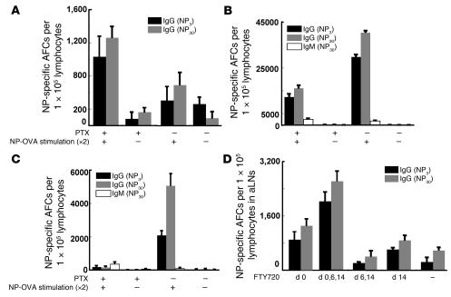
We previously demonstrated that artificial lymph nodes (aLNs) could be generated in mice by the implantation of stromal cell-embedded biocompatible scaffolds into their renal subcapsular spaces. T and B cell domains that form in aLNs have immune response functions similar to those of follicles of normal lymphoid tissue. In the present study, we show that the aLNs were transplantable to normal as well as SCID mice, where they efficiently induced secondary immune responses. Antigen-specific secondary responses were strongly induced in aLNs even 4 weeks after their transplantation. The antigen-specific antibody responses in lymphocyte-deficient SCID mice receiving transplanted aLNs were substantial. The cells from the aLNs migrated to the SCID mouse spleen and BM, where they expanded to generate large numbers of antigen-specific antibody-forming cells. Secondary responses were maintained over time after immunization (i.e., antigen challenge), indicating that aLNs can support the development of memory B cells and long-lived plasma cells. Memory CD4(+) T cells were enriched in the aLNs and spleens of aLN-transplanted SCID mice. Our results indicate that aLNs support strong antigen-specific secondary antibody responses in immunodeficient mice and suggest the possibility of future clinical applications.


Figures

Figure 1
Figure 1. Antigen-specific secondary IgG responses are induced in transplanted aLNs, but not in the spleens, of normal naive recipients even 4 weeks after transplantation.
The aLNs were formed first in NP-OVA–preimmunized BALB/c mice. After their formation, aLNs were removed and transplanted into renal subcapsular regions of naive, nonimmunized BALB/c mice. Mice were kept in cages for 3 weeks and then immunized i.v. with NP-OVA antigen (100 μg/mouse). Tissue sections of aLNs and sera of the recipient mice were collected 5 days after immunization. (AF) Immunohistochemical staining of aLNs (AC) and spleens of aLN-transplanted mice (DF) 4 weeks after transplantation. Antibodies used were as follows: FITC-labeled anti-mouse CD3, biotin-labeled anti-mouse B220, and Qdot 605 streptavidin conjugate (A and D); FITC-labeled anti-mouse CD3, biotin-labeled anti-mouse IgG1, and Qdot 605–labeled streptavidin (B and E); FITC-labeled anti-mouse IgM, biotin-labeled anti-mouse CD3, and Qdot 605 streptavidin conjugate (C and F). T, T cell; B, B cell; IgG1, IgG1 AFC; IgM, IgM AFC. Original magnification, ×100. (G) IgG1 NP-specific antibody titers in sera of aLN-transplanted BALB/c mice without immunization (Con) or immunized with NP-OVA. (H) Number of IgG1 NP-specific AFCs in aLNs and spleens of recipient BALB/c mice after immunization with NP-OVA.
Figure 2
Figure 2. Generation of aLNs.
Immunohistochemical staining shows that aLNs formed in NP-OVA–preimmunized EGFP transgenic BALB/c mice (AF). After their formation, aLNs were removed and transplanted into renal subcapsular regions of SCID mice, and then mice were immunized intravenously with NP-OVA. Immunohistochemical staining in the spleens of SCID mouse recipients carrying aLNs (G and H). Spleen cells were examined after the second immunization with NP-OVA. Increased numbers of IgG1 AFCs (G), compared with few IgG2 AFCs (H), were observed in SCID mouse spleens. Antibodies used were as follows: biotin-labeled anti-mouse B220 and Qdot 605 streptavidin conjugate (A); anti-mouse FDC-M1 and Alexa Fluor 594 conjugate (B); biotin-labeled anti-mouse CD11c and Qdot 605 streptavidin conjugate (C); biotin-labeled anti-mouse CD3 and Qdot 605 streptavidin conjugate; biotin-labeled anti-mouse CD4 and Qdot 605 streptavidin conjugate (E); biotin-labeled anti-mouse CD8 and Qdot 605 streptavidin conjugate (F); biotin-labeled anti-mouse IgG1 and Qdot 605 streptavidin conjugate (G); biotin-labeled anti-mouse IgG2b and Qdot 605 streptavidin conjugate (H). (C) The area circled in yellow shows the T cell area. Original magnification, ×100.
Figure 3
Figure 3. Kinetics of aLN formation.
Immunohistochemical staining of the cells in TEL-2–LTα stromal cell–embedded collagen sponges in BALB/c mice. The time course of aLN formation was examined 3, 9, 13, and 18 days after inoculation of collagen sponge into renal subcapsular space in BALB/c mice. Antibodies were as follows: FITC-labeled anti-mouse CD3, anti-mouse FDC-M1, and Alexa Fluor 594–conjugated anti-rat IgG (AD); FITC-labeled anti-mouse B220, anti-mouse FDC-M1, and Alexa Fluor 594–conjugated anti-rat IgG (EH).
Figure 4
Figure 4. Increased number of IgG1 NP-specific AFCs in aLN-transplanted SCID mice after immunization with NP-OVA.
(A and B) Number of NP-specific IgG AFCs in aLNs (A) or spleens (B) of aLN-transplanted SCID mice. Mice were immunized i.v. with NP-OVA on days 1 and 7 (100 μg/mouse and 10 μg/mouse, respectively) after transplantation of aLNs into SCID mice on day 0. The number of lymphocytes in spleens of aLN-transplanted SCID mice before and after NP-OVA immunization is denoted by the line (B). (C) Upon the second antigen stimulation, large numbers of IgG1 NP-specific AFCs were also detected in the BM of aLN-transplanted SCID mice. (D) Number of NP-specific IgG AFCs in spleens of recipient SCID and BALB/c mice. (E and F) Number of NP-specific IgG AFCs in aLNs (E) or spleens (F) of aLN-transplanted SCID mice immunized i.v. with NP-OVA on days 7 and 14 (100 μg/mouse and 10 μg/mouse, respectively) after transplantation of aLNs into SCID mice on day 0. The number of lymphocytes in spleens of aLN-transplanted SCID mice before and after NP-OVA immunization is denoted by the line (F).
Figure 5
Figure 5. VH gene sequence comparison of IgG1 antibodies (analyzed with IMGT/V-QUEST; see Methods) produced by hybridomas that were derived from the cell fusion of splenocytes with P3U1 plasmacytoma cells.
Alignment of VH gene sequences in group 1 of Table 3. CDRs are underlined. Blue, CDR1; red, CDR2; green, CDR3.
Figure 6
Figure 6. Enrichment of memory-type (CD44hiCD62Llo) CD4+ T cells in aLNs or spleens of aLN-transplanted SCID mice.
(A) FACS plots demonstrating an increase in the CD44hiCD62Llo memory-type CD4+ T cell population in aLNs compared with that in lymph nodes of recipient BALB/c mice. Downward pointing arrows indicate the CD44/CD62L profile of cells within the CD4+ T cell gate. (B) FACS plots demonstrating an increase in the CD44hiCD62Llo memory-type CD4+ T cell population in the spleens of aLN-transplanted SCID mice, regardless of antigen stimulation, compared with that of lymph nodes or spleens in normal BALB/c mouse preimmunized with the same doses of NP-OVA. Numbers in the plots in A and B indicate the percentage of cells within the CD62Llo/CD44+ gate. (C) Immunohistochemical staining of CD127+ T cells in aLNs. Three weeks after aLNs were formed in NP-OVA preimmunized BALB/c mice, aLNs and lymph nodes of recipient BALB/c mice were stained with FITC-labeled anti-mouse CD3, anti-mouse CD127 antibodies, and Alexa Fluor 594–conjugated anti-rat IgG. Most CD3+ T cells were also CD127+.
Figure 7
Figure 7. B cell profile in spleens of aLN-transplanted SCID mice.
FACS plots show germinal center B cells (R1), memory B cells (R2), and IgG1 NP-specific AFCs (R3) after the first and second immunizations. Numbers in the plots are the percentage of cells within the indicated gates.
Figure 8
Figure 8. Effect of PTX and FTY720 on the distribution of NP-specific AFCs after aLN transplant into SCID mice.
(AC) Number of NP-specific AFCs in aLNs (A), spleens (B), or BM (C) of recipient SCID mice transplanted with aLNs with or without PTX treatment. (D) Number of NP-specific AFCs in aLNs of SCID mice that received FTY720 administration on days 0, 6, and/or 14 relative to aLN transplantation (day 0). –, without PTX treatment and without NP-OVA stimulation.
Figure 9
Figure 9. Decreased expression of chemokine receptors on lymphocytes in aLNs (transplanted into SCID mice after the first immunization) by i.v. injection of NP-OVA.
(A) CCR7 and CXCR5 expression FACS profiles on lymphocytes in aLNs of recipient SCID or naive BALB/c mice. (B) Changes in chemokine receptor RNA levels as assessed by quantitative RT-PCR in aLNs of recipient SCID or naive BALB/c mice after antigen stimulation. –, no antigen stimulation; +, immunization with NP-OVA (100 μg/mouse). The y axis shows numbers of cells (A) and the relative RNA levels (B). The expression of each receptor RNA level was normalized to the expression of glyceradehyde-3-phosphate dehydrogenase (gapdh). *P < 0.1.

Similar articles

Cited by

References

    1. von Andrian U.H., Mempel T.R. Homing and cellular traffic in lymph nodes. Nat. Rev. Immunol. 2003;3:867–878. - PubMed
    1. Miyasaka M., Tanaka T. Lymphocyte trafficking across high endothelial venules: dogmas and enigmas. Nat. Rev. Immunol. 2004;4:360–370. - PubMed
    1. De Togni P., et al. Abnormal development of peripheral lymphoid organs in mice deficient in lymphotoxin. Science. 1994;264:703–707. - PubMed
    1. Banks T.A., et al. Lymphotoxin-alpha-deficient mice. Effects on secondary lymphoid organ development and humoral immune responsiveness. J. Immunol. . 1995;155:1685–1693. - PubMed
    1. Fu Y.X., Chaplin D.D. Development and maturation of secondary lymphoid tissues. Annu. Rev. Immunol. 1999;17:399–433. - PubMed

Publication types

Substances