. 2007 Apr;117(4):1029-36.
doi: 10.1172/JCI29472. Epub 2007 Mar 15.

Ablation of Cbl-b provides protection against transplanted and spontaneous tumors

Affiliations

Ablation of Cbl-b provides protection against transplanted and spontaneous tumors

Jeffrey Y Chiang et al. J Clin Invest. 2007 Apr.

Abstract

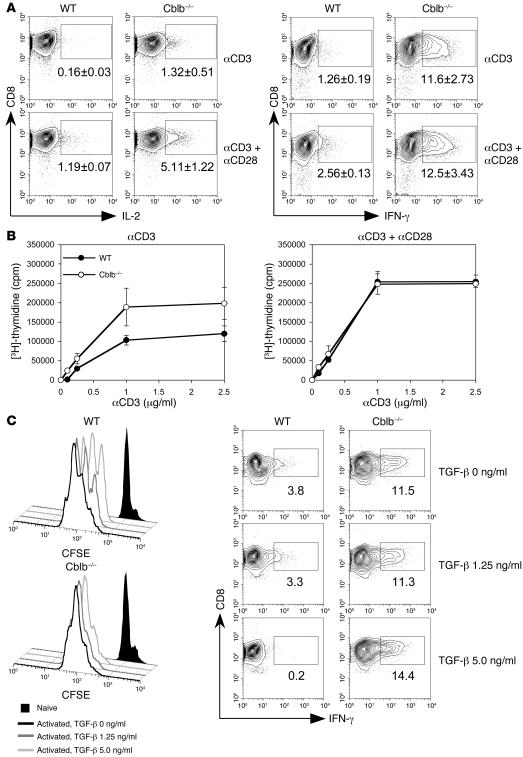
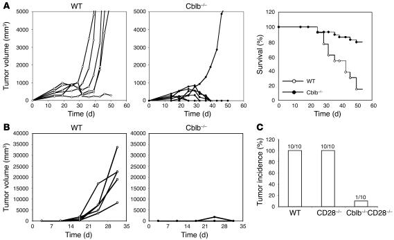
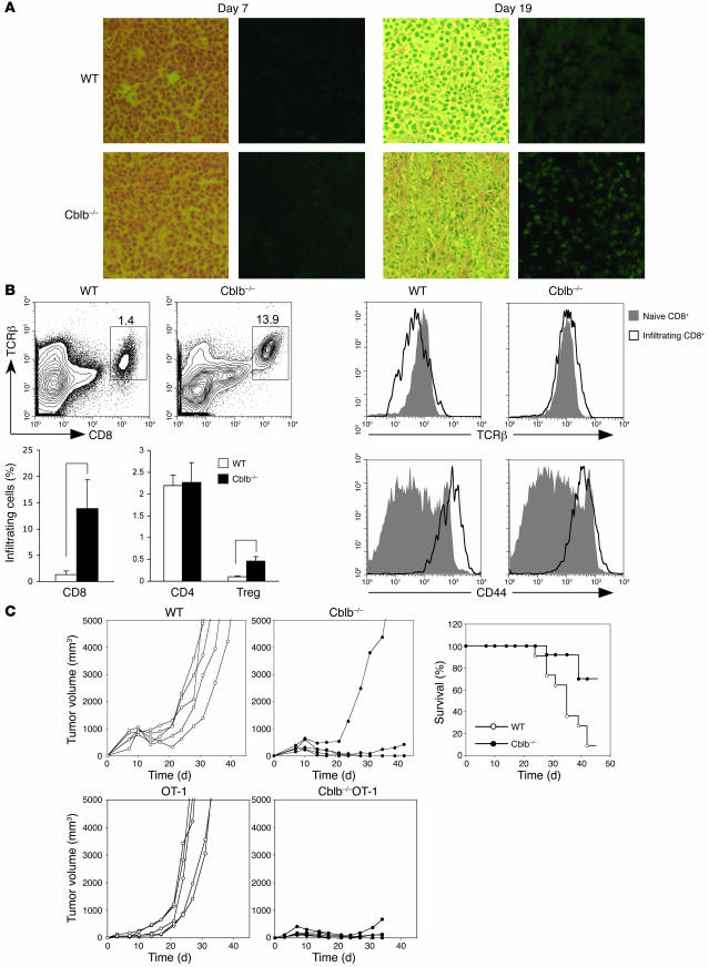
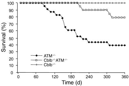
A significant challenge to efforts aimed at inducing effective antitumor immune responses is that CD8(+) T cells, which play a prominent role in these responses, may be unable to respond to tumors that lack costimulatory signals and that are protected by an immune suppressive environment such as that mediated by TGF-beta produced by tumor cells themselves or by infiltrating Tregs, often resulting in tolerance or anergy of tumor-specific T cells. Here we show that the in vitro activation of Cblb(-/-) CD8(+) T cells does not depend on CD28 costimulation and is resistant to TGF-beta suppression. In vivo studies further demonstrated that Cblb(-/-) mice, but not WT controls, efficiently rejected inoculated E.G7 and EL4 lymphomas that did not express B7 ligands and that introduction of the Cblb(-/-) mutation into tumor-prone ataxia telangiectasia mutated-deficient mice markedly reduced the incidence of spontaneous thymic lymphomas. Immunohistological study showed that E.G7 tumors from Cblb(-/-) mice contained massively infiltrating CD8(+) T cells. Adoptive transfer of purified Cblb(-/-) CD8(+) T cells into E.G7 tumor-bearing mice led to efficient eradication of established tumors. Thus, our data indicate that ablation of Cbl-b can be an efficient strategy for eliciting immune responses against both inoculated and spontaneous tumors.


Figures

Figure 1
Figure 1. CD28-independent proliferation and cytokine production by Cblb–/– CD8+ T cells.
(A) IL-2 and IFN-γ production. Purified CD8+ T cells from WT and Cblb–/– mice were stimulated with either plate-bound anti-CD3 or plate-bound anti-CD3 plus soluble anti-CD28 antibodies. IL-2– and IFN-γ–producing cells were visualized by intracellular staining and analyzed by flow cytometry. Shown are contour plots of intracellular staining for IL-2 and IFN-γ expression in CD8+ T cells. Percentages of IL-2– and IFN-γ–producing cells are indicated in the plots as mean ± SD from 3 independent experiments. The boxed regions indicate the gates used for calculation of the percentage of CD8+ T cells staining positive for IFN-γ. (B) TCR-induced proliferative response. Purified CD8+ T cells from WT and Cblb–/– mouse lymph nodes and spleens were stimulated with various concentrations of anti-CD3 antibodies in the presence or absence of anti-CD28 antibodies. Cell proliferation was determined by [3H]-thymidine incorporation and presented as mean ± SD for triplicate samples. Shown are representatives of 3 independent experiments. (C) Resistance of Cblb–/– CD8+ T cells to TGF-β suppression. Histograms (left) show CSFE intensities of labeled Cblb–/– and WT CD8+ T cells after 3 days of anti-CD3 and anti-CD28 stimulation. Cells were cultured in the absence or presence of different concentrations of TGF-β as indicated in the figure. Contour plots (bottom) show the IFN-γ production in the absence or presence of TGF-β. Percentages of IFN-γ+ cells are indicated in the plots.
Figure 2
Figure 2. Eradication of inoculated tumors in Cblb–/– mice.
(A) Growth rates of inoculated E.G7 tumors and survival of tumor-bearing mice. After 106 E.G7 cells were inoculated into the flanks of WT or Cblb–/– mice by s.c. injection, tumor growth was documented as total volume of tumor size. Left and middle: growth rates of E.G7 tumors in 1 representative experiment of 5 or more independent experiments. Each curve represents 1 mouse. Right: percentages of surviving mice (WT, n = 13; Cblb–/–, n = 29) during the course of tumor growth. When the tumor volume reached approximately 5,000 mm3, the mice were euthanized and recorded as dead. (B) Growth rates of EL4 tumors in Cblb–/– and WT mice. EL4 cells (5 × 10⁴ cells/mouse) were injected s.c. into the flanks of 5 WT and 5 Cblb–/– mice, and tumor growth was monitored over time. WT mice included both Cblb+/+ (n = 16) and Cblb+/– (n = 4) mice. The data presented are representative of 4 independent experiments. (C) Rejection of EL4 tumors by Cblb–/–CD28–/– mice. EL4 cells (5 × 10⁴ cells/mouse) were injected, and tumor growth was monitored as described in B. The number of mice with tumor growths is indicated at the top of each column.
Figure 3
Figure 3. Tumor rejection in Cblb–/– mice is mediated by Cblb–/– CD8+ T cells.
(A) Immunohistology of tumor-infiltrating CD8+ cells. Shown are sections of tumors stained with H&E (left) or anti-CD8 antibody (right). CD8+ cells are FITC positive (green). Tumors were from WT and Cblb–/– mice at the seventh or nineteenth day after inoculation. (B) Flow cytometric analysis of tumor infiltrates. Tumor infiltrates were prepared from tumors from WT or Cblb–/– mice. These cells were stained with anti-TCRβ, anti-CD8, anti-CD4, and anti-CD44 antibodies and analyzed on LSR II. Foxp3+ (Treg) cells were identified by intracellular staining with an anti-Foxp3 antibody according to manufacture protocol. Shown at the top left are contour plots of CD8 and TCRβ expression on tumor-infiltrating cells from 1 of 3 independent experiments. The percentages of CD8+ TCRβ+ cells are indicated in the plots. The percentages of CD8+ TCRβ+, CD4+, and Treg infiltrates in total tumor infiltrates are summarized and shown as bars (bottom left). *P < 0.001, **P < 0.005. Histograms show the levels of TCRβ and CD44 expression on the infiltrating CD8+ T cells (right). (C) Eradication of established E.G7 tumors by adoptively transferred Cblb–/– or Cblb–/– OT1 CD8+ T cells. 106 E.G7 cells were inoculated into C57BL/6 mice by s.c. injection. Seven days after the inoculation, 3 × 10⁶ purified WT or Cblb–/– CD8+ T cells (top) or WT OT1 or Cblb–/– OT1 CD8+ T cells (bottom) were transferred into the tumor-bearing mice by i.v. injection. Shown at the left are the tumor volumes at different time points after the CD8+ T cell transfer. Genotypes of the donor cells are indicated on the top of each plot. Results are from 1 of 5 or more independent experiments. The plot in the right panel shows percentages of surviving recipient mice (WT, n = 11; Cblb–/–, n = 13) that received WT or Cblb–/– CD8+ T cells. When the tumor volume reached approximately 5,000 mm3, the mice were euthanized and recorded as dead.
Figure 4
Figure 4. Tumor incidence in Cblb–/–ATM–/– double-mutant mice.
Numbers of surviving ATM–/– single-mutant (including 5 ATM–/–Cblb+/+ and 18 ATM–/–Cblb+/–) and Cblb–/–ATM–/– (n = 21) mice are shown. Cblb–/–ATM–/– double-mutant mice exhibited reduced incidence and delayed onset of spontaneous lymphoma.

Similar articles

Cited by

References

    1. Boon T., et al. Tumor antigens recognized by T lymphocytes. Annu. Rev. Immunol. 1994;12:337–365. - PubMed
    1. Houghton A.N., Gold J.S., Blachere N.E. Immunity against cancer: lessons learned from melanoma. Curr. Opin. Immunol. 2001;13:134–140. - PubMed
    1. Rosenberg S.A. Progress in human tumour immunology and immunotherapy. Nature. 2001;411:380–384. - PubMed
    1. Rosenberg S.A. A new era of cancer immunotherapy: converting theory to performance. CA Cancer J. Clin. 1999;49:70–73. - PubMed
    1. Boon T., van der Bruggen P. Human tumor antigens recognized by T lymphocytes. J. Exp. Med. 1996;183:725–729. - PMC - PubMed

Publication types

MeSH terms

Substances