

Case Reports
doi: 10.1080/13693780601045166.
Subcutaneous cryptococcosis due to Cryptococcus diffluens in a patient with sporotrichoid lesions case report, features of the case isolate and in vitro antifungal susceptibilities
Affiliations
- PMID: 17365654
- PMCID: PMC2714484
- DOI: 10.1080/13693780601045166
Item in Clipboard
Case Reports
Subcutaneous cryptococcosis due to Cryptococcus diffluens in a patient with sporotrichoid lesions case report, features of the case isolate and in vitro antifungal susceptibilities
Med Mycol.
2007 Mar.
Abstract
Environmental fungi, in particular primary pathogens and Cryptococcus spp. can be responsible for skin lesions mimicking sporotrichosis. In this paper, we report a case of subcutaneous cryptococcosis in an apparently healthy, young male patient due to a non-C. neoformans Cryptococcus species, C. diffluens. The isolate showed in vitro phenotypic switching that may affect virulence and host inflammatory and immune responses, and in vitro resistance to amphotericin B and 5-flucytosin. This species shares several phenotypic traits with C. neoformans, and, therefore, decisive diagnosis should be based on biopsy and culturing results followed by molecular identification.
Figures
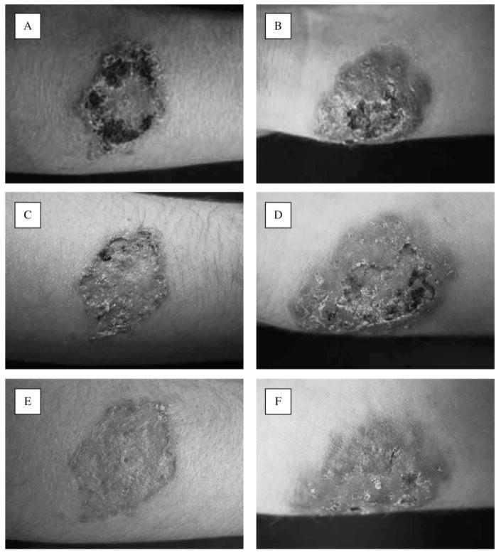
(A–F) Clinical appearance of the lesions, (A, B) before treatment, (C, D) at 10 days after treatment and (D, E) after 25 days of therapy.
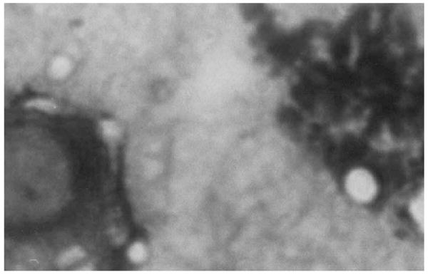
Imprinted tissue biopsy preparation showing encapsulated yeast cells (Giemsa stain, 100×).
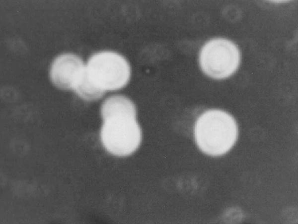
Microscopical morphology of the isolate: encapsulated yeast cells (India ink, 100×).
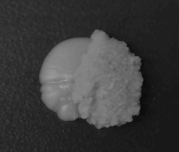
Sectored colony with mucoid (MC) and wrinkled (WR) colony parts after growth on SDA for 72 h.
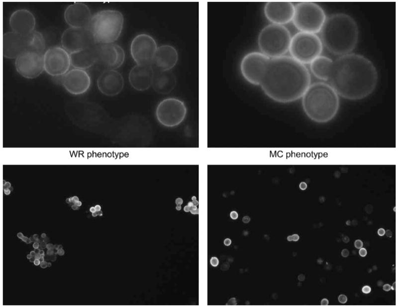
Indirect immunofluorescens with Mab 2H1 of wrinkled (WR) and mucoid (MC) cells respectively (upper panel: 100× and lower panel: 40×).
Similar articles
-
Susceptibility testing of Cryptococcus diffluens against amphotericin B, flucytosine, fluconazole, itraconazole, voriconazole and posaconazole.Med Mycol. 2009 Mar;47(2):169-76. doi: 10.1080/13693780802213407. Epub 2008 Jul 24. Med Mycol. 2009. PMID: 18654925
-
A dark strain in the Fusarium solani species complex isolated from primary subcutaneous sporotrichioid lesions associated with traumatic inoculation via a rose bush thorn.Med Mycol. 2010 Feb;48(1):103-9. doi: 10.3109/13693780902829250. Med Mycol. 2010. PMID: 20055744
-
Combination of amphotericin B with flucytosine is active in vitro against flucytosine-resistant isolates of Cryptococcus neoformans.Antimicrob Agents Chemother. 2007 Jan;51(1):383-5. doi: 10.1128/AAC.00446-06. Epub 2006 Oct 16. Antimicrob Agents Chemother. 2007. PMID: 17043122 Free PMC article.
-
Fungaemia due to Cryptococcus laurentii and a review of non-neoformans cryptococcaemia.Mycoses. 1998 Sep-Oct;41(7-8):277-80. doi: 10.1111/j.1439-0507.1998.tb00338.x. Mycoses. 1998. PMID: 9861831 Review.
-
Susceptibility profile of clinical isolates of non-Cryptococcus neoformans/non-Cryptococcus gattii Cryptococcus species and literature review.Med Mycol. 2010 Feb;48(1):90-6. doi: 10.3109/13693780902756073. Med Mycol. 2010. PMID: 19235546 Review.
Cited by
-
Infections due to Rare Cryptococcus Species. A Literature Review.J Fungi (Basel). 2021 Apr 7;7(4):279. doi: 10.3390/jof7040279. J Fungi (Basel). 2021. PMID: 33917243 Free PMC article. Review.
-
Presence of C. albidus, C. laurentii and C. uniguttulatus in crop and droppings of pigeon lofts (Columba livia).Mycopathologia. 2010 Apr;169(4):315-9. doi: 10.1007/s11046-009-9262-0. Epub 2009 Dec 10. Mycopathologia. 2010. PMID: 20012367
-
Cryptococcosis: epidemiology, fungal resistance, and new alternatives for treatment.Eur J Clin Microbiol Infect Dis. 2013 Nov;32(11):1377-91. doi: 10.1007/s10096-013-1915-8. Epub 2013 Jul 4. Eur J Clin Microbiol Infect Dis. 2013. PMID: 24141976 Review.
-
IMA Genome - F16 : Draft genome assemblies of Fusarium marasasianum, Huntiella abstrusa, two Immersiporthe knoxdaviesiana isolates, Macrophomina pseudophaseolina, Macrophomina phaseolina, Naganishia randhawae, and Pseudocercospora cruenta.IMA Fungus. 2022 Feb 23;13(1):3. doi: 10.1186/s43008-022-00089-z. IMA Fungus. 2022. PMID: 35197126 Free PMC article. No abstract available.
-
Phenotypic switching of Cryptococcus neoformans and Cryptococcus gattii.Mycopathologia. 2008 Oct;166(4):181-8. doi: 10.1007/s11046-008-9137-9. Epub 2008 Jun 21. Mycopathologia. 2008. PMID: 18568418 Free PMC article. Review.
References
-
- Boekhout T, Gueho E. Basidiomycetous yeasts. In: Howard Dexter H., editor. Pathogenic Fungi in Humans and Animals. 2. New York: Marcel Dekker; 2003. pp. 535–564.
-
- De Hoog GS, Guarro J, Gené JL, et al. Atlas of Clinical Fungi. 2. Utrecht/Reus: Centraalbureau voor Schimmelcultures, Universitat Rovira i Virgili; 2000. pp. 132–133.
-
- Bauters TGM, Swinne D, Boekhout T, et al. Repeated isolation of Cryptococcus laurentii in the oropharynx despite treatment with fluconazole. Mycopathologia. 2002;153:133–135. - PubMed
-
- Averbuch D, Boekhout T, Falk R, et al. Fungemia in a cancer patient caused by fluconazole resistant Cryptococcus laurentii. Med Mycol. 2002;40:479–484. - PubMed
Publication types
MeSH terms
Substances
Associated data
- Actions
- Actions
Grants and funding
LinkOut - more resources
Full Text Sources
Molecular Biology Databases
Miscellaneous
