. 2007 Mar;3(3):e35.
doi: 10.1371/journal.ppat.0030035.

Dynorphin activates quorum sensing quinolone signaling in Pseudomonas aeruginosa

Affiliations

Dynorphin activates quorum sensing quinolone signaling in Pseudomonas aeruginosa

Olga Zaborina et al. PLoS Pathog. 2007 Mar.

Erratum in

  • PLoS Pathog. 2007 May;3(5):e67. Petroff, Elaine [corrected to Petrof, Elaine O]

Abstract

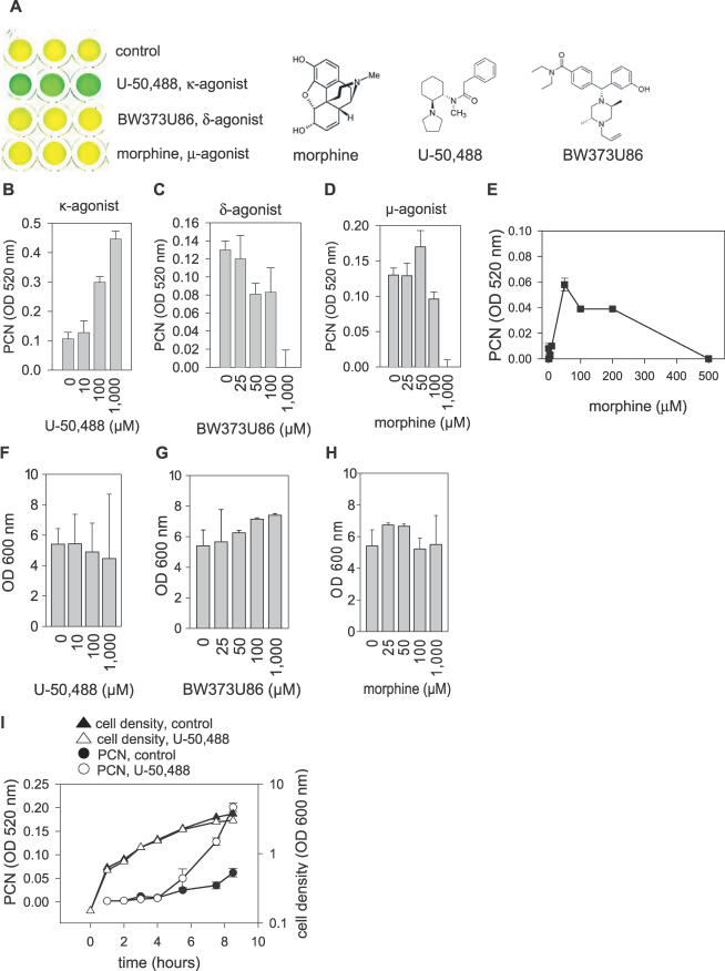
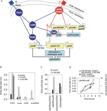
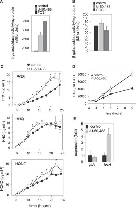
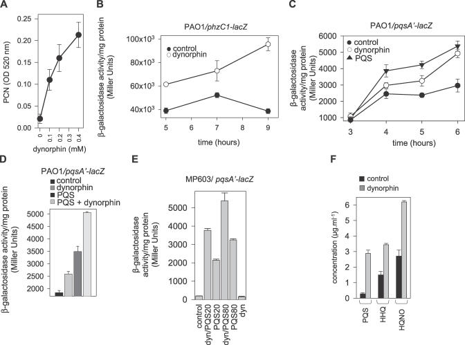
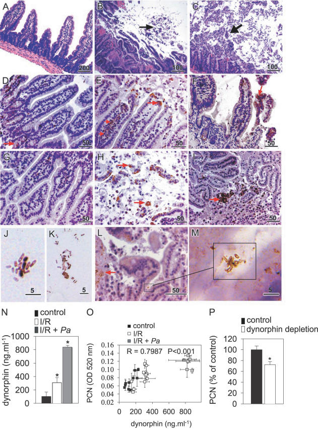
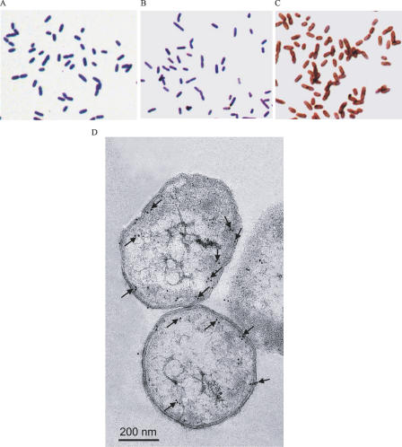
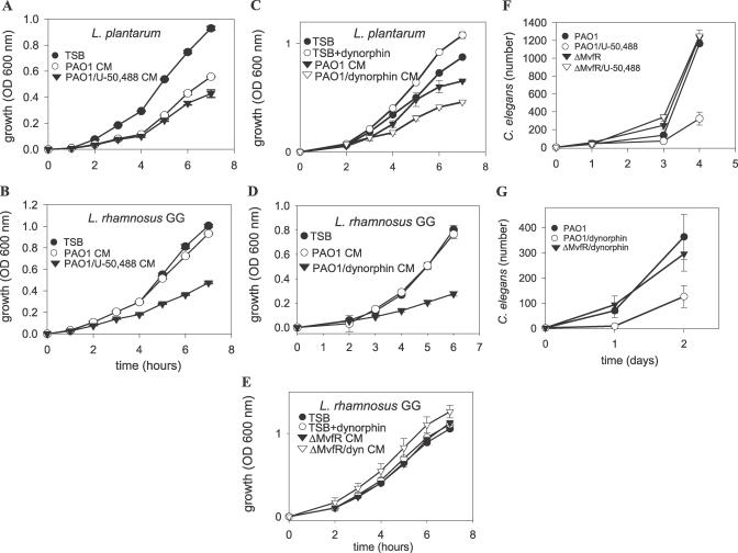
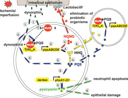
There is now substantial evidence that compounds released during host stress directly activate the virulence of certain opportunistic pathogens. Here, we considered that endogenous opioids might function as such compounds, given that they are among the first signals to be released at multiple tissue sites during host stress. We tested the ability of various opioid compounds to enhance the virulence of Pseudomonas aeruginosa using pyocyanin production as a biological readout, and demonstrated enhanced virulence when P. aeruginosa was exposed to synthetic (U-50,488) and endogenous (dynorphin) kappa-agonists. Using various mutants and reporter strains of P. aeruginosa, we identified involvement of key elements of the quorum sensing circuitry such as the global transcriptional regulator MvfR and the quorum sensing-related quinolone signaling molecules PQS, HHQ, and HQNO that respond to kappa-opioids. The in vivo significance of kappa-opioid signaling of P. aeruginosa was demonstrated in mice by showing that dynorphin is released from the intestinal mucosa following ischemia/reperfusion injury, activates quinolone signaling in P. aeruginosa, and enhances the virulence of P. aeruginosa against Lactobacillus spp. and Caenorhabditis elegans. Taken together, these data demonstrate that P. aeruginosa can intercept opioid compounds released during host stress and integrate them into core elements of quorum sensing circuitry leading to enhanced virulence.


Conflict of interest statement

Competing interests. The authors have declared that no competing interests exist.

Figures

Figure 1
Figure 1. U-50,488 Induces P. aeruginosa PAO1 to Produce PCN
Error bars are mean ± SD. (A) Changes in cell culture color in PAO1 following overnight exposure to 1 mM of κ- (U-50,488), δ- (BW373U86), and μ- (morphine) opioid receptor agonists. (B–D) Production of PCN in response to (B) κ-agonist U-50,488, (C) δ-agonist BW373U86, and (D) μ-agonist morphine. (E) Dose response curve of PCN production in PAO1 exposed to morphine. (F–H) Effect of opioids on growth of P. aeruginosa PAO1. (I) Dynamic tracking of PCN production in PAO1 exposed to 200 μM U-50,488.
Figure 2
Figure 2. Role of Proximal QS Regulatory Protein MvfR in Enhanced PCN Production in Response to U-50,488
Error bars, mean ± SD. (A) Schematic of PCN regulation in P. aeruginosa. (B) PCN production in ΔRhlI and ΔLasI mutants exposed to exogenous C4-HSL, 1 mM and U-50,488, 1 mM. (C) Production of PCN in ΔMvfR complemented with mvfR (ΔMvfR/mvfR) and ΔGacA complemented with gacA (ΔGacA/gacA) genes on a pUPC24 plasmid, or transformed with blank plasmid (ΔMvfR/pUCP24, ΔGacA/pUCP24) in the absence (control) or presence of U-50,488, 1 mM. (D) Dynamic tracking of PCN production in complemented mutant ΔMvfR/mvfR grown in the presence of 200 μM U-50,488.
Figure 3
Figure 3. U-50,488 Induces pqsABCDE Expression, Biosynthesis of HQNO, HHQ, and PQS, and Stimulates PA-IL Expression
Error bars, mean ± SD. (A) Effect of U-50,488, 200 μM and PQS, 100 μM on pqsA'-lacZ expression in P. aeruginosa strain PAO1/pGX5 following 5 h of incubation. (B) Effect of U-50,488, 200 μM and PQS, 100 μM on mvfR'-lacZ expression in P. aeruginosa strain PAO1/pGX1 following 5 h of incubation. (C) Effect of U-50,488, 200 μM on HQNO, HHQ, and PQS production by P. aeruginosa PAO1. * p < 0.01. (D) Dynamic tracking of PA-IL expression using PA-IL reporter strain P. aeruginosa 27853/PLL-EGFP. (E) Real-time PCR of lecA encoding PA-IL and the housekeeping gene gltA encoding citrate synthase in P. aeruginosa PAO1 grown to OD600nm = 3.0 in the presence of 200 μM of U-50,488. The graph was made based on the Ct levels for gltA, 20.26±0.81 (control) versus 20.78±0.26 (U-50,488); and for lecA, 29.53±0.43 (control) versus 27.42±0.97 (U-50,488). Ct levels for lecA blank control (no template) were ∼ 40.
Figure 4
Figure 4. Dynorphin Activates MvfR-Dependent Pathway in P. aeruginosa PAO1
Error bars, mean ± SD. (A) Dose-dependent effect of dynorphin on PCN production. (B) Expression of phzC1-lacZ in PAO1/pMW303 in the absence (control) or presence of 100 μM of dynorphin. (C) Dynamic tracking of expression of pqsA'-lacZ in PAO1/pGX5 grown in the presence of dynorphin, 100 μM, or PQS, 100 μM. (D) Expression of pqsA'-lacZ in PAO1/pGX5 in response to dynorphin, 100 μM, or PQS, 100 μM, or dynorphin plus PQS (100 μM each) determined after 5 h of incubation. (E) Expression of pqsA'-lacZ in MP603/pGX5 in response to dynorphin, 100 μM, or PQS (20 and 80 μM), or sum of dynorphin (100 μM) and PQS (20 μM); or sum of dynorphin (100 μM) and PQS (80 μM) determined after 5 h of incubation. (F) Concentration of HQNO, HHQ, and PQS in P. aeruginosa PAO1 after 8 h of growth in the absence (control) or presence of dynorphin, 100 μM.
Figure 5
Figure 5. In Vivo Production of Dynorphin in the Mouse Intestine during I/R
(A–C) Histology of small intestine from (A) control mice demonstrating intact mucosal epithelium, and (B) I/R and (C) I/R + Pa mice showing disruption of mucosal epithelium with desquamated epithelial cells inside the intestinal lumen (black arrows). (D–F, G–I) Immunohistochemistry of the small intestine from (D and G) control mice demonstrating scarce dynorphin localized to the epithelial crypt (brown staining, red arrow), and following (E and H) I/R and (F and I) I/R + Pa showing dynorphin migration up the lamina propria (red arrows, [E]) and its accumulation on villi and within the lumen. Scale bars are in μm. (J–M) Images of luminal bacteria from mouse small intestine subjected to I/R + Pa demonstrating (J) transfer of dynorphin to bacteria (brown-colored bacteria) and (K) positive dynorphin stained bacteria bound to desquamated epithelia; (I) abundant epithelial dynorphin staining (red arrow), and (M) co-localization of dynorphin stained luminal bacteria to sites of dynorphin accumulation at the epithelial surface. Scale bars are in μm. (N) Concentration of dynorphin in filtered luminal flushes isolated from intestine of control mice and mice subjected to I/R and I/R + Pa. n = 10/group, * p <0.001. (O) Correlation analysis between dynorphin concentration in luminal flushes and their ability to induce PCN production in PAO1. (P) Effect of dynorphin depletion with anti-dynorphin antibody on the ability of luminal flush samples to produce PCN in PAO1, n = 6/group, * p <0.005. Error bars, mean ± SD.
Figure 6
Figure 6. Dynorphin Binds to P. aeruginosa In Vitro, and Enters the Bacterial Cell Cytoplasm
(A–C) Binding of dynorphin to P. aeruginosa; (A) negative control demonstrating no dynorphin staining when cells were not incubated with dynorphin; (B) negative control demonstrating no dynorphin staining when cell were incubated with dynorphin but primary anti-dynorphin antibodies were omitted from staining procedure; and (C) positive staining (brown color) of P. aeruginosa incubated with dynorphin followed by whole procedure of immunostaining. (D) Immunoelectron microscopy of P. aeruginosa PAO1 cells incubated with dynorphin, 100 μM. Black arrows show 10-nm gold spots indicating the presence of dynorphin.
Figure 7
Figure 7. κ-Opioid Receptor Agonists Activate Virulence of P. aeruginosa against Probiotic Bacteria and C. elegans
Error bars, mean ± SD. (A and B) The exposure of P. aeruginosa PAO1 to U-50,488, 200 μM, increases the inhibiting effect of its extracellular milieu (conditioned media) on the growth of probiotic microorganisms (A) L. plantarum and (B) L. rhamnosus GG. (C and D) The exposure of P. aeruginosa PAO1 to dynorphin, 100 μM, increases the inhibiting effect of its extracellular milieu (conditioned media) on the growth of probiotic microorganisms (C) L. plantarum and (D) L. rhamnosus GG. (E) The extracellular milieu of P. aeruginosa PAO1 mutant ΔMvfR exposed to dynorphin, 100 μM, did not inhibit the growth of probiotic microorganism L. rhamnosus GG. (F and G) P. aeruginosa PAO1 but not mutant ΔMvfR exposed to (F) U-50,488, 200 μM, or (G) dynorphin, 100 μM, suppressed the production of new progeny in C. elegans.
Figure 8
Figure 8. Proposed Activation and Effectors Pathways of P. aeruginosa in Response to Host Stress (Intestinal I/R Injury)
(1) Dynorphin is released by intestinal tissues and accumulates in the lumen during ischemia/reperfusion and penetrates the plasma membrane of P. aeruginosa (dark green arrows). (2) Dynorphin synergizes with PQS via MvfR to increase the transcription of pqsABCDE leading to the production of HAQs, including HQNO and HHQ. (3) Increased HQNO production suppresses the growth of Lactobacillius spp., rendering the intestinal epithelium more vulnerable to invasion and the action of cytotoxins of P. aeruginosa (red arrows). (4) HHQ is the immediate precursor of PQS [23], and both compounds play an important role in bacterial cell-to-cell communication [23] (yellow and blue arrows). (5) PQS induces the expression of pqsABCDE [63], and is required for phzA1-G1expression, the gene responsible for PCN production (blue arrows). 6.) The release of PCN can induce neutrophils apoptosis and damage epithelial cells [64] (green arrows) allowing for immuno-evasion and deeper penetration of bacteria.

Similar articles

Cited by

References

    1. Dawkins R. The selfish gene. 3rd edition. New York: Oxford University Press; 2006. 384
    1. Camilli A, Bassler BL. Bacterial small-molecule signaling pathways. Science. 2006;311:1113–1116. - PMC - PubMed
    1. Wu L, Estrada O, Zaborina O, Bains M, Shen L, et al. Recognition of host immune activation by Pseudomonas aeruginosa . Science. 2005;309:774–777. - PubMed
    1. Luo G, Niesel DW, Shaban RA, Grimm EA, Klimpel GR. Tumor necrosis factor alpha binding to bacteria: Evidence for a high-affinity receptor and alteration of bacterial virulence properties. Infect Immun. 1993;61:830–835. - PMC - PubMed
    1. Porat R, Clark BD, Wolff SM, Dinarello CA. Enhancement of growth of virulent strains of Escherichia coli by interleukin-1. Science. 1991;254:430–432. - PubMed

Publication types

MeSH terms

Substances

Associated data