Transcriptional regulation by protein kinase A in Cryptococcus neoformans
- PMID: 17367210
- PMCID: PMC1828699
- DOI: 10.1371/journal.ppat.0030042
Transcriptional regulation by protein kinase A in Cryptococcus neoformans
Abstract
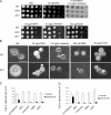
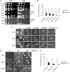
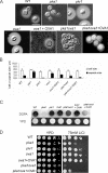
A defect in the PKA1 gene encoding the catalytic subunit of cyclic adenosine 5'-monophosphate (cAMP)-dependent protein kinase A (PKA) is known to reduce capsule size and attenuate virulence in the fungal pathogen Cryptococcus neoformans. Conversely, loss of the PKA regulatory subunit encoded by pkr1 results in overproduction of capsule and hypervirulence. We compared the transcriptomes between the pka1 and pkr1 mutants and a wild-type strain, and found that PKA influences transcript levels for genes involved in cell wall synthesis, transport functions such as iron uptake, the tricarboxylic acid cycle, and glycolysis. Among the myriad of transcriptional changes in the mutants, we also identified differential expression of ribosomal protein genes, genes encoding stress and chaperone functions, and genes for secretory pathway components and phospholipid synthesis. The transcriptional influence of PKA on these functions was reminiscent of the linkage between transcription, endoplasmic reticulum stress, and the unfolded protein response in Saccharomyces cerevisiae. Functional analyses confirmed that the PKA mutants have a differential response to temperature stress, caffeine, and lithium, and that secretion inhibitors block capsule production. Importantly, we also found that lithium treatment limits capsule size, thus reinforcing potential connections between this virulence trait and inositol and phospholipid metabolism. In addition, deletion of a PKA-regulated gene, OVA1, revealed an epistatic relationship with pka1 in the control of capsule size and melanin formation. OVA1 encodes a putative phosphatidylethanolamine-binding protein that appears to negatively influence capsule production and melanin accumulation. Overall, these findings support a role for PKA in regulating the delivery of virulence factors such as the capsular polysaccharide to the cell surface and serve to highlight the importance of secretion and phospholipid metabolism as potential targets for anti-cryptococcal therapy.
Conflict of interest statement
Figures


Similar articles
-
Regulated expression of cyclic AMP-dependent protein kinase A reveals an influence on cell size and the secretion of virulence factors in Cryptococcus neoformans.Mol Microbiol. 2012 Aug;85(4):700-15. doi: 10.1111/j.1365-2958.2012.08134.x. Epub 2012 Jul 13. Mol Microbiol. 2012. PMID: 22717009
-
Cyclic AMP-dependent protein kinase controls virulence of the fungal pathogen Cryptococcus neoformans.Mol Cell Biol. 2001 May;21(9):3179-91. doi: 10.1128/MCB.21.9.3179-3191.2001. Mol Cell Biol. 2001. PMID: 11287622 Free PMC article.
-
Cyclic AMP-dependent protein kinase catalytic subunits have divergent roles in virulence factor production in two varieties of the fungal pathogen Cryptococcus neoformans.Eukaryot Cell. 2004 Feb;3(1):14-26. doi: 10.1128/EC.3.1.14-26.2004. Eukaryot Cell. 2004. PMID: 14871933 Free PMC article.
-
The cAMP/Protein Kinase a Pathway Regulates Virulence and Adaptation to Host Conditions in Cryptococcus neoformans.Front Cell Infect Microbiol. 2019 Jun 18;9:212. doi: 10.3389/fcimb.2019.00212. eCollection 2019. Front Cell Infect Microbiol. 2019. PMID: 31275865 Free PMC article. Review.
-
Signal transduction pathways regulating differentiation and pathogenicity of Cryptococcus neoformans.Fungal Genet Biol. 1998 Oct;25(1):1-14. doi: 10.1006/fgbi.1998.1079. Fungal Genet Biol. 1998. PMID: 9806801 Review.
Cited by
-
The endosomal sorting complex required for transport machinery influences haem uptake and capsule elaboration in Cryptococcus neoformans.Mol Microbiol. 2015 Jun;96(5):973-92. doi: 10.1111/mmi.12985. Epub 2015 Mar 28. Mol Microbiol. 2015. PMID: 25732100 Free PMC article.
-
Pathogenic Delivery: The Biological Roles of Cryptococcal Extracellular Vesicles.Pathogens. 2020 Sep 16;9(9):754. doi: 10.3390/pathogens9090754. Pathogens. 2020. PMID: 32948010 Free PMC article. Review.
-
PKR Protects the Major Catalytic Subunit of PKA Cpk1 from FgBlm10-Mediated Proteasome Degradation in Fusarium graminearum.Int J Mol Sci. 2022 Sep 6;23(18):10208. doi: 10.3390/ijms231810208. Int J Mol Sci. 2022. PMID: 36142119 Free PMC article.
-
Role for Golgi reassembly and stacking protein (GRASP) in polysaccharide secretion and fungal virulence.Mol Microbiol. 2011 Jul;81(1):206-18. doi: 10.1111/j.1365-2958.2011.07686.x. Epub 2011 May 18. Mol Microbiol. 2011. PMID: 21542865 Free PMC article.
-
An encapsulation of iron homeostasis and virulence in Cryptococcus neoformans.Trends Microbiol. 2013 Sep;21(9):457-65. doi: 10.1016/j.tim.2013.05.007. Epub 2013 Jun 25. Trends Microbiol. 2013. PMID: 23810126 Free PMC article. Review.
References
-
- Casadevall A, Perfect JR. Cryptocococcus neoformans. Washington (D. C.): ASM Press; 1998. 541
-
- Perfect JR. Cryptococcus neoformans: A sugar-coated killer with designer genes. FEMS Immun Med Microbiol. 2005;45:395–404. - PubMed
-
- Janbon G. Cryptococcus neoformans capsule biosynthesis and regulation. FEMS Yeast Research. 2004;4:765–771. - PubMed
Publication types
MeSH terms
Substances
Associated data
- Actions
- Actions
- Actions
- Actions
- Actions
Grants and funding
LinkOut - more resources
Full Text Sources
Other Literature Sources
Molecular Biology Databases

