. 2007 May 4;368(3):729-41.
doi: 10.1016/j.jmb.2007.02.057. Epub 2007 Feb 22.

A conformational switch in the ligand-binding domain regulates the dependence of the glucocorticoid receptor on Hsp90

Affiliations

A conformational switch in the ligand-binding domain regulates the dependence of the glucocorticoid receptor on Hsp90

D Ricketson et al. J Mol Biol. .

Abstract

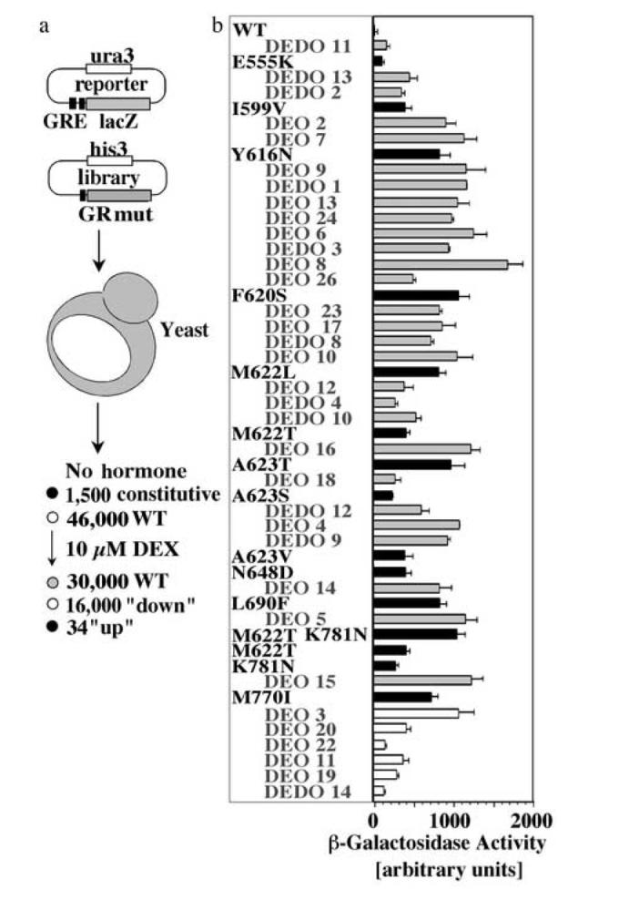
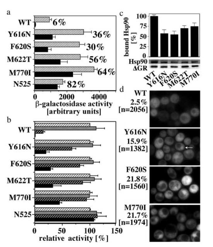
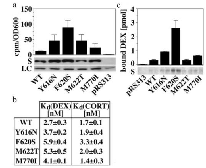
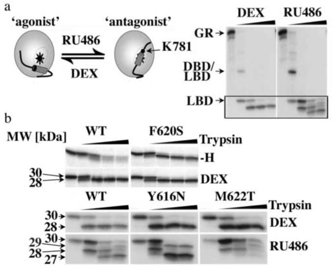
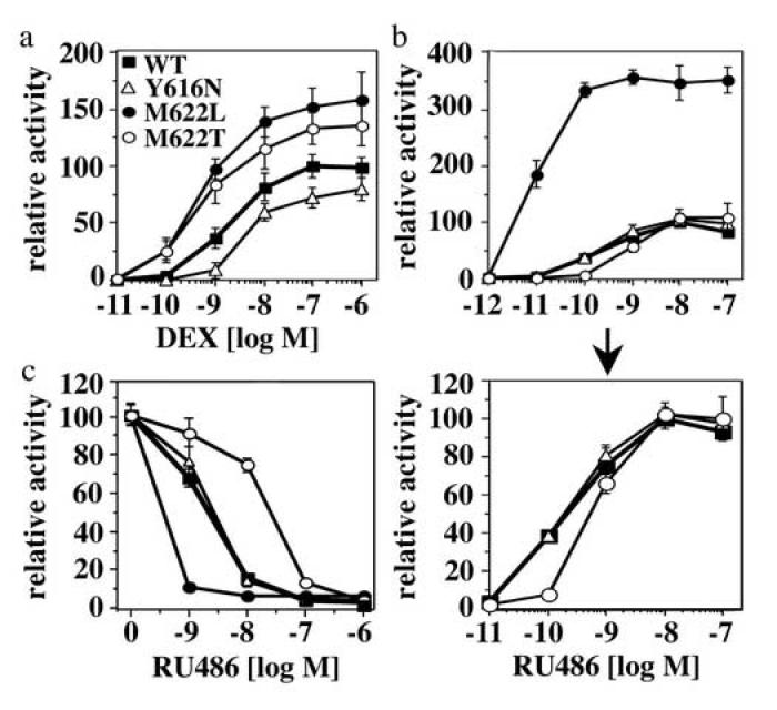
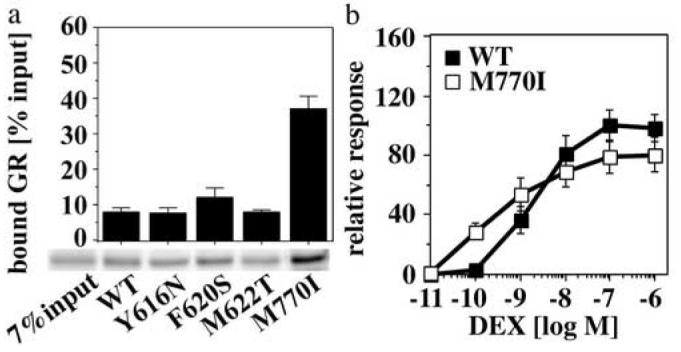
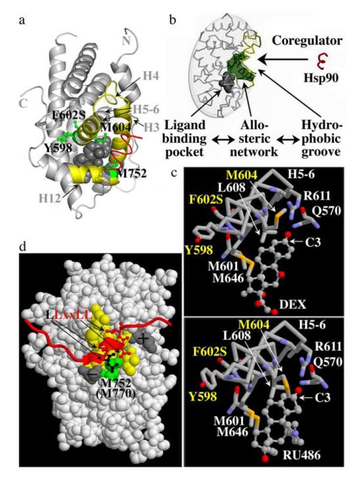
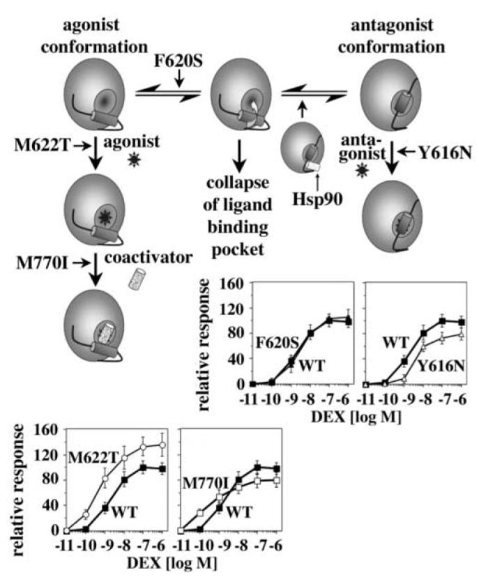
Steroid hormone receptors (SRs) are transcription factors that act as regulatory switches by altering gene expression in response to ligands. The highly conserved ligand-binding domain of SRs is a precise but versatile molecular switch that can adopt distinct conformations. Differential stabilization of these conformations by ligands, DNA response elements and transcriptional coregulators controls the activity of SRs in a gene-specific and cell-specific manner. In the case of the glucocorticoid receptor (GR), high-affinity ligand binding requires the interaction of the LBD with the heat shock protein 90 (Hsp90). Here, we show that the dependence of the ligand binding ability of GR on Hsp90 can be modified by the replacement of single amino acids within an allosteric network that connects the buried ligand-binding pocket and a solvent-exposed coregulator interaction surface. Each of the identified mutations altered the equilibrium between alternative GR conformations distinctively, indicating that the Hsp90 dependence of SRs may correlate with differences in the conformational dynamics of these receptors. Our results suggest that Hsp90 stabilizes the GR ligand-binding pocket indirectly by utilizing the allosteric network, while allowing the receptor to remain structurally uncommitted. Thus, in addition to ensuring the accessibility of the GR ligand-binding pocket to ligands, Hsp90 seems to enable hormones and coregulators to act as allosteric effectors, which forms the basis for gene-specific and cell-specific responses of GR to ligands.


Figures

Fig. 1
Fig. 1
Identification of GR mutants with increased hormone responsiveness (a) After transformation with an expression library of random GR mutants and a GR-controlled β-galactosidase reporter, about 47,500 W303 yeast transformants were screened for increased β-galactosidase activity in the presence of 10 μM DEX. This screen identified 1,500 constitutive mutants, 16,000 mutants with impaired response to DEX and 34 GR mutants with increased responsiveness to DEX. (b) β-Galactosidase activity of yeast expressing GR WT, the original isolates (e.g. DEDO11; grey bars) or GR mutants containing single site replacements identified in the original isolates (e.g. E555K; black bars). 28 of the original 34 isolates with increased DEX responsiveness were due to a single amino acid change in the receptor LBD. Most of these mutations were identified in multiple isolates. DEO3, DEO11, DEO19, DEO 20, DEO22 and DEDO14 contained mutations in either the GR N-terminal domain or DBD that were not further characterized in the context of this study. These functional analyses were conducted in the yeast strain YNK410 in the presence of 10 μM DEX. Shown are the averages and standard deviations of three independent experiments each performed with at least three transformants of each GR variant.
Fig. 2
Fig. 2
Four of the GR LBD mutants with increased DEX responsiveness are less dependent on Hsp90 (a) Activation of a GR-controlled β-galactosidase reporter gene by GR WT and mutants in the presence of normal Hsp90 levels (striped bars) or low Hsp90 levels (∼5% of normal levels; black bars). These analyses were conducted in the yeast strain GRS4 whose sole source of Hsp90 is a plasmid born human Hsp90 gene expressed under the control of a leaky GAL1 promoter. N525 is a constitutively active GR deletion mutant that lacks the LBD and does not bind Hsp90. Cultures were grown in the presence of 50 μM CORT. The given percentages identify the fraction of β-galactosidase activity retained upon lowering Hsp90 levels. Shown are only mutants that retained at least 30% of the activity at normal Hsp90 levels. (b) Relative activity of GR WT and mutants in the presence of either vehicle (DMSO; stripped white background) or the Hsp90 inhibitor GA (10 μM-grey bars, 50 μM-stripped black background, 100 μM-black bars) in the yeast strain YNK410. Cultures were grown at RT in the presence of 50 μM CORT. Shown are the averages and standard deviations of three independent experiments each performed with at least three transformants of each GR variant (a,b). (c) Co-immunoprecipitation of in vitro expressed, 35S-labeled Hsp90 and ΔGR WT and mutants using a GR-specific antibody (P20, Santa Cruz). The ΔGR deletion mutant lacks the N-terminal AF1 domain (amino acid 108-317). This deletion does not affect the interaction of Hsp90 with GR. For isolation, ΔGR: Hsp90 heterocomplexes were stabilized with molybdate. Co-immunoprecipitating proteins were separated by SDS PAGE and the relative amounts of co-immunoprecipitating Hsp90 and ΔGR were quantified by phosphoimaging. Shown are the averages and standard deviations of three experiments, as well as a representative autoradiogram. (d) Quantification of nuclear localization of GFP-GR WT and mutants in yeast (YNK410) in the absence of hormone. Examples for nuclear GFP-GR are marked by arrows. The percentage of yeast displaying nuclear GFP-GR and the number of evaluated cells are indicated.
Fig. 3
Fig. 3
The identified mutations stabilize soluble and hormone-binding competent conformations of GR (a) Expression of soluble GR protein (S) and in vivo accumulation of 3H-DEX (2 h incubation) in yeast (YNK410) expressing either GR WT or mutants. Shown are the averages and standard deviations of three independent experiments performed in duplicates as described in the Materials and Method section. Protein expression was monitored by immunoblot analysis using the monoclonal GR antibody BUGR2. A selected band of the Ponceau Red-stained transblot is shown as loading control (LC). (b) Dissociation constants of DEX and CORT binding reactions using GR WT and mutants expressed in reticulocyte lysate. Binding curves were measured as described in the Materials and Methods section and fit by nonlinear regression using a single site saturation binding model. Shown are the averages and standard deviations of three binding experiments per GR variant. (c) Soluble expression and 3H-DEX (15 nM) binding of recombinantly expressed and Talon purified GR WT and mutant LBDs corresponding to 100 μl bacterial culture (∼1-10 pmol). The Coomassie-stained polyacrylamide gel monitors the yield of soluble, Talon purified WT/mutant GR LBDs in two independent purifications each.
Fig. 4
Fig. 4
The Y616N, F620S and M622T replacements alter the equilibrium between different GR conformations (a) Schematic representation of the “agonist” and “antagonist” structure of the GR LBD as defined by ref. . The autoradiogram shows the fragment patterns of in vitro expressed, 35S-labeled GR WT bound to either DEX or RU486 (10 μM each) after 1 h incubation with trypsin (0, 37.5, 75, 150, or 300 μg/ml) at 4°C. (b) Comparison of the trypsin fragment patterns of GR WT and F620S LBDs in the absence (-H) and presence of DEX, as well as of GR WT, Y616N and M622T bound to either DEX or RU486 (displayed are fragments within the boxed part of the autoradiograms shown in (a)). Trypsin digests were conducted as in (a). The 29 and 27 kDa fragments of RU486-bound GR are generated by cleavage at K781 at the C-terminus of H12, which is accessible in the “antagonist conformation” but not in the “agonist conformation” of the GR LBD.
Fig. 5
Fig. 5
Replacements of M622 and Y616 differentially alter the response of GR to DEX and RU486 in F9 carcinoma cells (a,b) Comparison of DEX (a) and RU486 (b) dose response curves of WT and mutant GR in transiently transfected F9 carcinoma cells using a TAT3 luciferase reporter. Luciferase units were normalized relative to the GR WT activity in the presence of 0.1 μM DEX or 10 nM RU486. At saturation, in F9 cells the activity of DEX-bound GR is about 100-fold higher than the activity of RU486-bound GR. Shown are the averages and standard deviations of three experiments performed in triplicates. (c) RU486 competition of DEX-bound GR WT and mutants (10 nM DEX). These experiments were performed as described in (a) and (b).
Fig. 6
Fig. 6
The M770I mutation alters the interaction of GR with coactivators (a) GST-pulldown assay comparing the interaction of equimolar concentrations of in vitro expressed, 35S-labeled GR WT and mutants with a glutathione agarose-bound GST-GRIP1 fragment (aa 567-1121; 2 μM). GST-GRIP1: GR complexes were separated by SDS-PAGE. Total and bound receptor concentrations were determined by phosphoimaging. Shown are the averages and standard deviations of three binding experiments performed in duplicates, as well as a representative autoradiogram. (b) DEX dose response curves for GR WT and M770I in transiently transfected F9 carcinoma cells using a TAT3 luciferase reporter. Relative luciferase units were normalized with respect to the GR WT activity in the presence of 0.1 μM DEX. Shown are the averages and standard deviations of three experiments performed in triplicates.
Fig. 7
Fig. 7
Structural interpretation of the isolated GR LBD mutations (a) Structure of the DEX-bound (grey sphere) human GR LBD mutant F602S bound to the amphipathic α-helix of an interacting coregulator (red) based on ref. . Regions of α-helices H3, H4, H5-6, H6 and connecting loops that form the solvent exposed hydrophobic groove are outlined in yellow. Residues corresponding to rat Y616 (hGR Y598), F620 (hGR F602), M622 (hGR M604) reside in H5-6, and M770 (hGR M752) in H12 of the GR LBD. Note that the particular conformation of Y598 in this structure is likely induced by the F602S mutation. (b) Schematic presentation of the allosteric network (green) connecting the buried ligand-binding pocket (grey sphere) and the exposed hydrophobic groove (yellow) that is bound by coregulators (red) (reprinted from ref. with permission of the Proc. Natl. Acad. Sci. U S A). (c) Interactions of H5-6 with DEX and RU486 based on a structure of the human GR mutant F602S. While M601 (rat GR M619), L608 (rat GR L626), R611 (rat GR R629), Q570 (rat GR Q588) and M646 (rat GR M664) show similar interactions with both ligands, the conformation of M604 (rat GR M622) changes in a ligand-dependent manner. Contrary to M604, which is part of the ligand-binding pocket, Y598 and F602 (rat GR Y616 and F620) face away from the ligand-binding pocket. Oxygen atoms are red, nitrogens blue and sulphur yellow. (d) Space-filled presentation of the human GR LBD bound to the NR-box 3 binding site of the coactivator GRIP1 (red). The hydrophobic groove is shown in yellow; charged residues that stabilize the interaction of coactivators with GR are shown in grey. The residue corresponding to rat GR M770 (human GR M752, green) interacts with a leucine residue (black) that precedes the conserved coactivator “LxxLL”motif.
Fig. 8
Fig. 8
A Conformational switch in the LBD regulates the dependence of GR on Hsp90 Model for the stabilization of the GR LBD by Hsp90 and the identified GR mutations. The GR LBD appears to be in an equilibrium between multiple conformations including the “agonist conformation” (left) and the “antagonist conformation” (right). Stabilization of any of these conformations reduces the risk that the ligand-binding pocket collapses during the transitioning between these conformations. In a previous study we provided evidence that Hsp90 stabilizes the antagonist conformation of GR. The GR mutations identified in this study appear to use a similar mechanism and stabilize either the antagonist or the agonist conformation of GR. Contrary to the stabilizing effect of Hsp90, which is transient, the mutation-induced changes in the equilibrium between the different GR conformations are permanent resulting in distinctive and in most cases unfavorable changes for the hormone responsiveness of GR in F9 carcinoma cells.

Similar articles

Cited by

References

    1. Mangelsdorf DJ, Thummel C, Beato M, Herrlich P, Schutz G, Umesono K, Blumberg B, Kastner P, Mark M, Chambon P, Evans RM. The nuclear receptor superfamily: the second decade. Cell. 1995;83:835–9. - PMC - PubMed
    1. Aranda A, Pascual A. Nuclear hormone receptors and gene expression. Physiol Rev. 2001;81:1269–304. - PubMed
    1. Renaud JP, Moras D. Structural studies on nuclear receptors. Cell Mol Life Sci. 2000;57:1748–69. - PMC - PubMed
    1. Robin-Jagerschmidt C, Wurtz JM, Guillot B, Gofflo D, Benhamou B, Vergezac A, Ossart C, Moras D, Philibert D. Residues in the ligand binding domain that confer progestin or glucocorticoid specificity and modulate the receptor transactivation capacity. Mol Endocrinol. 2000;14:1028–37. - PubMed
    1. Nettles KW, Sun J, Radek JT, Sheng S, Rodriguez AL, Katzenellenbogen JA, Katzenellenbogen BS, Greene GL. Allosteric control of ligand selectivity between estrogen receptors alpha and beta: implications for other nuclear receptors. Mol Cell. 2004;13:317–27. - PubMed

Publication types

MeSH terms

LinkOut - more resources