. 2007 May;33(3):140-54.
doi: 10.1016/j.jchemneu.2007.02.002. Epub 2007 Feb 16.

The microtubule-associated protein doublecortin is broadly expressed in the telencephalon of adult canaries

Affiliations

The microtubule-associated protein doublecortin is broadly expressed in the telencephalon of adult canaries

Géraldine Boseret et al. J Chem Neuroanat. 2007 May.

Abstract

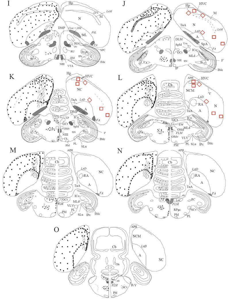
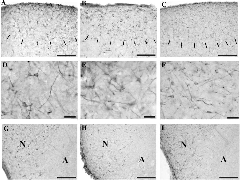
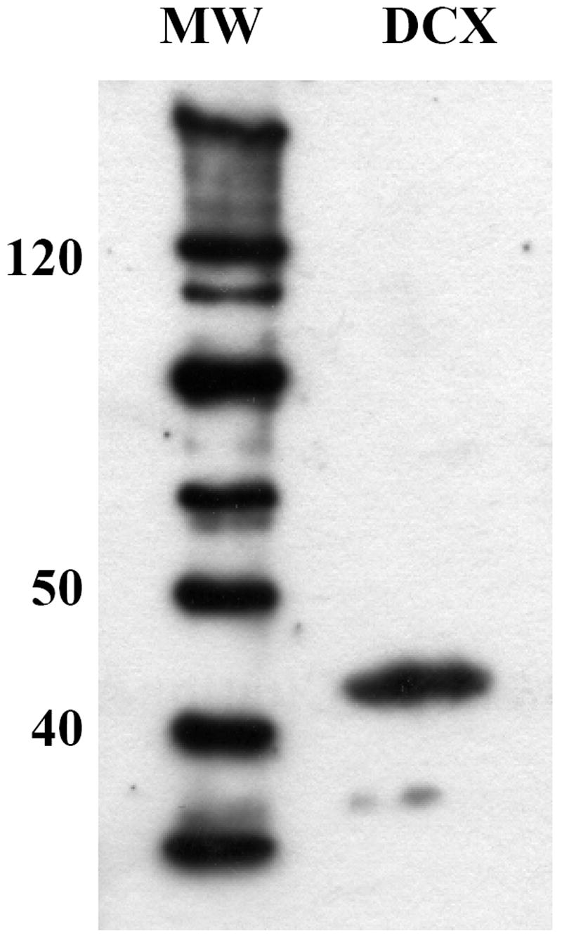
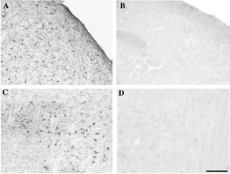
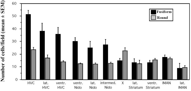
The protein doublecortin (DCX) is expressed in post-mitotic migrating and differentiating neurons in the developing vertebrate brain and, as a part of the microtubule machinery, is required for neuronal migration. DCX expression is generally maximal during embryonic and early post-natal life but decreases markedly and almost disappears in older animals in parallel with the major decrease or cessation of neurogenesis. In several seasonally breeding songbird species such as canaries, the volume of several song control nuclei in the brain varies seasonally such that the largest nuclei are observed in the late spring and early summer. This variation is based on changes in cell size, dendritic branching, and, in nucleus HVC, on the incorporation of neurons newly born in adulthood. Because songbirds maintain an active neurogenesis and neuronal incorporation in their telencephalon throughout their lives, we investigated here the distribution of DCX-immunoreactive (ir) structures in the brain of adult male canaries. Densely stained DCX-ir cells were found exclusively in parts of the telencephalon that are known to incorporate new neurons in adulthood, in particular the nidopallium. Within this brain region, the boundaries of the song control nucleus HVC could be clearly distinguished from surrounding structures by a higher density of DCX-ir structures. In most telencephalic areas, about two thirds of these cells displayed a uni- or bipolar fusiform morphology suggesting they were migrating neurons. The rest of the DCX-ir cells in the telencephalon were larger and had a round multipolar morphology. No such staining was found in the rest of the brain. The broad expression of DCX specifically in adult brain structures that exhibit the characteristic of active incorporation of new neurons suggests that DCX plays a key role in the migration of new neurons in the brain of adult songbirds as it presumably does during ontogeny.


Figures

Figure 1
Figure 1
Schematic drawings of coronal sections through the canary brain illustrating the distribution of DCX-ir structures in a rostral to caudal order (from A to O). Densely stained DCX-ir perikarya are represented by dots (.) and weakly stained cells are represented by small open circles (○). Cells associated with immunoreactive DCX-ir small vesicles resembling punctate structures are represented by stars. DCX-ir fibers that are not obviously associated with immunoreactive perykarya are represented by wavy lines. The density of symbols has been adjusted within each type of DCX-ir cells to give a qualitative estimate of the number of the immunoreactive structures. The densities of symbols should, however, not be compared across cell types; the densely stained DCX-ir cells are indeed by far more numerous than the two other types of staining. The nomenclature is based on the canary brain atlas (Stokes et al 1974) but including recent modifications recommended by the avian nomenclature forum (Reiner et al., 2003) (see text for additional explanations). Abbreviations: A: Arcopallium; APH: Area parahippocampalis; Area X: Area X; BC: Brachium conjunctivum; BSTL: Nucleus striae terminalis lateralis; Cb: Cerebellum; Cbi: Nucleus cerebellum internum; CO: Chiasma opticum; CoA: Commissura anterior; CoS: Nucleus commissuralis septi; CP: Commissura posterior; CS: Nucleus centralis superior; DLL: Nucleus dorsolateralis anterior thalami, pars lateralis; DLM: Nucleus dorsolateralis anterior thalami, pars medialis; DMP: Nucleus dorsomedialis posterior; DSV: Decussatio supraoptica ventralis; E: Entopallium; EW: Edinger-Westphal Nucleus; FA: Tractus fronto-arcopallialis; FLM: Fasciculus longitudinalis medialis; FPL: Fasciculus prosencephali lateralis; GCt: Griseum centrale; GLv: Nucleus geniculatus lateralis, pars ventralis; GP: Globus pallidus; HA: Hyperpallium apicale; HD: Hyperpallium densocellulare; Hp: Hippocampus; HVC: HVC; ICo: Nucleus Intercollicularis; IMc:Nucleus isthmi, pars magnocellularis; INP: Nucleus intrapeduncularis; IP: Nucleus Interpeduncularis; IPc: Nucleus isthmi, pars parvocellularis; IS: Nucleus Interstitialis; LAD: Lamina arcopallialis dorsalis; LaM: Lamina mesopallialis; LFM: Lamina frontalis suprema; LMAN: Nucleus lateralis magnocellularis nidopallii anterioris; LoC: Locus Coeruleus; LPS: Lamina pallio-subpallialis; LFS: Lamina frontalis superior; M: Mesopallium; MLd: Nucleus mesencephalicus lateralis, pars dorsalis; MMAN: Nucleus medialis magnocellularis nidopallii anterioris; N: Nidopallium; NC: Nidopallium caudale; NCM: Nidopallium caudomediale; NDB: Nucleus diagonalis Brocae; NIII: Nervus oculomotorius; nIV: Nucleus nervi trochlealis; OM: tractus occipito-mesencephalicus; OMd: Nucleus nervi oculomotorii, pars dorsalis; OMv: Nucleus nervi oculomotorii, pars ventralis; OV: Nucleus ovoidalis; PL: Nucleus pontis lateralis; PM: Nucleus pontis medialis; POM: Nucleus preopticus medialis; Pr V: Nucleus sensorius principalis nervi trigemini; Pt: Nucleus pretectalis; PVN: Nucleus paraventricularis; RA: Nucleus robustus arcopallialis; RPgc: Nucleus reticularis pontis caudalis, pars gigantocellularis; Rt: Nucleus rotundus; Ru: Nucleus ruber; SCv: Nucleus subcoeruleus ventralis; SGP: Substantia grisea et fibrosa periventricularis; SL: Nucleus septalis lateralis; SLu: Nucleus semi-lunaris; SM: Nucleus septalis medialis; SN: Substantia Nigra; SP: Nucleus subpretectalis; SpA: Area subpallialis amygdalae; SpL: Nucleus spiriformis lateralis; SpM: Nucleus spiriformis medialis; SRt: Nucleus subrotundus; StL: Striatum laterale; StM: Striatum mediale; TeO: Tectum opticum; TIO: Tractus isthmo-opticus; TnA: Nucleus taeniae amygdalae; TrO: Tractus opticus; TSM: tractus septopallio-mesencephalicus; Tu: Nucleus tuberis; Uva: Nucleus uvaeformis; V: Ventriculus; VLV: Nucleus ventralis lemnisci lateralis; VMN: Nucleus ventromedialis hypothalami; VTA: Area ventralis tegmenti.
Figure 1
Figure 1
Schematic drawings of coronal sections through the canary brain illustrating the distribution of DCX-ir structures in a rostral to caudal order (from A to O). Densely stained DCX-ir perikarya are represented by dots (.) and weakly stained cells are represented by small open circles (○). Cells associated with immunoreactive DCX-ir small vesicles resembling punctate structures are represented by stars. DCX-ir fibers that are not obviously associated with immunoreactive perykarya are represented by wavy lines. The density of symbols has been adjusted within each type of DCX-ir cells to give a qualitative estimate of the number of the immunoreactive structures. The densities of symbols should, however, not be compared across cell types; the densely stained DCX-ir cells are indeed by far more numerous than the two other types of staining. The nomenclature is based on the canary brain atlas (Stokes et al 1974) but including recent modifications recommended by the avian nomenclature forum (Reiner et al., 2003) (see text for additional explanations). Abbreviations: A: Arcopallium; APH: Area parahippocampalis; Area X: Area X; BC: Brachium conjunctivum; BSTL: Nucleus striae terminalis lateralis; Cb: Cerebellum; Cbi: Nucleus cerebellum internum; CO: Chiasma opticum; CoA: Commissura anterior; CoS: Nucleus commissuralis septi; CP: Commissura posterior; CS: Nucleus centralis superior; DLL: Nucleus dorsolateralis anterior thalami, pars lateralis; DLM: Nucleus dorsolateralis anterior thalami, pars medialis; DMP: Nucleus dorsomedialis posterior; DSV: Decussatio supraoptica ventralis; E: Entopallium; EW: Edinger-Westphal Nucleus; FA: Tractus fronto-arcopallialis; FLM: Fasciculus longitudinalis medialis; FPL: Fasciculus prosencephali lateralis; GCt: Griseum centrale; GLv: Nucleus geniculatus lateralis, pars ventralis; GP: Globus pallidus; HA: Hyperpallium apicale; HD: Hyperpallium densocellulare; Hp: Hippocampus; HVC: HVC; ICo: Nucleus Intercollicularis; IMc:Nucleus isthmi, pars magnocellularis; INP: Nucleus intrapeduncularis; IP: Nucleus Interpeduncularis; IPc: Nucleus isthmi, pars parvocellularis; IS: Nucleus Interstitialis; LAD: Lamina arcopallialis dorsalis; LaM: Lamina mesopallialis; LFM: Lamina frontalis suprema; LMAN: Nucleus lateralis magnocellularis nidopallii anterioris; LoC: Locus Coeruleus; LPS: Lamina pallio-subpallialis; LFS: Lamina frontalis superior; M: Mesopallium; MLd: Nucleus mesencephalicus lateralis, pars dorsalis; MMAN: Nucleus medialis magnocellularis nidopallii anterioris; N: Nidopallium; NC: Nidopallium caudale; NCM: Nidopallium caudomediale; NDB: Nucleus diagonalis Brocae; NIII: Nervus oculomotorius; nIV: Nucleus nervi trochlealis; OM: tractus occipito-mesencephalicus; OMd: Nucleus nervi oculomotorii, pars dorsalis; OMv: Nucleus nervi oculomotorii, pars ventralis; OV: Nucleus ovoidalis; PL: Nucleus pontis lateralis; PM: Nucleus pontis medialis; POM: Nucleus preopticus medialis; Pr V: Nucleus sensorius principalis nervi trigemini; Pt: Nucleus pretectalis; PVN: Nucleus paraventricularis; RA: Nucleus robustus arcopallialis; RPgc: Nucleus reticularis pontis caudalis, pars gigantocellularis; Rt: Nucleus rotundus; Ru: Nucleus ruber; SCv: Nucleus subcoeruleus ventralis; SGP: Substantia grisea et fibrosa periventricularis; SL: Nucleus septalis lateralis; SLu: Nucleus semi-lunaris; SM: Nucleus septalis medialis; SN: Substantia Nigra; SP: Nucleus subpretectalis; SpA: Area subpallialis amygdalae; SpL: Nucleus spiriformis lateralis; SpM: Nucleus spiriformis medialis; SRt: Nucleus subrotundus; StL: Striatum laterale; StM: Striatum mediale; TeO: Tectum opticum; TIO: Tractus isthmo-opticus; TnA: Nucleus taeniae amygdalae; TrO: Tractus opticus; TSM: tractus septopallio-mesencephalicus; Tu: Nucleus tuberis; Uva: Nucleus uvaeformis; V: Ventriculus; VLV: Nucleus ventralis lemnisci lateralis; VMN: Nucleus ventromedialis hypothalami; VTA: Area ventralis tegmenti.
Figure 2
Figure 2
Photomicrographs illustrating the different types of DCX-immunoreactive material observed in the canary brain. A. Densely stained round multipolar cells found in the telencephalon. B. Fusiform elongated cells (arrows) found in the same telencephalic areas as round cells illustrated in A. One round imunoreactive cell (asterisk) is also seen in this field. C. Weakly stained round cells with few immunostained processes detected mainly in di- and mesencephalic areas (here in the Nucleus dorsolateralis anterior thalami, pars medialis). D. Weakly stained cells found in some di- and mesencephalic areas (here in the Substantia nigra) that are associated with small immunoreactive dots reminiscent of punctate structures. The insert illustrates a higher magnification of such a cell. Panel D was photographed with Nomarski interferential contrast. Magnification bar: 20 μm for panels A-D, 10 μm in the insert of panels B and D.
Figure 3
Figure 3
Photomicrographs of HVC at low (panels A-C) and high (panels D-F) magnification and of the border between the Nidopallium (N) and Arcopallium (A) (G-I) in sections that had been stained either with the anti-DCX pep antibody (A, D, G) or the rabbit Nterm DCX antibody (B, E, H) or the goat Cterm DCX antibody (C, F, I). Similar immunoreactive structures are identified by three different antibodies directed against different parts of the DCX protein. Arrows in panels A-C indicate the border of HVC. Panels G-I correspond to the medial border of the telencephalon at the junction between N and A at a rostro-caudal level corresponding to panel M in Figure 1. Magnification bars: 100 μm in A-C and G-I, 20 μm in D-F.
Figure 4
Figure 4
Western blot demonstrating that the anti-DCX pep antibody recognizes a protein with a molecular weight slightly higher than 40 KDa consistent with the size of DCX as identified in mammals. MW: molecular weight markers; DCX: doublecortin in a canary brain extract.
Figure 5
Figure 5
Photomicrographs of sections through HVC (A, B) or through the Substantia nigra (C, D) that were stained with the anti-DCX pep antibody (A, C) or with the antibody that had been preadsorbed with the three peptides used as antigen (B, D). Both the strong (A) and the weak (C) immunoreactive signals are completely or almost completely blocked by predsorbtion (B, D). Magnification bar: 100 μm.
Figure 6
Figure 6
Photomicrographs illustrating the different types of DCX-ir material identified in key regions of the canary brain. A. HVC and adjacent Nidopallium (N), B. Nucleus robustus archistriatalis (RA) and adjacent Arcopallium (A) just ventral to the Lamina arcopallialis dorsalis, C. Area X and surrounding Striatum mediale, D. Weakly labeled cells in the mesencephalic Griseum centrale (GCt = periaqueductal gray) located ventrally to the Aqueductus cerebri (Aq) and the Commissura posterior (CP), E. Weakly labeled cells in the Substantia nigra (SN), F. Weakly labeled cells in the Nucleus dorsolateralis anterior thalami, pars medialis (DLM), G. Bundle of parallel fibers originating in the subventricular zone (SVZ) and crossing a large area of the Striatum mediale (MSt), H. Fibers intermingled with parallel fusiform cells in the dorsal part of the Mesopallium (M) just ventral to the Lamina frontalis superior (LFS) and the Hyperpallium densocellulare (HD), I. Parallel fibers crossing the Lamina arcopallialis dorsalis (LAD) from the Nidopallium (N) to the Arcopallium (A), J-K-L. views of the subventricular zome (SVZ) at the level of Nucleus taeniae amygdalae (level L in Fig. 1; J), of the Commissura anterior (level G in Fig. 1; K) and of the Area X (level B in Fig. 1; L), M. Low magnification of the optic lobes showing the stratified imunoreactive signal that is not generally associated with clear cellular structures. At higher magnification a few weakly labeled cells are however detected. Magnification bar shown in M represents 200 μm in A-D and F, 100 μm in E, G-I, M and 50 μm in J-L.
Figure 7
Figure 7
Average numbers of round or fusiform DCX-ir cells in 4 photosensitive male canaries that were castrated, treated with exogenous testosterone and exposed to a female partner. Cells were counted in 200 by 200 μm fields that were placed in standardized locations within the telencephalon, namely in the song control nucleus HVC, just lateral (lat. HVC) or ventral (ventr. HVC) to this nucleus, in the ventral (ventr.), lateral (lat.) or intermediate (intermed.) Nidopallium (Nido), in Area X (X) or in the Striatum lateral (lat.) or ventral (ventr.) to this nucleus, or finally in LMAN or just lateral to this nucleus (lat. LMAN. See methods and Fig. 1 for a more precise description of these areas).

References

    1. Alvarez-Buylla A, Kirn JR, Nottebohm F. Birth of projection neurons in adult avian brain may be related to perceptual or motor learning. Science. 1990;249:1444–1446. - PubMed
    1. Alvarez-Buylla A, Theelen M, Nottebohm F. Birth of projection neurons in the higher vocal center of the canary forebrain before, during, and after song learning. Proc Natl Acad Sci USA. 1988;85:8722–8726. - PMC - PubMed
    1. Bai J, Ramos RL, Ackman JB, Thomas AM, Lee RV, LoTurco JJ. RNAi reveals doublecortin is required for radial migration in rat neocortex. Nat Neurosci. 2003;6:1277–83. - PubMed
    1. Ball GF. Neuroendocrine basis of seasonal changes in vocal behavior among songbirds. In: Hauser M, Konishi M, editors. The design of animal communication. MIT Press; Cambridge,MA: 1999. pp. 213–253.
    1. Ball GF, Auger CJ, Bernard DJ, Charlier TD, Sartor JJ, Riters LV, Balthazart J. Seasonal Plasticity in the song control system. Multiple brain sites of steroid hormone action and the importance of variation in song behavior. Ann NY Acad Sci. 2004;1016:1–25. - PubMed

Publication types

MeSH terms

Substances

LinkOut - more resources