Review
. 2007 Apr;14(4):398-405.
doi: 10.1016/j.acra.2007.01.005.

Molecular imaging research in the outcomes era: measuring outcomes for individualized cancer therapy

Affiliations
Review

Molecular imaging research in the outcomes era: measuring outcomes for individualized cancer therapy

David A Mankoff et al. Acad Radiol. 2007 Apr.

Abstract

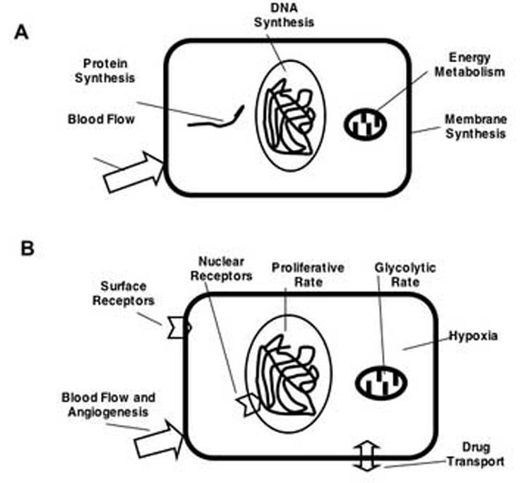
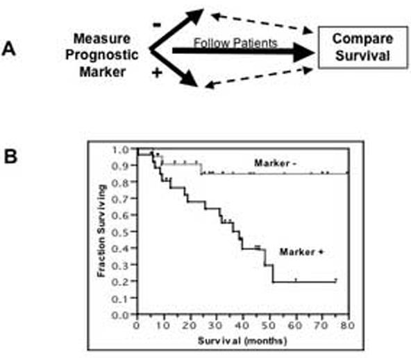
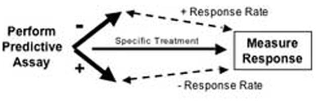
Advances in molecular imaging, combined with the goal of personalized cancer therapy, call for new approaches to clinical study design for trials testing imaging to guide therapy. The role of cancer imaging must expand and move beyond tumor detection and localization to incorporate quantitative evaluation of regional tumor phenotype. Imaging study design and outcome analysis must move beyond metrics designed to measure the performance for detection to include measures of prognosis, prediction of therapeutic success, and early therapy response. This implies changes in how studies are carried and out, and importantly in the regulatory oversight of cancer imaging. Demonstration that a biochemical or molecular imaging method correctly and accurately measures a specific biologic feature should be sufficient for approval for clinical trials. It may be possible that a combination of imaging procedures known to accurately depict tumor phenotype may be prognostic, even if the individual study cannot be directly validated against patient outcomes. Therefore, it will be important to be able to apply a range of possible imaging studies to different targeted cancer therapy trials. Academia and industry must work together with regulatory agencies and payers to facilitate well designed clinical studies, with appropriate outcome measures, to test the effectiveness of imaging in helping to direct cancer therapy. These will assure the appropriate use of imaging to direct treatment and make an important step towards individualized cancer therapy.


Figures

Figure 1
Figure 1
Illustration of cancer cell targets for cancer imaging. Targets for tumor detection (A) must be process present in high levels in the tumor, but absent or present in low levels in normal tissue. There are a greater number of possible targets for cancer imaging to guide tumor therapy (B), where measuring both increased and decreased levels is important in choosing and monitoring treatment.
Figure 2
Figure 2
Diagram for clinical study design to test a prognostic marker. The prognostic marker may be a tissue assay, an imaging measure, or a combination of tissue and imaging results.
Figure 3
Figure 3
Diagram for clinical study design to test a predictive assay.
Figure 4
Figure 4
Coronal PET images of FDG uptake (left) and 18F-fluoroestradiol (FES) uptake (right) are shown for two patients with recurrent and metastatic disease from estrogen receptor positive (ER+) breast cancers (30). The top patient (Patient 1) showed sternal metastases that were metabolically active by FDG PET (2nd column) with matched uptake of FES (1st column), indicated preserved ER expression (arrow). The bottom patient (Patient 2) showed a site of bone metastasis by FDG PET (arrow) but no corresponding uptake by FES, suggesting a loss of ER expression. Both patients were treated with hormonal therapy subsequent to PET imaging. Patient 1 had an excellent objective response while Patient 2 had disease progression, both indicated by post therapy FDG (right column). Normal liver and kidney uptake is also seen in the images for both radiopharmaceuticals. This example illustrates how uptake on FES PET can be predictive for response of breast cancer to endocrine therapy.
Figure 5
Figure 5
Diagram of biologic processes involved in cancer response to successful therapy.
Figure 6
Figure 6
Study design for testing a measure of cancer response. The change in the response measure is compared to the “gold standard” response measure to determine the accuracy of predicting a response and, importantly, to patient outcome including time-toprogression (TTP) and survival.
Figure 7
Figure 7
Illustration of a study testing a novel imaging procedure as a measure of response to therapy. The change in uptake of 99mTc-sestamibi (MIBI) in locally advanced breast cancer was compared to the gold standard response (post-therapy histopathology) (40) and post-therapy MIBI uptake was compared to patient outcome (41). Images (A) show the ability to distinguish pathologic complete response (CR) versus partial response (PR), and a plot of the change in MIBI uptake ratios (lesion-to-normal breast uptake (L:N ratio)) shows that values for CR versus PR are nearly completely separated (B). ROC analysis depicts the ability to classify CR versus PR based upon the change in MIBI uptake on a plot of the sensitivity for CR versus 1-specificty (C). Comparison of residual MIBI uptake post-therapy to overall survival (D) demonstrates that low post-therapy MIBI uptake is predictive of survival.

Similar articles

Cited by

References

    1. Kaklamani V, O′Regan RM. New targeted therapies in breast cancer. Semin Oncol. 2004;31(2 Suppl 4):20–5. - PubMed
    1. Sawyers CL. Making progress through molecular attacks on cancer. Cold Spring Harb Symp Quant Biol. 2005;70:479–82. - PubMed
    1. Beckman RA, Loeb LA. Genetic instability in cancer: theory and experiment. Semin Cancer Biol. 2005;15(6):423–35. - PubMed
    1. Aboagye EO, Price PM. Use of positron emission tomography in anticancer drug development. Invest New Drugs. 2003;21(2):169–81. - PubMed
    1. Kelloff GJ, Hoffman JM, Johnson B, Scher HI, Siegel BA, Cheng EY, Cheson BD, O′Shaughnessy J, Guyton KZ, Mankoff DA, Shankar L, Larson SM, Sigman CC, Schilsky RL, Sullivan DC. Progress and promise of FDG-PET imaging for cancer patient management and oncologic drug development. Clin Cancer Res. 2005;11(8):2785–808. - PubMed

Publication types

Substances