. 2007 Jun 1;74(3):506-14.
doi: 10.1016/j.cardiores.2007.02.014. Epub 2007 Feb 20.

Laminar shear stress up-regulates the expression of stearoyl-CoA desaturase-1 in vascular endothelial cells

Affiliations

Laminar shear stress up-regulates the expression of stearoyl-CoA desaturase-1 in vascular endothelial cells

Xiaomei Qin et al. Cardiovasc Res. .

Abstract

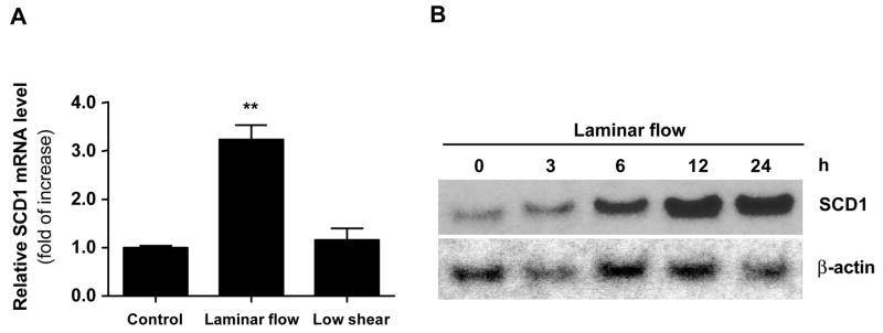
Objective: Laminar shear stress plays critical roles in vascular homeostasis and exerts various metabolic effects on endothelial cells (ECs). Stearoyl-CoA desaturase-1 (SCD1), which catalyzes the biosynthesis of monounsaturated fatty acids, affects the lipid composition and fluidity of the cell membrane. Thus, we examined the effect of laminar flow on SCD1 expression in ECs.

Methods: A flow chamber was used to impose a laminar shear stress on a confluent monolayer of human vascular ECs. The expression of SCD1 was examined using real-time RT-PCR and Northern and Western blotting. Immunohistochemical staining was used to assess the expression of SCD1 in Sprague-Dawley rat arteries, including the sites of arterial bifurcation.

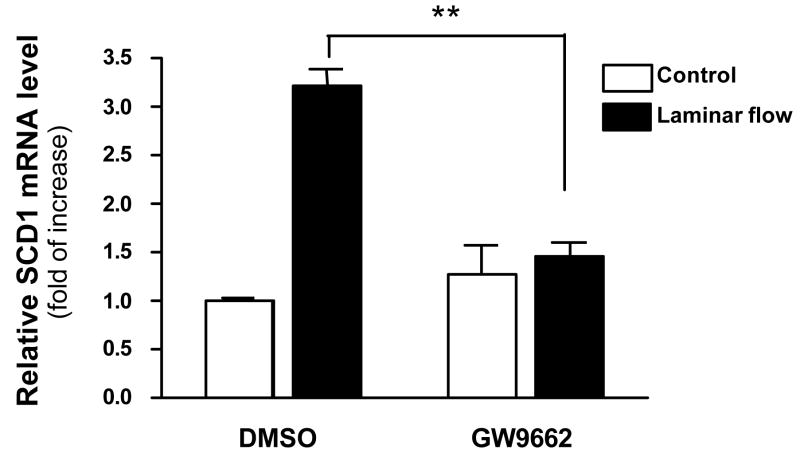
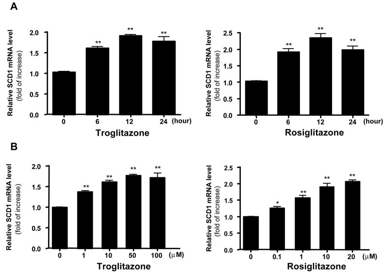
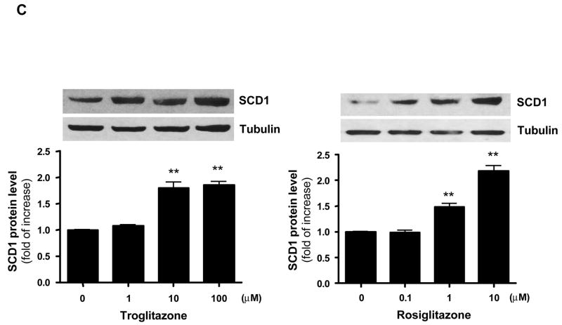
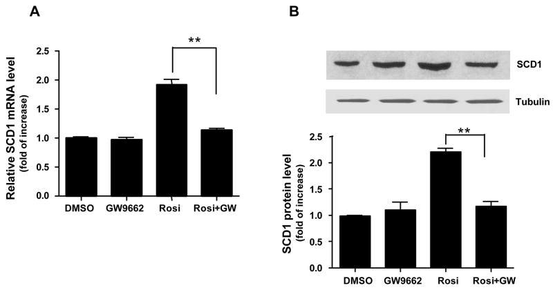
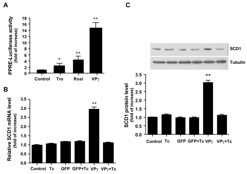
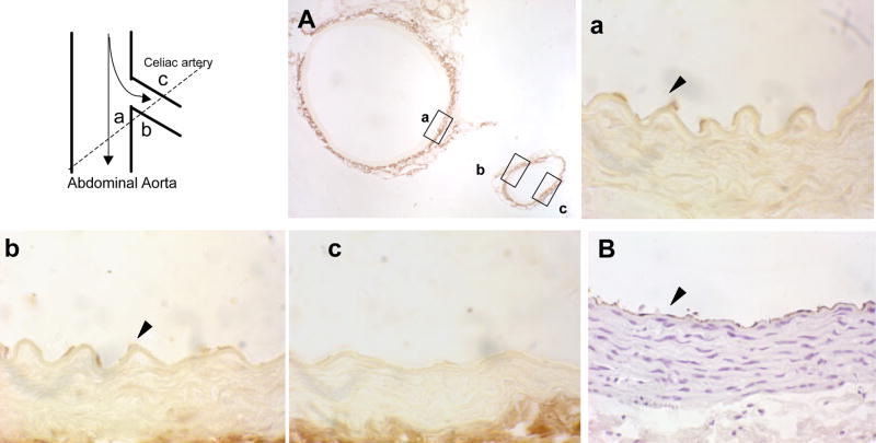
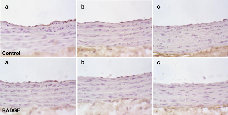
Results: Laminar shear stress (12 dyn/cm2, 12 h) markedly increased the gene expression of SCD1 in ECs. The flow-induced SCD1 expression was attenuated by peroxisome proliferator-activated receptor (PPAR)-gamma antagonists both in vitro and in vivo. Troglitazone and rosiglitazone significantly increased the gene expression of SCD1. Furthermore, overexpression of a constitutively active PPARgamma induced the expression of SCD1 in ECs. Immunohistochemical study of cross-sections from rat celiac arteries revealed that endothelial expression of SCD1 was substantially higher on the medial division apex, where the shear stress is high and more laminar, than the lateral aspect, where the shear stress is low and unsteady.

Conclusion: These in vitro and in vivo results demonstrate that laminar flow increased the expression of SCD1 in endothelium through a PPARgamma-specific mechanism, which may contribute to the shear stress-mediated protective roles in ECs.


Figures

Fig. 1
Fig. 1. Laminar flow induced SCD1 mRNA expression in ECs
(A) ECs were exposed to laminar flow (12 dyn/cm2), low shear (2 dyn/cm2) or kept under a static condition for 12 h. SCD1 gene expression was analyzed with qRT-PCR. The levels of SCD1 mRNA were normalized to the β-actin level and expressed as fold of increase in relation to the control. The data represent means ± SEM of 3 independent experiments. **P<0.01 vs. control. (B) ECs were exposed to laminar flow (12 dyn/cm2) for 0, 3, 6, 12 or 24 h. Northern blots were hybridized with 32P-labeled cDNA probes for human SCD1 and β-actin.
Fig. 2
Fig. 2. PPARγ Antagonist GW9662 Inhibited the Laminar Flow-induced SCD1 Gene Expression
HUVECs were pretreated with GW9662 (10 μM for 30 min) or control (DMSO 1:1,000) before the exposure to laminar flow or static condition. The SCD1 mRNA expression was analyzed with the use of real-time RT-PCR. The data represent the means ± SEM of 3 independent experiments. **P<0.01 vs. DMSO.
Fig. 3
Fig. 3. Troglitazone and Rosiglitazone Increased the Expression of SCD1 in ECs
(A) HUVECs were treated with troglitazone (50 μM) or rosiglitazone (10 μM) for 0, 6, 12 and 24 h. (B) HUVECs were treated with various concentrations of troglitazone or rosiglitazone for 24 h. Total RNA was isolated and examined with real-time RT-PCR for SCD1 mRNA expression. (C) HUVECs were treated with troglitazone or rosiglitazone at the indicated concentrations for 24 h. Western blotting were performed with primary antibodies against SCD1 or tubulin. Data represent mean ± SEM of 3 independent experiments and are expressed as fold of increase compared to controls. * P<0.05, **P<0.01 vs. control.
Fig. 4
Fig. 4. PPARγ antagonist blocked TZD-induced SCD1 expression in ECs
(A) HUVECs were pretreated with GW9662 (10 μM) or solvent for 30 min before exposure to rosiglitazone (Rosi) for 24 h. Total RNA was extracted and examined for SCD1 mRNA expression with the use of real-time RT-PCR. (B) GW9662 blocked rosiglitazone-induced SCD protein expression. Data represent mean ± SEM of 3 independent experiments. **P<0.01
Fig. 5
Fig. 5. Constitutive activation of PPARγ induced the expression of SCD1 in ECs
(A) VP-PPARγ constitutively activated the PPRE-driven reporter. HUVECs were co-transfected with plasmids encoding PPRE-TK-luciferase and β-galactosidase before exposure to DMSO, troglitazone (Tro) or rosiglitazone (Rosi). Alternatively, the cells were co-transfected with VP-PPARγ plasmid (VPγ). Luciferase activity was measured and normalized to β-galactosidase activity. (B) VP-PPARγ induced the expression of SCD1 mRNA. ECs were infected with Ad-VP-PPARγ and Ad-tTA, or Ad-GFP and Ad-tTA for 24 h in the presence or absence of tetracycline (Tc). SCD1 mRNA was analyzed with qRT-PCR. (C) VP-PPARγ induced the expression of SCD1 protein. Data represent mean ± SEM of 3 independent experiments. * P<0.05, **P<0.01
Fig. 6
Fig. 6. Immunohistochemical Detection of SCD1 in Rat Arteries
(A) Cross section of rat celiac artery branch points, including the abdominal aorta and celiac artery, immunohistochemically stained with anti-SCD1. Enlarged views are shown for areas in the abdominal aorta (a) and the medial (b) and lateral (c) aspects of the celiac artery. Dotted line in drawing shows the plane at which the vessels were sectioned. (B) Antibody against von Willebrand factor (vWF) was used to confirm the endothelial integrity throughout the lumen areas. The sections were counterstained with hematoxylin. The arrows indicate the endothelium of the vessel. (C) SCD1 expression in abdominal aorta (a) and the medial (b) and lateral (c) aspects of the celiac arteries from rats treated with BADGE (lower panel) or control (upper panel) for 7 days. The results shown are representative of 3 animals in each group.

Similar articles

Cited by

References

    1. Chien S, Li S, Shyy YJ. Effects of mechanical forces on signal transduction and gene expression in endothelial cells. Hypertension. 1998;31:162–9. - PubMed
    1. Garcia-Cardena G, Comander J, Anderson KR, Blackman BR, Gimbrone MA., Jr Biomechanical activation of vascular endothelium as a determinant of its functional phenotype. Proc Natl Acad Sci USA. 2001;98:4478–85. - PMC - PubMed
    1. Haidekker MA, L’Heureux N, Frangos JA. Fluid shear stress increases membrane fluidity in endothelial cells: a study with DCVJ fluorescence. Am J Physiol Heart Circ Physiol. 2000;278:H1401–6. - PubMed
    1. Butler PJ, Norwich G, Weinbaum S, Chien S. Shear stress induces a time- and position-dependent increase in endothelial cell membrane fluidity. Am J Physiol Cell Physiol. 2001;280:C962–9. - PubMed
    1. Flowers MT, Miyazaki M, Liu X, Ntambi JM. Probing the role of stearoyl-CoA desaturase-1 in hepatic insulin resistance. J Clin Invest. 2006;116:1478–81. - PMC - PubMed

Publication types

MeSH terms