

Familial cavitary optic disk anomalies: identification of a novel genetic locus
- PMID: 17368552
- PMCID: PMC3684050
- DOI: 10.1016/j.ajo.2007.01.042
Familial cavitary optic disk anomalies: identification of a novel genetic locus
Abstract
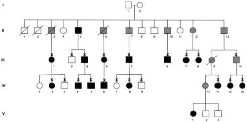
Purpose: To identify the chromosomal location of the gene involved in the pathogenesis of cavitary optic disk anomalies in a large pedigree with autosomal dominant inheritance of disease.
Design: Linkage analysis of a pedigree affected with cavitary optic disk anomalies.
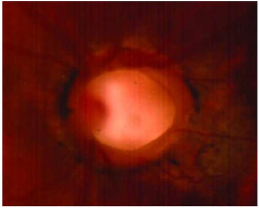
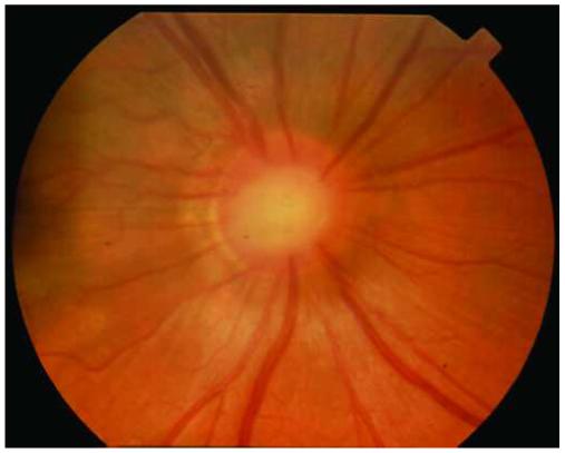
Methods: Optic disk photographs were examined for the presence of cavitary optic disk anomalies. Sixteen affected family members and one obligate carrier were identified and studied with linkage analysis using both microarrays of single nucleotide polymorphisms (SNPs) and short tandem repeat polymorphism (STRP) markers.
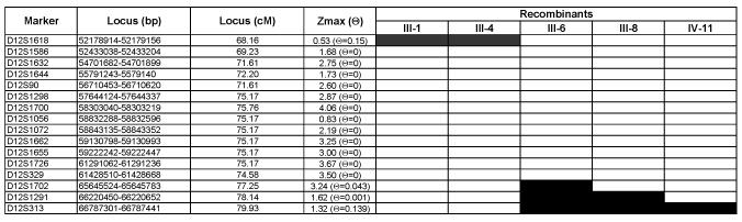
Results: Multipoint linkage analysis of SNP genotypes yielded a maximum nonparametric logarithm of the odds (LOD) score of 21.7 with markers located on chromosome 12q. Linkage was confirmed with 16 STRP markers in the 12q region. A maximum two-point LOD score of 4.06 (theta = 0) was obtained with marker D12S1700. The disease interval defined by observed recombinants is 9.1 cM, which corresponds to 13.5 Mbp. Three candidate genes (GDF-11, NEUROD4, and WIF1) in the chromosome 12q locus were evaluated as possible disease-causing genes. No mutations were detected in the coding sequence of these genes.
Conclusions: The discovery of the chromosomal location of a gene responsible for cavitary optic disk anomalies is a key step in identifying the genetic basis of this condition and ultimately may provide important insight into the pathogenesis of more common optic nerve diseases such as normal-tension glaucoma and primary open-angle glaucoma (POAG).
Figures
Similar articles
-
Congenital cavitary optic disc anomaly and Axenfeld's anomaly in Wolf-Hirschhorn syndrome: A case report and review of the literature.Ophthalmic Genet. 2018 Apr;39(2):271-274. doi: 10.1080/13816810.2017.1408850. Epub 2017 Dec 4. Ophthalmic Genet. 2018. PMID: 29199884 Free PMC article. Review.
-
A large family with inherited optic disc anomalies: a correlation between a new genetic locus and complex ocular phenotypes.Sci Rep. 2017 Aug 10;7(1):7799. doi: 10.1038/s41598-017-07730-7. Sci Rep. 2017. PMID: 28798362 Free PMC article.
-
Familial cavitary optic disk anomalies: clinical features of a large family with examples of progressive optic nerve head cupping.Am J Ophthalmol. 2007 May;143(5):788-794. doi: 10.1016/j.ajo.2007.01.029. Epub 2007 Mar 23. Am J Ophthalmol. 2007. PMID: 17362864
-
Refinement of the MYP3 locus on human chromosome 12 in a German family with Mendelian autosomal dominant high-grade myopia by SNP array mapping.Int J Mol Med. 2008 Apr;21(4):429-38. Int J Mol Med. 2008. PMID: 18360688
-
Cavitary anomalies of the optic disc: neurologic significance.Curr Neurol Neurosci Rep. 2008 Sep;8(5):409-13. doi: 10.1007/s11910-008-0063-5. Curr Neurol Neurosci Rep. 2008. PMID: 18713577 Review.
Cited by
-
GDF11 Antagonizes Psoriasis-like Skin Inflammation via Suppression of NF-κB Signaling Pathway.Inflammation. 2019 Feb;42(1):319-330. doi: 10.1007/s10753-018-0895-3. Inflammation. 2019. PMID: 30259241
-
Heterozygous triplication of upstream regulatory sequences leads to dysregulation of matrix metalloproteinase 19 in patients with cavitary optic disc anomaly.Hum Mutat. 2015 Mar;36(3):369-78. doi: 10.1002/humu.22754. Hum Mutat. 2015. PMID: 25581579 Free PMC article.
-
Congenital cavitary optic disc anomaly and Axenfeld's anomaly in Wolf-Hirschhorn syndrome: A case report and review of the literature.Ophthalmic Genet. 2018 Apr;39(2):271-274. doi: 10.1080/13816810.2017.1408850. Epub 2017 Dec 4. Ophthalmic Genet. 2018. PMID: 29199884 Free PMC article. Review.
-
Comparison of an expanded ataxia interactome with patient medical records reveals a relationship between macular degeneration and ataxia.Hum Mol Genet. 2011 Feb 1;20(3):510-27. doi: 10.1093/hmg/ddq496. Epub 2010 Nov 15. Hum Mol Genet. 2011. PMID: 21078624 Free PMC article.
-
A large family with inherited optic disc anomalies: a correlation between a new genetic locus and complex ocular phenotypes.Sci Rep. 2017 Aug 10;7(1):7799. doi: 10.1038/s41598-017-07730-7. Sci Rep. 2017. PMID: 28798362 Free PMC article.
References
-
- Corbett JJ, Savino PJ, Schatz NJ, Orr LS. Cavitary developmental defects of the optic disc. Visual loss associated with optic pits and colobomas. Arch Neurol. 1980;37:210–213. - PubMed
-
- Slusher MM, Weaver RG, Jr., Greven CM, Mundorf TK, Cashwell LF. The spectrum of cavitary optic disc anomalies in a family. Ophthalmology. 1989;96:342–347. - PubMed
-
- Stefko ST, Campochiaro P, Wang P, Li Y, Zhu D, Traboulsi EI. Dominant inheritance of optic pits. Am J Ophthalmol. 1997;124:112–113. - PubMed
Publication types
MeSH terms
Substances
Grants and funding
LinkOut - more resources
Full Text Sources
Medical
Miscellaneous
