

Recent progress in computer-aided diagnosis of lung nodules on thin-section CT
- PMID: 17369020
- PMCID: PMC1948076
- DOI: 10.1016/j.compmedimag.2007.02.005
Recent progress in computer-aided diagnosis of lung nodules on thin-section CT
Abstract
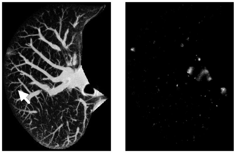
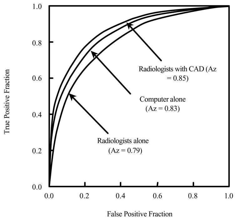
Computer-aided diagnosis (CAD) provides a computer output as a "second opinion" in order to assist radiologists in the diagnosis of various diseases on medical images. Currently, a significant research effort is being devoted to the detection and characterization of lung nodules in thin-section computed tomography (CT) images, which represents one of the newest directions of CAD development in thoracic imaging. We describe in this article the current status of the development and evaluation of CAD schemes for the detection and characterization of lung nodules in thin-section CT. We also review a number of observer performance studies in which it was attempted to assess the potential clinical usefulness of CAD schemes for nodule detection and characterization in thin-section CT. Whereas current CAD schemes for nodule characterization have achieved high performance levels and would be able to improve radiologists' performance in the characterization of nodules in thin-section CT, current schemes for nodule detection appear to report many false positives, and, therefore, significant efforts are needed in order further to improve the performance levels of current CAD schemes for nodule detection in thin-section CT.
Figures
Similar articles
-
Computer-aided diagnosis in thoracic CT.Semin Ultrasound CT MR. 2005 Oct;26(5):357-63. doi: 10.1053/j.sult.2005.07.001. Semin Ultrasound CT MR. 2005. PMID: 16274004 Review.
-
High performance lung nodule detection schemes in CT using local and global information.Med Phys. 2012 Aug;39(8):5157-68. doi: 10.1118/1.4737109. Med Phys. 2012. PMID: 22894441 Free PMC article.
-
Neural network-based computer-aided diagnosis in distinguishing malignant from benign solitary pulmonary nodules by computed tomography.Chin Med J (Engl). 2007 Jul 20;120(14):1211-5. Chin Med J (Engl). 2007. PMID: 17697569
-
Computerized detection of lung nodules in thin-section CT images by use of selective enhancement filters and an automated rule-based classifier.Acad Radiol. 2008 Feb;15(2):165-75. doi: 10.1016/j.acra.2007.09.018. Acad Radiol. 2008. PMID: 18206615 Free PMC article.
-
Current status and future potential of computer-aided diagnosis in medical imaging.Br J Radiol. 2005;78 Spec No 1:S3-S19. doi: 10.1259/bjr/82933343. Br J Radiol. 2005. PMID: 15917443 Review.
Cited by
-
Novel inhibitors of AKT: assessment of a different approach targeting the pleckstrin homology domain.Curr Med Chem. 2011;18(18):2727-42. doi: 10.2174/092986711796011292. Curr Med Chem. 2011. PMID: 21649580 Free PMC article. Review.
-
Computer-aided detection (CADe) and diagnosis (CADx) system for lung cancer with likelihood of malignancy.Biomed Eng Online. 2016 Jan 6;15(1):2. doi: 10.1186/s12938-015-0120-7. Biomed Eng Online. 2016. PMID: 26759159 Free PMC article.
-
Comparing the performance of trained radiographers against experienced radiologists in the UK lung cancer screening (UKLS) trial.Br J Radiol. 2016 Oct;89(1066):20160301. doi: 10.1259/bjr.20160301. Epub 2016 Jul 27. Br J Radiol. 2016. PMID: 27461068 Free PMC article. Clinical Trial.
-
Hybrid method for the detection of pulmonary nodules using positron emission tomography/computed tomography: a preliminary study.Int J Comput Assist Radiol Surg. 2014 Jan;9(1):59-69. doi: 10.1007/s11548-013-0910-y. Epub 2013 Jun 23. Int J Comput Assist Radiol Surg. 2014. PMID: 23793722
-
Feature Selection for Automatic Tuberculosis Screening in Frontal Chest Radiographs.J Med Syst. 2018 Jun 29;42(8):146. doi: 10.1007/s10916-018-0991-9. J Med Syst. 2018. PMID: 29959539
References
-
- Doi K. Overview on research and development of computer-aided diagnostic schemes. Seminars in Ultrasound, CT, and MRI. 2004;25:404–410. - PubMed
-
- Giger ML. Computerized analysis of images in the detection and diagnosis of breast cancer. Seminars in Ultrasound, CT, and MRI. 2004;25:411–418. - PubMed
-
- Li Q, Li F, Suzuki K, Shiraishi J, Abe H, Engelmann R, Nie YK, MacMahon H, Doi K. Computer-Aided Diagnosis in Thoracic CT. Seminars in US, CT, and MRI. 2005;26:357–363. - PubMed
-
- Yoshida H, Dachman AH. Computer-aided diagnosis for CT colonography. Seminars in Ultrasound, CT, and MRI. 2004;25:419–431. - PubMed
-
- Greenlee RT, Murray T, Bolden S, Wingo PA. Cancer statistics, 2000. CA Cancer Journal of Clinicians. 2000;50:7–33. - PubMed
Publication types
MeSH terms
Grants and funding
LinkOut - more resources
Full Text Sources
Medical
Miscellaneous
