. 2007 May;189(10):3846-54.
doi: 10.1128/JB.00027-07. Epub 2007 Mar 16.

Glycine residues in the hydrophobic core of the GspB signal sequence route export toward the accessory Sec pathway

Affiliations

Glycine residues in the hydrophobic core of the GspB signal sequence route export toward the accessory Sec pathway

Barbara A Bensing et al. J Bacteriol. 2007 May.

Abstract

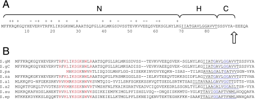
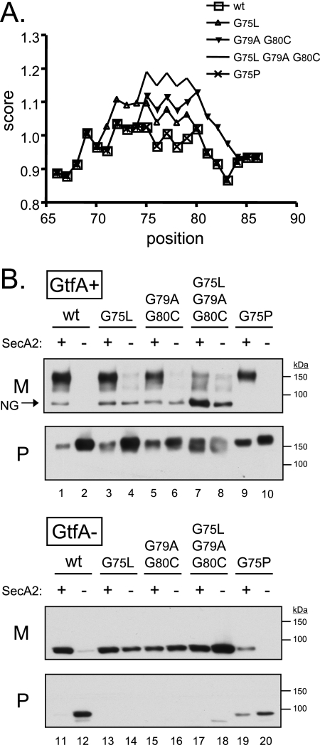
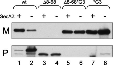
The Streptococcus gordonii cell surface glycoprotein GspB mediates high-affinity binding to distinct sialylated carbohydrate structures on human platelets and salivary proteins. GspB is glycosylated in the cytoplasm of S. gordonii and is then transported to the cell surface via a dedicated transport system that includes the accessory Sec components SecA2 and SecY2. The means by which the GspB preprotein is selectively recognized by the accessory Sec system have not been characterized fully. GspB has a 90-residue amino-terminal signal sequence that displays a traditional tripartite structure, with an atypically long amino-terminal (N) region followed by hydrophobic (H) and cleavage regions. In this report, we investigate the relative importance of the N and H regions of the GspB signal peptide for trafficking of the preprotein. The results show that the extended N region does not prevent export by the canonical Sec system. Instead, three glycine residues in the H region not only are necessary for export via the accessory Sec pathway but also interfere with export via the canonical Sec route. Replacement of the H-region glycine residues with helix-promoting residues led to a decrease in the efficiency of SecA2-dependent transport of the preprotein and a simultaneous increase in SecA2-independent translocation. Thus, the hydrophobic core of the GspB signal sequence is responsible primarily for routing towards the accessory Sec system.


Figures

FIG. 1.
FIG. 1.
Accessory sec locus of S. gordonii M99 and domain organization of GspB. Upper diagram, map of the 23-kb M99 chromosomal locus containing gspB and the accessory sec genes. Asp1-5, SecY2, and SecA2 are components of the accessory Sec system. Gly, Nss, GtfA, and GtfB affect the glycosylation of GspB. Lower diagram, GspB polypeptide domains. SS, signal sequence; SRR1, first serine-rich region; BR, basic region; SRR2, second serine-rich region; CWA, cell wall-anchoring domain. GspB736flag is a variant of GspB that is truncated at residue 736 of 3,072 residues and has a C-terminal 3XFLAG tag.
FIG. 2.
FIG. 2.
(A) Characteristics of the GspB signal sequence. N, H, and C refer to the amino-terminal, hydrophobic, and cleavage regions, respectively. Basic (+) and acidic (−) amino acid residues in the N region are indicated. Residues comprising the H region are underlined. The arrow indicates the location of an experimentally determined cleavage point. (B) Comparison of the amino-terminal ends of selected GspB homologues. The sequences were aligned with the ClustalW algorithm (www.ebi.ac.uk/clustalw), using the default parameters. S.gM, S. gordonii M99 (GenBank accession number AAL13053); S.gC, S. gordonii Challis (GenBank accession number BAA97453); S.pa, S. parasanguinis (GenBank accession number AAC79868); S.sa, S. sanguinis (obtained from the Virginia Commonwealth University sequencing project at www.sanguinis.mic.vcu.edu); S.a1, S. agalactiae COH1 (GenBank accession number EAO76299); S.a2, S. agalactiae 2603V/R (GenBank accession number AAN00330); S.au, Staphylococcus aureus (GenBank accession number ABD31977); S.ep, Staphylococcus epidermidis (GenBank accession number AAO05891).
FIG. 3.
FIG. 3.
(A) Predicted effect of amino acid substitutions on alpha-helix formation within the H region of the GspB signal sequence (positions 65 through 86). Data were obtained from the ExPASy ProtScale analysis website (http://www.expasy.org/tools/protscale.html), using the Chou and Fasman scale (13). (B) Export of GspB736flag variants carrying substitutions in the H region of the signal peptide. Proteins were detected by using an anti-FLAG monoclonal antibody. The lanes contain proteins present in 75 μl of cultures grown for 6 h. The arrow indicates a nonglycosylated (NG) form of GspB736flag secreted by the GtfA+ strains (determined by migration under SDS-PAGE with an apparent molecular mass that is equivalent to the predicted mass of 80 kDa and by failure to react with sWGA; data not shown). M, medium fraction; P, protoplast fraction.
FIG. 4.
FIG. 4.
(A) Construction of the merodiploid strain PS1144. The dashed line in the PS1144 chromosomal locus represents the plasmid backbone (not drawn to scale). (B) Export of GspB and GspB736flag by merodiploid strains. Lane 1, PS946 (gspB736flag gspB); lane 2, PS1144 (gspB736flag gspB*G3). The lanes contain proteins present in 75 μl of cultures grown for 6 h. Proteins present in the protoplast (P), cell wall (W), or culture medium (M) were detected by Western blotting with the indicated antibodies.
FIG. 5.
FIG. 5.
Effect of sodium azide on export of GspB736flag*G3 from secA2 mutant strain PS1146. The lanes contain protein from 75 μl of cultures grown at high density for 90 min. Proteins were detected by using an anti-FLAG monoclonal antibody. M, medium fraction; P, protoplast fraction.
FIG. 6.
FIG. 6.
Effect of mutations in the signal sequence on export of nonglycosylated GspB736flag variants from gtfA mutant strains. Proteins were detected by using an anti-FLAG monoclonal antibody. The lanes contain proteins present in 75 μl of cultures grown for 6 h. M, medium fraction; P, protoplast fraction. Lane 1, PS1179; lane 2, PS1177; lane 3, PS1033; lane 4, PS1172; lane 5, PS1171; lane 6, PS1139; lane 7, PS1136; lane 8, PS1137.

References

    1. Adams, H., P. A. Scotti, H. De Cock, J. Luirink, and J. Tommassen. 2002. The presence of a helix breaker in the hydrophobic core of signal sequences of secretory proteins prevents recognition by the signal-recognition particle in Escherichia coli. Eur. J. Biochem. 269:5564-5571. - PubMed
    1. Arbely, E., Z. Granot, I. Kass, J. Orly, and I. T. Arkin. 2006. A trimerizing GxxxG motif is uniquely inserted in the severe acute respiratory syndrome (SARS) coronavirus spike protein transmembrane domain. Biochemistry 45:11349-11356. - PubMed
    1. Ausubel, F. M., R. Brent, R. E. Kingston, D. D. Moore, J. G. Seidman, J. A. Smith, and K. Struhl. 1997. Current protocols in molecular biology. John Wiley & Sons, New York, NY.
    1. Bae, T., and O. Schneewind. 2003. The YSIRK-G/S motif of staphylococcal protein A and its role in efficiency of signal peptide processing. J. Bacteriol. 185:2910-2919. - PMC - PubMed
    1. Bendtsen, J. D., H. Nielsen, G. von Heijne, and S. Brunak. 2004. Improved prediction of signal peptides: SignalP 3.0. J. Mol. Biol. 340:783-795. - PubMed

Publication types

MeSH terms

LinkOut - more resources