. 2007 Mar 27;104(13):5395-400.
doi: 10.1073/pnas.0608219104. Epub 2007 Mar 16.

A role for a complex between activated G protein-coupled receptors in yeast cellular mating

Affiliations

A role for a complex between activated G protein-coupled receptors in yeast cellular mating

Chunhua Shi et al. Proc Natl Acad Sci U S A. .

Abstract

Cell-cell fusion is a fundamental process that facilitates a wide variety of biological events in organisms ranging from yeast to humans. However, relatively little is actually understood with respect to fusion mechanisms. In the model organism Saccharomyces cerevisiae, mating of opposite-type cells is triggered by pheromone activation of the G protein-coupled receptors, alpha-factor receptor (Ste2p) and a-factor receptor (Ste3p), leading to mitogen-activated protein kinase signaling, growth arrest, and cellular fusion events. Herein we now provide evidence of a role for these receptors in the later cell fusion stage of mating. In vitro assays demonstrated the ability of the receptors to promote mixing of proteoliposomes containing phosphatidylserine, potentially based on a pheromone-dependent interaction between Ste2p and Ste3p that was confirmed by tandem affinity purification and cellular pull-down assays. The cellular mating activity of Ste2p was subsequently probed in vivo. Notably, a receptor-null yeast strain expressing N-terminally truncated Ste2p yielded a phenotype demonstrating wild-type signaling but arrested mating. The arrested prezygotes showed evidence of some cell wall erosion but no membrane juxtaposition at the fusion site. Further, in vitro analyses correlated this mutation with loss of the interaction between Ste2p and Ste3p and inhibition of related lipid mixing. Overall, these results support a role for a complex between activated yeast pheromone receptors in later cell fusion stages of mating, possibly mediating events at the level of cell wall digestion and membrane juxtaposition before membrane fusion.


Conflict of interest statement

The authors declare no conflict of interest.

Figures

Fig. 1.
Fig. 1.
Activated Ste2p and Ste3p mediate lipid mixing in vitro. (A) Fluorescent liposomes containing Ste2p-(His)8 were mixed with nonfluorescent liposomes containing Ste3p-(His)8 in the presence (red star) or absence (black hexagon) of a- and α-factor. 7-Nitro-2–1,3-benzoxadiazol-4-yl dequenching was detected at 2-min intervals. Fusion in the absence of Ste2p-(His)8 and Ste3p-(His)8 was carried out in the presence (blue triangle) or absence (green square) of a- (125 μM) and α-factor (500 μM) to exclude the possibility of lipid mixing mediated by pheromones alone. (B) Fluorescent liposomes containing Ste2p-(His)8 were mixed with nonfluorescent liposomes containing Ste3p-(His)8 in the presence (red star) or absence (black hexagon) of a- and α-factor with 10 mM Ca2+ included in the system.
Fig. 2.
Fig. 2.
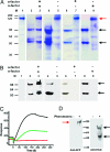
Characterization of a ligand-dependent interaction between Ste2p and Ste3p. (A) SDS/PAGE analysis of TAP elution fractions. Lane 1, elution of Fc-Ste2p-GFP; lanes 2, 4, 6, and 8, control samples of Ste3p-(His)8 purified from Ni-NTA for TAP. Lanes 3, 5, 7, and 9, elution of Fc-Ste2p-GFP after incubation with purified Ste3p-(His)8 in the presence or absence of a- and α-factor. The arrows indicate the expected positions of the Fc-Ste2p-GFP (≈125-kDa) band and the monomer (≈52 kDa) and dimer (≈100 kDa) Ste3p-(His)8 bands. (B) Western blot of A probing for the presence of Ste3p-(His)8 by using Anti-HisG HRP as the primary antibody. (C) Characterization of the interaction between Ste2p and Ste3p by surface plasmon resonance. Purified, recombinant detergent-solubilized a-factor-Ste3p-(His)8 complex was immobilized on an Ni-NTA biosensor chip surface and Fc-Ste2p-GFP passed over the chip at 800- (black line) and 200-nM (green line) concentrations in the presence of both pheromones or at 800-nM concentration without pheromones (red line). (D) Characterization of the interaction between Ste2p and Ste3p by cellular pull-down assay. (Left) Western blot showing detergent solubilized Fc-Ste2p-GFP pulled down by transfected 293E cells presenting Ste3p-(His)8 in the presence (+) or absence (−) of a- and α-factor. Anti-GFP was used as the primary antibody. (Right) Western blot showing detergent solubilized Ste3p-(His)8 pulled down by transfected 293E cells presenting Fc-Ste2p-GFP in the presence (+) or absence (−) of a- and α-factor. Anti-HisG HRP was used as the primary antibody.
Fig. 3.
Fig. 3.
The extracellular N-terminal region of Ste2p mediates cellular fusion activities in vivo. (A) Halo assay determination of α-factor stimulated signaling and cell cycle arrest activity in a receptor null MATa yeast strain (YFL026W) overexpressing (a) Ste2p-GFP, (b) Ste2pΔN20-GFP, and (c) Ste2pΔN30-GFP. 5, 2, 1, and 0.5-μl volumes of a 10-mM α-factor stock solution were spotted in positions 1–4 respectively. (B) Auxotrophic MATα SCY060 was mated with receptor null MATa yeast strains (YFL026W) expressing Ste2p-GFP (lane 1), Ste2pΔN20-GFP (lane 2), Ste2pΔN30-GFP (lane 3), and Ste4p (lane 4). (C) Representative matings costained with FM4-64 and DAPI to simultaneous visualize membranes and DNA. (Scale bars, 5 μm.) (D) Representative Ste2pΔN20-GFP arrested prezygotes probed by EM. CW, cell wall; CM, cell membrane. (Scale bars, 1 μm.)
Fig. 4.
Fig. 4.
The Ste2pΔN20 mutant does not support Ste3p complex formation or lipid mixing. (A) Deletion of the N-terminal 20 amino acids of Ste2p eliminates interaction with activated Ste3p in vitro. (Left) SDS/PAGE analysis of TAP elution fractions. (Right) Western blot of Left probing for the presence of Ste3p-(His)8 by using anti-HisG HRP as the primary antibody. Lanes 1, control samples of Ste3p-(His)8 purified from Ni-NTA for TAP. Lanes 2, elution of Fc-Ste2pΔN20-GFP after incubation with purified Ste3p-(His)8 in the presence of a- and α-factor. The arrows indicate the expected positions of the Fc-Ste2pΔN20-GFP (≈120 kDa) band and the monomer (≈52 kDa) and dimer (≈100 kDa) Ste3p-(His)8 bands. (B) Deletion of the N-terminal 20 amino acids of Ste2p eliminates lipid mixing activity. Fluorescent liposomes containing Ste2pΔN20-(His)8 were mixed with nonfluorescent liposomes containing Ste3p-(His)8 in the presence (blue hexagon) or absence (green inverted square) of a- and α-factor. 7-Nitro-2–1,3-benzoxadiazol-4-yl dequenching was detected at 3-min intervals. A positive-control experiment for the liposome fusion with Ste2p-(His)8 and Ste3p-(His)8 in the presence of pheromones (red star) was repeated for comparison.

References

    1. Shemer G, Podbilewicz B. BioEssays. 2003;25(7):672–682. - PubMed
    1. Chen EH, Olson EN. Science. 2005;308:369–373. - PubMed
    1. Jahn R, Lang T, Sudhof TC. Cell. 2003;112:519–533. - PubMed
    1. White JM, Rose MD. Curr Biol. 2001;11:R16–R20. - PubMed
    1. Bartwell L. Peptides. 2005;26:339–350. - PMC - PubMed

Publication types

MeSH terms