. 2007 Mar 15;21(6):655-67.
doi: 10.1101/gad.1511107.

Mad3p, a pseudosubstrate inhibitor of APCCdc20 in the spindle assembly checkpoint

Affiliations

Mad3p, a pseudosubstrate inhibitor of APCCdc20 in the spindle assembly checkpoint

Janet L Burton et al. Genes Dev. .

Abstract

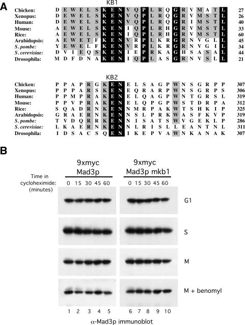
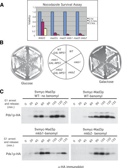
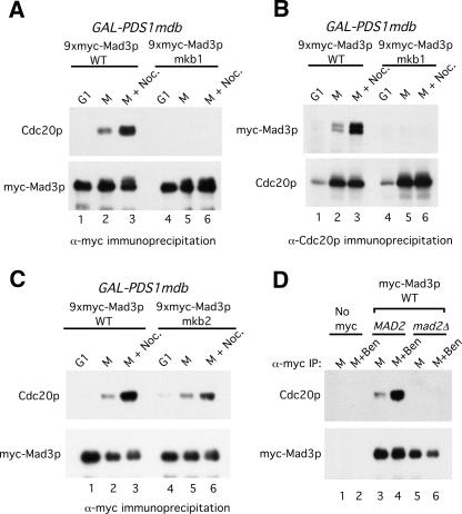
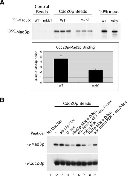
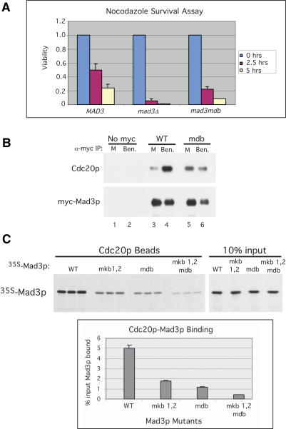
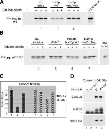
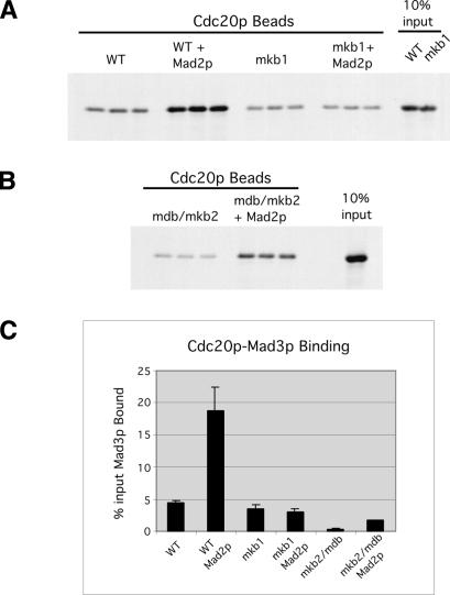
Inappropriate attachment/tension between chromosomal kinetochores and the kinetochore microtubules activates the spindle assembly checkpoint, which delays anaphase by blocking the ubiquitin-mediated degradation of securin/Pds1p by APCCdc20. The checkpoint proteins Mad2 and Mad3/BubR1 bind to Cdc20, although how they inhibit APCCdc20 is unclear. We investigated the roles of two evolutionarily conserved KEN boxes and a D box within Mad3/BubR1. Although such motifs usually mediate APC-substrate recognition and ubiquitination, they have no apparent role in Mad3p turnover in Saccharomyces cerevisiae. Instead, these motifs are important for Mad3p function in the checkpoint and for binding to Cdc20p. We show that the Mad3p D box and KEN boxes function together to mediate Cdc20p-Mad3p interaction and that Mad3p and an anaphase-promoting complex (APC) substrate, Hsl1p, compete for Cdc20p binding in a D-box- and KEN-box-dependent manner. In vivo, we observed an increased binding of Cdc20p to Mad3p and decreased binding to Hsl1p upon checkpoint activation. Furthermore, we demonstrate that Mad2p stimulates the association between Mad3p and Cdc20p and that this stimulated binding requires KEN box 1 within Mad3p. These findings implicate Mad3p as a pseudosubstrate inhibitor of APCCdc20, competing with APC substrates for Cdc20p binding. We present a model aimed at unifying previous analyses of checkpoint function by focusing on the Mad3-Cdc20 interaction.


Figures

Figure 1.
Figure 1.
The conserved KB1 has no apparent role in Mad3p turnover. (A) Alignment of the Mad3/BubR1 proteins from various species is shown with invariant residues shaded in black and highly conserved residues shaded in gray. KB1 and KB2 motifs are labeled. Sequences were aligned using the Clustal W program. Numbers at the right indicate the last residue shown on each line. (B) Both Mad3p WT and mkb1 are stable proteins. Strains containing Mad3p WT (YJB800) and Mad3p-mkb1 (YJB801) were arrested in G1 with α-factor (100 ng/mL), in S phase with hydroxyurea (100 mM), in M phase by expression of GAL-PDS1mdb, or in M phase with an activated spindle checkpoint by expression of GAL-PDS1mdb and the addition of benomyl (50 μg/mL). Samples were taken at the indicated times after cycloheximide addition to inhibit protein synthesis. Mad3p was detected by immunoblotting with anti-Mad3p antibodies.
Figure 2.
Figure 2.
The KEN boxes of Mad3p are required for spindle checkpoint function. (A) Nocodazole survival assay. Wild-type (YJB15), mad3Δ (YJB522), mad3mkb1 (YJB804), and mad3mkb2 (YJB932) strains were exposed to nocodazole (15 μg/mL) for 0, 2.5, and 5 h and then plated on YPD plates lacking the drug. Viability was calculated by dividing the number of colonies formed by cells treated with nocodazole for 2.5 and 5 h by the number formed by untreated cells. Error bars depict the standard deviations from three independent experiments. (B) Mps1p overexpression assay. Wild-type, mad3mkb1, and mad3mkb2 strains either with (YJB900, YJB901, and YJB923) or without (YJB894, YJB895, and YJB919) a GAL-myc-MPS1 plasmid (E591) were plated on glucose- and galactose-containing plates. Unlike mad3mkb1 and mad3mkb2 strains, wild-type cells overexpressing Mps1p underwent checkpoint arrest as shown by their inability to form colonies in the presence of galactose. Schematic indicates the positions of the different strains on the plates. (C) mad3mkb1 and mad3mkb2 cells fail to arrest in metaphase in response to benomyl. Wild-type (YJB894), mad3mkb1 (YJB895), and mad3mkb2 (YJB919) strains were arrested in G1 with α-factor (100 ng/mL) and then released synchronously into medium either lacking or containing benomyl (50 μg/mL) to induce the spindle checkpoint. (Top right panel) Pds1p is stabilized in wild-type cells in the presence of benomyl, reflecting a mitotic arrest and indicating the presence of a functional spindle checkpoint. In contrast, Pds1p levels fluctuated in mad3mkb1 and mad3mkb2 cells, which entered and then exited mitosis (bottom panels), similar to wild-type cells in the absence of benomyl (top left panel).
Figure 3.
Figure 3.
Mad3p-mkb1 is defective in its association with Cdc20p. (A) Mad3p was immunoprecipitated from extracts of strains expressing wild-type myc-Mad3p (YJB800) or myc-Mad3p-mkb1 (YJB801) that were arrested in G1, M, or M plus nocodazole (see Materials and Methods). Mad3p and coimmunoprecipitating Cdc20p were detected by immunoblotting. (B) Cdc20p was immunoprecipitated from extracts of strains (YJB800 and YJB801) arrested as described in A. Cdc20p and coimmunoprecipitated Mad3p were detected by immunoblotting as in A. (C) As in A using strains expressing myc-Mad3p wild type (YJB800) or mkb2 (YJB924). (D) Efficient Mad3p association with Cdc20p requires Mad2p. myc-Mad3p was immunoprecipitated using anti-myc antibodies from extracts of M-phase or M-phase plus benomyl arrested cells in wild-type (YJB800) or mad2Δ (YJB905) strains. Mad3p and coimmunoprecipitating Cdc20p were detected by immunoblotting. No Cdc20p was detected from a yeast strain lacking myc-Mad3p (YJB793).
Figure 4.
Figure 4.
KEN-box- and D-box-dependent binding of Mad3p to Cdc20p in vitro. (A) Wild-type Mad3p and Mad3p-mkb1 were translated in vitro in the presence of 35S-methionine and then mixed with control or GST-Cdc20p glutathione beads. The beads were washed, and bound 35S-Mad3p was detected by autoradiography. Ten percent of the input Mad3p is shown in the lanes at the right. The percentages of input Mad3p and Mad3p-mkb1 bound to GST-Cdc20p were quantitated by PhosphorImage analysis using the three independent binding events for each sample. (B) KEN-box and D-box peptides dramatically reduce Mad3p binding to Cdc20p. Wild-type Mad3p was purified from baculovirus-infected cells and incubated with Cdc20p beads that had been preincubated with 500 μM KB1 peptide (“Mad3p KEN”), Hsl1p D-box peptide (“D-box”), Hsl1p KEN-box peptide (“Hsl1p KEN”), combinations of these peptides, or scrambled versions of these peptides (“scr.”), as indicated. The beads were washed, and bound Mad3p and Cdc20p were detected by immunoblotting.
Figure 5.
Figure 5.
Mad3p has a D-box motif important for the spindle checkpoint and Cdc20p binding. (A) Nocodazole survival assay of MAD3 (YJB697), mad3Δ (YJB522), and mad3mdb (YJB926) strains. Viability was calculated as in Figure 2A. (B) Mad3p-mdb binds Cdc20p with reduced efficiency. Mad3p (YJB800) and Mad3p-mdb (YJB928) were immunoprecipitated from extracts of cells arrested in M or M plus benomyl (see Materials and Methods). Mad3p and coimmunoprecipitating Cdc20p were detected by immunoblotting. No Mad3p or Cdc20p was immunoprecipitated from a “no myc” control strain extract (YJB793). (C) The Mad3p D box and KEN boxes function together to bind Cdc20p. 35S-methionine labeled wild type (WT), Mad3p-mkb1mkb2, Mad3p-mdb, and Mad3p-mkb1mkb2mdb were produced by in vitro translation and incubated in triplicate with GST-Cdc20p glutathione beads. The beads were washed, and bound proteins were detected by autoradiography. (Top panels) Ten percent of the input Mad3p is shown in the lanes at right. (Bottom panel) The percentage of input Mad3p bound to GST-Cdc20p for each sample was quantitated by PhosphorImage analysis for the three independent binding events.
Figure 6.
Figure 6.
Mad3p and Hsl1p compete for Cdc20p binding. (A) Hsl1p wild type but not mdb/mkb can compete with Mad3p for binding to Cdc20p. GST-Cdc20p beads were preincubated in the absence or the presence of wild-type or mdb/mkb MBP-Hsl1p followed by incubation with wild-type 35S-Mad3p as in Figure 4A. Bound 35S-Mad3p was detected by autoradiography. (B) Mad3p WT but not Mad3p-Mut. can compete with Hsl1p667–872 for binding to Cdc20p. Cdc20p beads were preincubated with Mad2p alone or with Mad2p and wild-type or triply mutated Mad3p followed by incubation with 35S-Hsl1p667–872. Bound 35S-Hsl1p667–872 was detected by autoradiography. (C) The relative levels of wild-type 35S-Mad3p (left bars 1–3) or 35S-Hsl1p667–872 (right bars 4–7) bound to Cdc20p were quantitated by PhosphorImage analysis of the samples shown in A and B. The bindings of 35S-Mad3p and 35S-Hsl1p667–872 to Cdc20p in the absence of competitor proteins were given relative values of 100. Error bars represent the standard deviations from the three independent binding events shown for each sample. (D) Cell extracts were prepared from a strain (YJB864) constitutively expressing Hsl1p667–872-HA that was arrested in M phase or M phase plus benomyl to induce the spindle checkpoint. Cdc20p was immunoprecipitated, and coimmunoprecipitating Mad3p and Hsl1p-HA levels were assessed with anti-Mad3p and anti-HA antibodies, respectively. (Lanes 3,4) Checkpoint activation increased the amount of Mad3p associated with Cdc20p and decreased the amount of Hsl1p-HA associated with Cdc20p. (Lanes 1,2) None of these proteins was detected using a control preimmune serum. Asterisk denotes IgG heavy chain.
Figure 7.
Figure 7.
Mad2p stimulates Mad3p binding to Cdc20p in vitro in a KB1-dependent manner. GST-Cdc20p beads were preincubated with or without Mad2p followed by the addition of either wild-type or mkb1 35S-labeled Mad3p (A) or 35S-labeled Mad3p-mkb2mdb (B). (C) The amount of bound Mad3p was detected by PhosphorImage analysis. Error bars represent the standard deviations for the three independent binding events shown for each sample.

References

    1. Ausubel F.M., Brent R., Kingston R.E., Moore D.D., Seidman J.G., Smith J.A., Struhl K., Brent R., Kingston R.E., Moore D.D., Seidman J.G., Smith J.A., Struhl K., Kingston R.E., Moore D.D., Seidman J.G., Smith J.A., Struhl K., Moore D.D., Seidman J.G., Smith J.A., Struhl K., Seidman J.G., Smith J.A., Struhl K., Smith J.A., Struhl K., Struhl K. Current protocols in molecular biology. Wiley; New York: 1995.
    1. Burton J.L., Solomon M.J., Solomon M.J. Hsl1p, a Swe1p inhibitor, is degraded via the anaphase-promoting complex. Mol. Cell. Biol. 2000;20:4614–4625. - PMC - PubMed
    1. Burton J.L., Solomon M.J., Solomon M.J. D box and KEN box motifs in budding yeast Hsl1p are required for APC-mediated degradation and direct binding to Cdc20p and Cdh1p. Genes & Dev. 2001;15:2381–2395. - PMC - PubMed
    1. Burton J.L., Tsakraklides V., Solomon M.J., Tsakraklides V., Solomon M.J., Solomon M.J. Assembly of an APC–Cdh1–substrate complex is stimulated by engagement of a destruction box. Mol. Cell. 2005;18:533–542. - PubMed
    1. Carroll C.W., Enquist-Newman M., Morgan D.O., Enquist-Newman M., Morgan D.O., Morgan D.O. The APC subunit Doc1 promotes recognition of the substrate destruction box. Curr. Biol. 2005;15:11–18. - PubMed

Publication types

MeSH terms