

X-ray microbeams: Tumor therapy and central nervous system research
- PMID: 17369874
- PMCID: PMC1828126
- DOI: 10.1016/j.nima.2005.03.062
X-ray microbeams: Tumor therapy and central nervous system research
Abstract
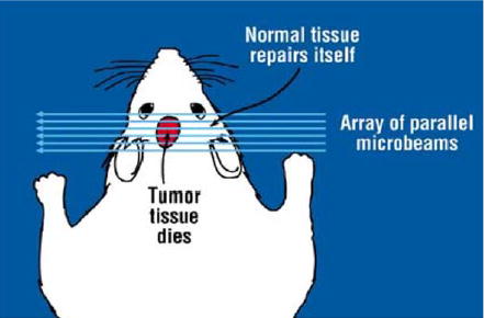
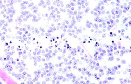
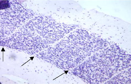
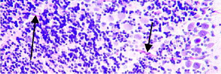
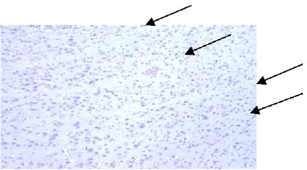
Irradiation with parallel arrays of thin, planar slices of X-ray beams (microplanar beams, or microbeams) spares normal tissue, including the central nervous system (CNS), and preferentially damages tumors. The effects are mediated, at least in part, by the tissue's microvasculature that seems to effectively repair itself in normal tissue but fails to do so in tumors. Consequently, the therapeutic index of single-fraction unidirectional microbeam irradiations has been shown to be larger than that of single-fraction unidirectional unsegmented beams in treating the intracranial rat 9L gliosarcoma tumor model (9LGS) and the subcutaneous murine mammary carcinoma EMT-6. This paper presents results demonstrating that individual microbeams, or arrays of parallel ones, can also be used for targeted, selective cell ablation in the CNS, and also to induce demyelination. The results highlight the value of the method as a powerful tool for studying the CNS through selective cell ablation, besides its potential as a treatment modality in clinical oncology.
Figures
Similar articles
-
Murine EMT-6 carcinoma: high therapeutic efficacy of microbeam radiation therapy.Radiat Res. 2003 May;159(5):632-41. doi: 10.1667/0033-7587(2003)159[0632:mechte]2.0.co;2. Radiat Res. 2003. PMID: 12710874
-
Response of rat intracranial 9L gliosarcoma to microbeam radiation therapy.Neuro Oncol. 2002 Jan;4(1):26-38. doi: 10.1093/neuonc/4.1.26. Neuro Oncol. 2002. PMID: 11772430 Free PMC article.
-
Response of rat skin to high-dose unidirectional x-ray microbeams: a histological study.Radiat Res. 2003 Aug;160(2):133-42. doi: 10.1667/3033. Radiat Res. 2003. PMID: 12859223
-
Effects of pulsed, spatially fractionated, microscopic synchrotron X-ray beams on normal and tumoral brain tissue.Mutat Res. 2010 Apr-Jun;704(1-3):160-6. doi: 10.1016/j.mrrev.2009.12.003. Epub 2009 Dec 23. Mutat Res. 2010. PMID: 20034592 Review.
-
Synchrotron-generated microbeam radiosurgery: a novel experimental approach to modulate brain function.Neurol Res. 2011 Oct;33(8):825-31. doi: 10.1179/016164111X13123658647445. Neurol Res. 2011. PMID: 22004705 Review.
Cited by
-
Synchrotron-generated microbeam sensorimotor cortex transections induce seizure control without disruption of neurological functions.PLoS One. 2013;8(1):e53549. doi: 10.1371/journal.pone.0053549. Epub 2013 Jan 14. PLoS One. 2013. PMID: 23341950 Free PMC article.
-
Animal Models in Microbeam Radiation Therapy: A Scoping Review.Cancers (Basel). 2020 Feb 25;12(3):527. doi: 10.3390/cancers12030527. Cancers (Basel). 2020. PMID: 32106397 Free PMC article.
-
In Vivo Microbeam Radiation Therapy at a Conventional Small Animal Irradiator.Cancers (Basel). 2024 Jan 30;16(3):0. doi: 10.3390/cancers16030581. Cancers (Basel). 2024. PMID: 38339332 Free PMC article.
-
Image-guided microbeam irradiation to brain tumour bearing mice using a carbon nanotube x-ray source array.Phys Med Biol. 2014 Mar 7;59(5):1283-303. doi: 10.1088/0031-9155/59/5/1283. Epub 2014 Feb 20. Phys Med Biol. 2014. PMID: 24556798 Free PMC article.
-
Sparing of tissue by using micro-slit-beam radiation therapy reduces neurotoxicity compared with broad-beam radiation therapy.J Radiat Res. 2017 Jan;58(1):17-23. doi: 10.1093/jrr/rrw065. Epub 2016 Jul 15. J Radiat Res. 2017. PMID: 27422939 Free PMC article.
References
-
- Slatkin DN, Spanne P, Dilmanian FA, Sandborg M. Med Phys. 1992;19:1395. - PubMed
-
- Thomlinson W, et al. Cell Mol Biol. 2000;46(6):1053. - PubMed
-
- Laissue J, Spanne PO, Dilmanian FA, Gebbers JO, Slatkin DN. Schweiz med Wschr. 1992;122:1627.
-
- Slatkin DN, Dilmanian FA, Nawrocky NM, Spanne P, Gebbers JO, Archer DW, Laissue JA. Rev Sci Instrum. 1995;66:1459.
Grants and funding
LinkOut - more resources
Full Text Sources
Other Literature Sources