

Lewis X oligosaccharides targeting to DC-SIGN enhanced antigen-specific immune response
- PMID: 17371544
- PMCID: PMC2265933
- DOI: 10.1111/j.1365-2567.2007.02554.x
Lewis X oligosaccharides targeting to DC-SIGN enhanced antigen-specific immune response
Abstract
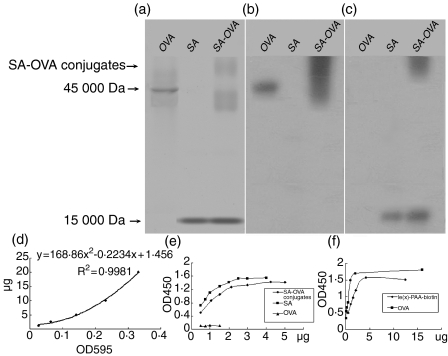
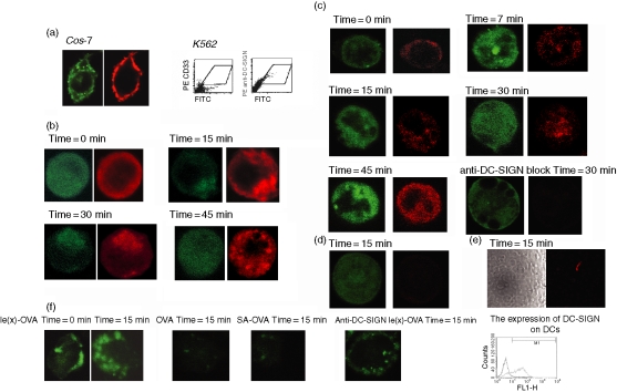
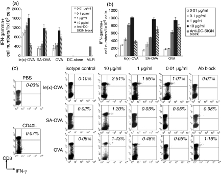
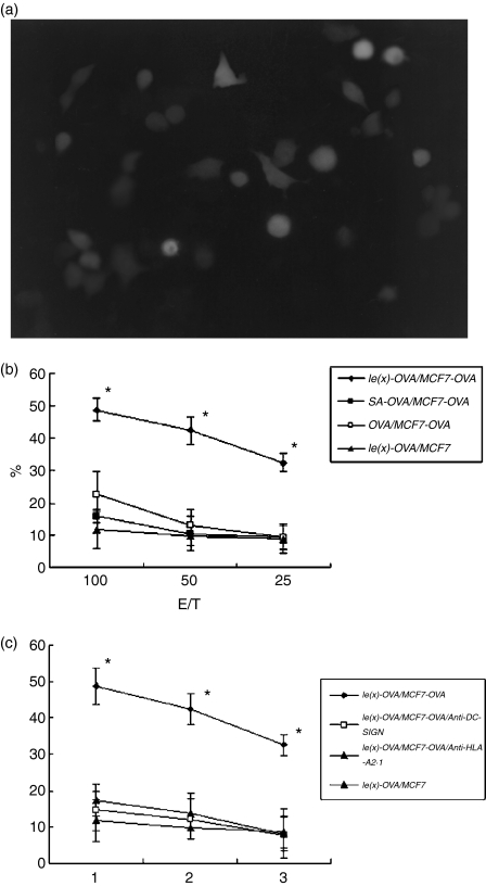
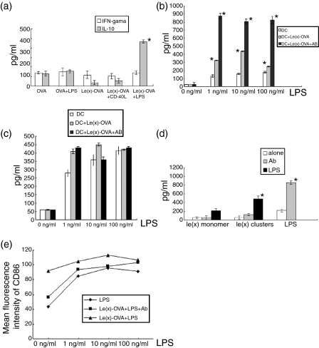
Dendritic cell-specific intercellular-adhesion-molecule-grabbing non-integrin (DC-SIGN) is a potential target receptor for vaccination purposes. In the present study, we employed Lewis X (Le(x)) oligosaccharides, which mimic natural ligands, to target ovalbumin (OVA) to human dendritic cells (DCs) via DC-SIGN, to investigate the effect of this DC-SIGN-targeting strategy on the OVA-specific immune response. We demonstrated that Le(x) oligosaccharides could enhance the OVA-specific immune response as determined by enzyme-linked immunospot assay (ELISPOT), intracellular interferon-gamma staining and (51)Cr-release assay. An almost 300-fold lower dose of Le(x)-OVA induced balanced interferon-gamma-secreting cells compared to OVA alone. Furthermore, secretion of interleukin-10, a reported mediator of immune suppression related to DC-SIGN, was not increased by Le(x)-OVA, either alone or together with sCD40L-stimulated groups. A blocking antibody against DC-SIGN (12507) reduced the numbers of interferon-gamma-secreting cells during Le(x)-OVA stimulation, yet it did not prevent Le(x) oligosaccharides from promoting the secretion of interleukin-10 that was induced by ultra-pure lipopolysaccharide. These results suggested that the strategy of DC-SIGN targeting mediated by Le(x) oligosaccharides could promote a T-cell response. This DC-targeting may imply a novel vaccination strategy.
Figures
Similar articles
-
Targeting glycan modified OVA to murine DC-SIGN transgenic dendritic cells enhances MHC class I and II presentation.Mol Immunol. 2009 Dec;47(2-3):164-74. doi: 10.1016/j.molimm.2009.09.026. Epub 2009 Oct 8. Mol Immunol. 2009. PMID: 19818504
-
Glycan-modified liposomes boost CD4+ and CD8+ T-cell responses by targeting DC-SIGN on dendritic cells.J Control Release. 2012 May 30;160(1):88-95. doi: 10.1016/j.jconrel.2012.02.007. Epub 2012 Feb 15. J Control Release. 2012. PMID: 22366522
-
In vivo targeting of human DC-SIGN drastically enhances CD8⁺ T-cell-mediated protective immunity.Eur J Immunol. 2013 Oct;43(10):2543-53. doi: 10.1002/eji.201343429. Epub 2013 Jul 22. Eur J Immunol. 2013. PMID: 23784881
-
Glycan-based DC-SIGN targeting vaccines to enhance antigen cross-presentation.Mol Immunol. 2013 Sep;55(2):143-5. doi: 10.1016/j.molimm.2012.10.031. Epub 2012 Nov 14. Mol Immunol. 2013. PMID: 23158834 Review.
-
DC-SIGN (dendritic cell-specific ICAM-grabbing non-integrin) and DC-SIGN-related (DC-SIGNR): friend or foe?Clin Sci (Lond). 2003 Apr;104(4):437-46. Clin Sci (Lond). 2003. PMID: 12653690 Review.
Cited by
-
Glycomaterials for probing host-pathogen interactions and the immune response.Exp Biol Med (Maywood). 2016 May;241(10):1042-53. doi: 10.1177/1535370216647811. Epub 2016 May 4. Exp Biol Med (Maywood). 2016. PMID: 27190259 Free PMC article. Review.
-
Immunological and Toxicological Assessment of Triterpenoid Saponins Bearing Lewis-X- and QS-21-Based Trisaccharides.Chemistry. 2025 May 19;31(28):e202500994. doi: 10.1002/chem.202500994. Epub 2025 Apr 21. Chemistry. 2025. PMID: 40192644 Free PMC article.
-
Targeted glycoproteomic analysis reveals that kappa-5 is a major, uniquely glycosylated component of Schistosoma mansoni egg antigens.Mol Cell Proteomics. 2011 May;10(5):M110.005710. doi: 10.1074/mcp.M110.005710. Epub 2011 Mar 3. Mol Cell Proteomics. 2011. PMID: 21372247 Free PMC article.
-
Targeting antigens to dendritic cell receptors for vaccine development.J Drug Deliv. 2013;2013:869718. doi: 10.1155/2013/869718. Epub 2013 Oct 8. J Drug Deliv. 2013. PMID: 24228179 Free PMC article. Review.
-
In situ Delivery of Antigen to DC-SIGN(+)CD14(+) Dermal Dendritic Cells Results in Enhanced CD8(+) T-Cell Responses.J Invest Dermatol. 2015 Sep;135(9):2228-2236. doi: 10.1038/jid.2015.152. Epub 2015 Apr 14. J Invest Dermatol. 2015. PMID: 25885805
References
-
- Bonifaz L, Bonnyay D, Mahnke K, Rivera M, Nussenzweig MC, Steinman RM. Efficient targeting of protein antigen to the dendritic cell receptor DEC-205 in the steady state leads to antigen presentation on major histocompatibility complex class I products and peripheral CD8+ T cell tolerance. J Exp Med. 2002;196:1627–38. - PMC - PubMed
-
- Ramakrishna V, Treml JF, Vitale L, Connolly JE, et al. Mannose receptor targeting of tumor antigen pmel17 to human dendritic cells directs anti-melanoma T cell responses via multiple HLA molecules. J Immunol. 2004;172:2845–52. - PubMed
-
- Geijtenbeek TB, Kwon DS, Torensma R, et al. DC-SIGN, a dendritic cell-specific HIV-1-binding protein that enhances trans-infection of T cells. Cell. 2000;100:587–97. - PubMed
-
- Soilleux EJ, Morris LS, Leslie G, et al. Constitutive and induced expression of DC-SIGN on dendritic cell and macrophage subpopulations in situ and in vitro. J Leukoc Biol. 2002;71:445–57. - PubMed
-
- Engering A, Geijtenbeek TB, van Vliet SJ, et al. The dendritic cell-specific adhesion receptor DC-SIGN internalizes antigen for presentation to T cells. J Immunol. 2002;168:2118–26. - PubMed
Publication types
MeSH terms
Substances
LinkOut - more resources
Full Text Sources
