. 2007 Mar 19:4:19.
doi: 10.1186/1742-4690-4-19.

Mutation in the loop C-terminal to the cyclophilin A binding site of HIV-1 capsid protein disrupts proper virus assembly and infectivity

Affiliations

Mutation in the loop C-terminal to the cyclophilin A binding site of HIV-1 capsid protein disrupts proper virus assembly and infectivity

Samir Abdurahman et al. Retrovirology. .

Abstract

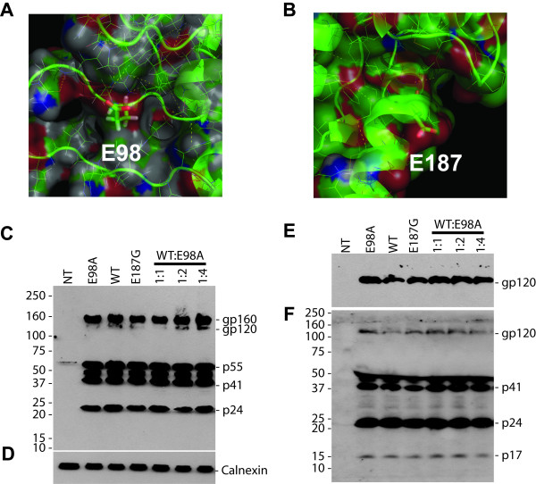
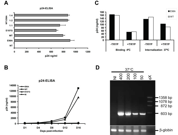
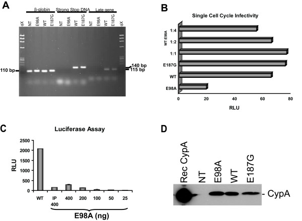
We have studied the effects associated with two single amino acid substitution mutations in HIV-1 capsid (CA), the E98A and E187G. Both amino acids are well conserved among all major HIV-1 subtypes. HIV-1 infectivity is critically dependent on proper CA cone formation and mutations in CA are lethal when they inhibit CA assembly by destabilizing the intra and/or inter molecular CA contacts, which ultimately abrogate viral replication. Glu98, which is located on a surface of a flexible cyclophilin A binding loop is not involved in any intra-molecular contacts with other CA residues. In contrast, Glu187 has extensive intra-molecular contacts with eight other CA residues. Additionally, Glu187 has been shown to form a salt-bridge with Arg18 of another N-terminal CA monomer in a N-C dimer. However, despite proper virus release, glycoprotein incorporation and Gag processing, electron microscopy analysis revealed that, in contrast to the E187G mutant, only the E98A particles had aberrant core morphology that resulted in loss of infectivity.


Figures

Figure 1
Figure 1
Structural view and Western blot analysis of capsid mutants. A close view of the structure of the cyclophilin A binding loop in the N-terminal (A) and the position of E187 in the C-terminal (B) HIV-1CA domains. The two residues in this study, E98 and E187, are being explicitly highlighted. The figure was produced with PyMOL [27] and the structure was obtained from the Protein Data Bank (cf PDB entry 1E6J [3]). (C to F) Western blot analysis of mutant and wild-type pNL4-3 transfected cells (C and D), and viral lysates (E and F). HeLa-tat cells were transfected as indicated with 2 μg of proviral DNAs using the non-liposomal FuGENE transfection reagent (Roche) as recommended by the manufacturer. Cells were also co-transfected with mutant and wild-type pNL4-3 as indicated. Forty-eight to 72 hrs post-transfection, cells were harvested and proteins were separated by SDS-PAGE in 4–12% gels and transferred to a nitrocellulose membrane. The membranes were initially probed with HIV+ patient serum (C and F) and were then reprobed with rabbit anti-calnexin antibody (D) or mouse monoclonal anti-V3 antibody (E) using horseradish peroxidase-conjugated secondary antibodies raised against mouse (DAKO, 1:4000), human (Pierce, 1:20,000), or rabbit (Sigma, 1:4,000) IgG. The protein bands were visualized by chemiluminescence. The positions of specific viral proteins are indicated to the right. Numbers to the left depict positions of molecular mass markers in kDa.
Figure 2
Figure 2
Virus release and internalization studies. p24-ELISA of transfected 293T cell (A) and infected H9 cell (B) culture supernatants. (A) 293T cells were transfected or co-transfected with mutant and wild-type pNL4-3 (2 μg) as indicated using the non-liposomal FuGENE transfection reagent (Roche) as recommended by the manufacturer. Culture supernatants were then assayed for p24 antigen contents 72 hrs post-transfection using an in-house p24 antigen ELISA [28]. Similar results were also obtained with transfected HeLa-tat cells. Virus stocks were then prepared from cleared and filtered culture supernatants (pre-cleared by centrifugation at 1,200 rpm for 7 min and filtered through a 0.45-μm-pore-size membrane) treated with DNase I (Roche) at 20 μg/ml final concentration at 37°C for 1 h. Aliquots in 300-μl fractions of the virus stocks were saved at -80°C until needed. (B) H9 cells (2 × 10⁵ cells) were infected with the X4 NL4-3 strain of mutant or wild type HIV-1 stocks using 200 ng of p24 antigen per well in 24-well plates. Three hours after infection, unbound viruses were removed by centrifugation, washed and resuspended in 1 ml complete RPMI medium per well. The infections were performed in triplicates and supernatants were collected at days 1, 4, 8, 12 and 16 post-infection and tested for p24 antigen contents by p24-ELISA. NI, non-infected control. (C) For virus binding and internalization assay, monolayered TZM-bl cells were seeded one day before infection and following day, medium was removed and cells were inoculated with equal amounts (400 ng of p24 antigen) of mutant or wild type NL4-3 virus stocks (treated with DNase I) with 20 μg/ml DEAE-dextran (in a total volume of 300 μl to 60,000 cells per well in 12-well plates). After adsorption period of 2 hrs, input viruses were removed and cells were treated with trypsin (+TRYP) or not (-TRYP) and the amount of cell associated p24 was measured using the p24-ELISA. (D) TZM-bl cells were also infected as described above with the amount virus indicated and after adsorption period of 2 hrs, input viruses were removed and cells were fed with 1 ml of complete DMEM with 5 μM indinavir and cultured for 24 hrs. Equal amounts of total RNA isolated from E98A infected TZM-bl cells were subjected to nested RT-PCR using specific primers that amplified a 593 bp fragment of the p17 viral RNA. The outer primer pair 5'-GCA GTG GCG CCC GAA CAG and 5'-TTCTGA TAA TGC TGA AAA CAT GGG TAT and inner primer pair 5'-CTC TCG ACG CAG GAC TC and 5'-ACC CAT GCA TTT AAA GTT CTA G was used. As an internal control, the human β-globin RNA was amplified using the primers described elsewhere [29].
Figure 3
Figure 3
Viral infectivity assay. (A) Detection of proviral DNA. H9 cells were infected as above and total cellular DNA was prepared 16 days post-infection using Qiagen's DNA isolation kit and analyzed by PCR using a set of primers specific for negative strand strong-stop DNA and a conserved region of the gag, described previously [30, 31]. Early gene products were amplified using the forward primer Ra 5'-TCT CTG GTT AGA CCA GAT CTG-3' (459–479) and the reverse primer U5a 5'-GTC TGA GGG ATC TCT AGT TAC-3' (584–604). Late gene products representing a conserved region of the HIV-1 gag was amplified with the forward primer SK-38 5'-ATC CAC CTA TCC CAG TAG GAG AAA T-3' (1090–1117) and the reverse primer SK-39 5'-TTT GGT CCT TGT CTT ATG TCC AGA ATG C-3' (1177–1204) that amplified a 115-bp fragment. We also examined the viral cDNA production at 16 hrs post-infection and been able to detect in all cells infected with mutant and wild-type virions (data not shown). To normalize for the quantity of total cellular DNA present in each sample, human β-globin DNA was amplified [29]. (B) Single cell cycle infectivity of mutant and wild-type virus particles on TZM-bl reporter cell lines. Cells (2 × 10⁴) were infected as described above with equal amounts (25 ng p24 antigen) of mutant and wild-type virus or chimeric virus stock prepared by co-transfection of mutant and wild-type pNL4-3 at a ratio of 1:1, 2:1, and 4:1. Infected cells were then cultured in the presence of 5 μM indinavir. Twenty-four hours post-infection, cells were harvested in 200 μl Glo lysis buffer (Promega) and assayed for luciferase activity with the luciferase assay kit obtained from Promega. RLU, relative light unit. (C) TZM-bl cells (8 × 10⁴) were infected as described above with 400 ng of wild-type NL4-3 virus or with E98A virus that was first immunoprecipitated with anti-Tat monoclonal antibody (indicated with IP 400). Cells were also infected with E98A virus stock that had been two-fold serially diluted. After 48 hrs, culture supernatants were removed and cells were assayed for luciferase activity. (D) Detection of virion associated cyclophilin A (CypA) by Western blot analysis. Cell free culture supernatants from 293T cells transfected with mutant and wild-type pNL4-3 were equilibrated for p24 antigen concentration and equal amounts of virus was precipitated with Viraffinity (CPG Inc) as recommended by the manufacturer. Culture supernatants were mixed (4:1) with Viraffinity and the mixture was incubated at room temperature for 5 min and centrifuged at 1000 × g for 10 min. The viral pellets were washed and dissolved in 1× RIPA buffer [50 mM Tris/HCl (pH 7.4), 150 mM NaCl, 1% Triton X-100, 1% sodium deoxycholate and 0.1% SDS, supplemented with a complete protease inhibitor cocktail from Roche]. The viral proteins were finally separated by SDS-PAGE, transferred onto a nitrocellulose membrane and probed with rabbit anti-CypA antibody (Calbiochem, 1:2,000) and as secondary antibody horseradish peroxidase-conjugated anti-rabbit IgG. Rec CypA, recombinant CypA; NT, non-transfected control.
Figure 4
Figure 4
Transmission electron microscopy analysis of mutant and wild-type virions as described previously [4]. (A) With the control virus, a dense core material was shown inside the envelope of immature virus (left panel) and mature virus with dense conical core structure (right panel). (B) Many particles produced by cells transfected with the E98A mutant had either virions with an immature structure or abnormal core morphology (left panel) and a very few detectable cones. Under higher magnification, the E98A virions were observed to be a heterogeneous population of particles (right panel) with varying size and conical core structures, where a number of virions with an electron-lucent centre and aberrant cores were detected (lower panel). (C) E187G virions with a characteristic dense conical core material. Bars, 100 nm. (D) Numerical (%) analysis of 372 wild type NL4-3 and 798 E98A virus particles with respective morphology.

References

    1. Pettit SC, Moody MD, Wehbie RS, Kaplan AH, Nantermet PV, Klein CA, Swanstrom R. The p2 domain of human immunodeficiency virus type 1 Gag regulates sequential proteolytic processing and is required to produce fully infectious virions. J Virol. 1994;68:8017–8027. - PMC - PubMed
    1. Wiegers K, Rutter G, Kottler H, Tessmer U, Hohenberg H, Krausslich HG. Sequential steps in human immunodeficiency virus particle maturation revealed by alterations of individual Gag polyprotein cleavage sites. J Virol. 1998;72:2846–2854. - PMC - PubMed
    1. Monaco-Malbet S, Berthet-Colominas C, Novelli A, Battai N, Piga N, Cheynet V, Mallet F, Cusack S. Mutual conformational adaptations in antigen and antibody upon complex formation between an Fab and HIV-1 capsid protein p24. Structure. 2000;8:1069–1077. doi: 10.1016/S0969-2126(00)00507-4. - DOI - PubMed
    1. Abdurahman S, Hoglund S, Goobar-Larsson L, Vahlne A. Selected amino acid substitutions in the C-terminal region of human immunodeficiency virus type 1 capsid protein affect virus assembly and release. J Gen Virol. 2004;85:2903–2913. doi: 10.1099/vir.0.80137-0. - DOI - PubMed
    1. Wang CT, Barklis E. Assembly, processing, and infectivity of human immunodeficiency virus type 1 gag mutants. J Virol. 1993;67:4264–4273. - PMC - PubMed

Publication types

MeSH terms

LinkOut - more resources