

Step-down infusions of Gd-DTPA yield greater contrast-enhanced magnetic resonance images of BBB damage in acute stroke than bolus injections
- PMID: 17371719
- PMCID: PMC1965264
- DOI: 10.1016/j.mri.2006.09.003
Step-down infusions of Gd-DTPA yield greater contrast-enhanced magnetic resonance images of BBB damage in acute stroke than bolus injections
Abstract
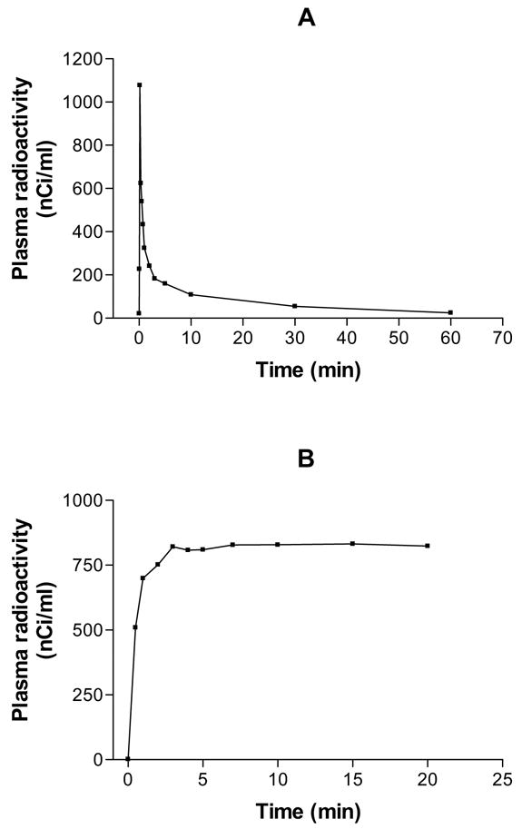
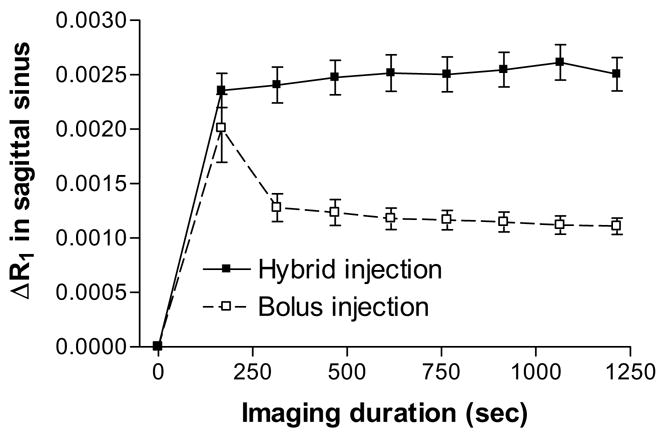
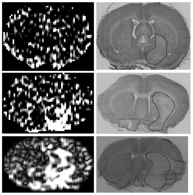
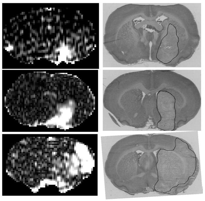
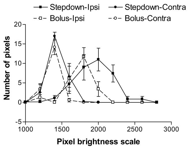
A rat model of transient suture occlusion of one middle cerebral artery (MCA) was used to create a unilateral reperfused cerebral ischemic infarct with blood-brain barrier (BBB) opening. Opening of the BBB was visualized and quantitated by magnetic resonance (MR) contrast enhancement with a Look-Locker T(1)-weighted sequence either following an intravenous bolus injection (n=7) or during a step-down infusion (n=7) of gadolinium-diethylenetriaminepentaacetic acid (Gd-DTPA). Blood levels of Gd-DTPA after either input were monitored via changes in sagittal sinus relaxation rate. Blood-to-brain influx constants (K(i)) were calculated by Patlak plots. On the basis of the MRI parameters and lesion size, the ischemic injury was determined to be similar in the two groups. The bolus injection input produced a sharp rise in blood levels of Gd-DTPA that declined quickly, whereas the step-down infusion led to a sharp rise that was maintained relatively constant for the period of imaging. Visual contrast enhancement and signal-to-noise (S/N) ratios were better with the step-down method (S/N=1.8) than with bolus injection (S/N=1.3). The K(i) values were not significantly different between the two groups (P>.05) and were around 0.005 ml/(g min). The major reason for the better imaging of BBB opening by the step-down infusion was the higher amounts of Gd-DTPA in plasma and tissue during most of the experimental period. These results suggest that step-down MR contrast agent (MRCA) administration schedule may be more advantageous for detection and delineation of acute BBB injury than the usually used bolus injections.
Figures
Similar articles
-
Estimating blood and brain concentrations and blood-to-brain influx by magnetic resonance imaging with step-down infusion of Gd-DTPA in focal transient cerebral ischemia and confirmation by quantitative autoradiography with Gd-[(14)C]DTPA.J Cereb Blood Flow Metab. 2009 May;29(5):1048-58. doi: 10.1038/jcbfm.2009.20. Epub 2009 Mar 25. J Cereb Blood Flow Metab. 2009. PMID: 19319145 Free PMC article.
-
Quantitation and localization of blood-to-brain influx by magnetic resonance imaging and quantitative autoradiography in a model of transient focal ischemia.Magn Reson Med. 2005 Oct;54(4):813-21. doi: 10.1002/mrm.20629. Magn Reson Med. 2005. PMID: 16142715
-
MRI and quantitative autoradiographic studies following bolus injections of unlabeled and (14)C-labeled gadolinium-diethylenetriaminepentaacetic acid in a rat model of stroke yield similar distribution volumes and blood-to-brain influx rate constants.NMR Biomed. 2011 Jun;24(5):547-58. doi: 10.1002/nbm.1625. Epub 2010 Dec 12. NMR Biomed. 2011. PMID: 21674656 Free PMC article.
-
Neuroinflammatory imaging biomarkers: relevance to multiple sclerosis and its therapy.Neurotherapeutics. 2013 Jan;10(1):111-23. doi: 10.1007/s13311-012-0155-4. Neurotherapeutics. 2013. PMID: 23132327 Free PMC article. Review.
-
CSF enhancement on post-contrast fluid-attenuated inversion recovery images; a systematic review.Neuroimage Clin. 2020;28:102456. doi: 10.1016/j.nicl.2020.102456. Epub 2020 Oct 2. Neuroimage Clin. 2020. PMID: 33053497 Free PMC article.
Cited by
-
Multimodal MRI for ischemic stroke: from acute therapy to preventive strategies.J Clin Neurol. 2009 Sep;5(3):107-19. doi: 10.3988/jcn.2009.5.3.107. Epub 2009 Sep 30. J Clin Neurol. 2009. PMID: 19826561 Free PMC article.
-
Multiparametric magnetic resonance imaging and repeated measurements of blood-brain barrier permeability to contrast agents.Methods Mol Biol. 2011;686:193-212. doi: 10.1007/978-1-60761-938-3_8. Methods Mol Biol. 2011. PMID: 21082372 Free PMC article.
-
The quest for a better insight into physiology of fluids and barriers of the brain: the exemplary career of Joseph D. Fenstermacher.Fluids Barriers CNS. 2015 Jan 12;12:1. doi: 10.1186/2045-8118-12-1. eCollection 2015. Fluids Barriers CNS. 2015. PMID: 25745556 Free PMC article. Review.
-
Quantification of Blood-Brain-Barrier Permeability Dysregulation and Inflammatory Activity in MS Lesions by Dynamic-Contrast Enhanced MR Imaging.Basic Clin Neurosci. 2022 Jan-Feb;13(1):117-128. doi: 10.32598/bcn.2022.575.1. Epub 2022 Jan 1. Basic Clin Neurosci. 2022. PMID: 36589018 Free PMC article.
-
Estimating blood and brain concentrations and blood-to-brain influx by magnetic resonance imaging with step-down infusion of Gd-DTPA in focal transient cerebral ischemia and confirmation by quantitative autoradiography with Gd-[(14)C]DTPA.J Cereb Blood Flow Metab. 2009 May;29(5):1048-58. doi: 10.1038/jcbfm.2009.20. Epub 2009 Mar 25. J Cereb Blood Flow Metab. 2009. PMID: 19319145 Free PMC article.
References
-
- Weinmann H-J, Brasch RC, Press W-R, Wesbey GE. Characteristics of gadolinium-DTPA complex: a potential NMR contrast agent. Am J Radiol. 1984;142:619–624. - PubMed
-
- Niendorf HP, Felix R, Laniado M, Schorner W, Claussen C, Wienmann H-J. Gadolinium-DTPA: a new contrast agent for magnetic resonance imaging. Radiation Med. 1985;3:7–12. - PubMed
-
- Virapongse C, Mancuso A, Quisling R. Human brain infarcts: Gd-DTPA-enhanced MR imaging. Radiology. 1986;161:785–794. - PubMed
-
- Elster AD, Moody DM. Early cerebral infarction: gadopentedate dimeglumine enhancement. Radiology. 1990;177:627–632. - PubMed
Publication types
MeSH terms
Substances
Grants and funding
LinkOut - more resources
Full Text Sources
Medical
