. 2007 Mar 26;176(7):1061-71.
doi: 10.1083/jcb.200611026. Epub 2007 Mar 19.

Fibulin-5/DANCE has an elastogenic organizer activity that is abrogated by proteolytic cleavage in vivo

Affiliations

Fibulin-5/DANCE has an elastogenic organizer activity that is abrogated by proteolytic cleavage in vivo

Maretoshi Hirai et al. J Cell Biol. .

Abstract

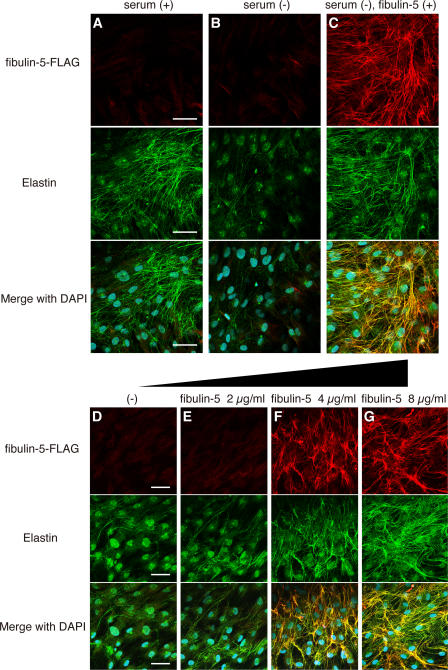
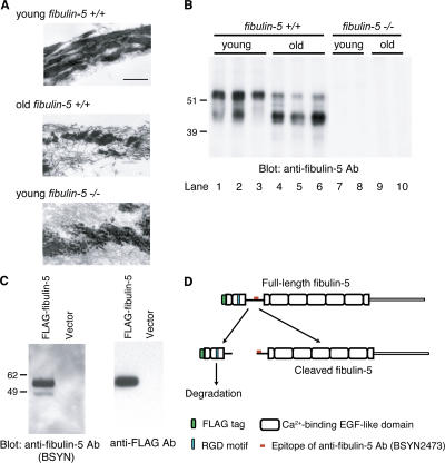
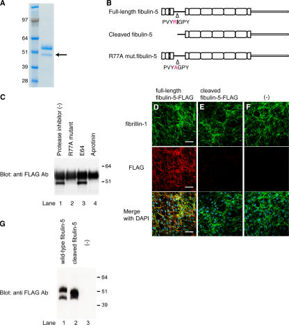
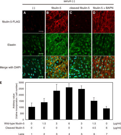
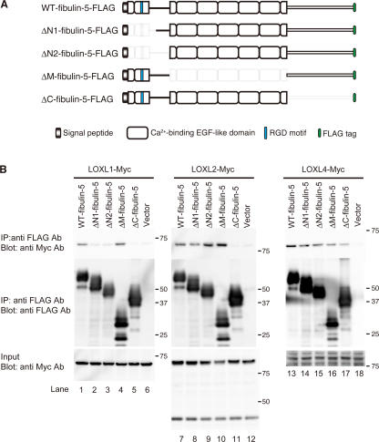
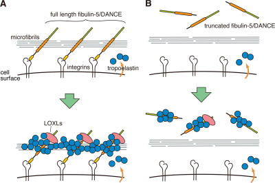
Elastic fibers are required for the elasticity and integrity of various organs. We and others previously showed that fibulin-5 (also called developing arteries and neural crest EGF-like [DANCE] or embryonic vascular EGF-like repeat-containing protein [EVEC]) is indispensable for elastogenesis by studying fibulin-5-deficient mice, which recapitulate human aging phenotypes caused by disorganized elastic fibers (Nakamura, T., P.R. Lozano, Y. Ikeda, Y. Iwanaga, A. Hinek, S. Minamisawa, C.F. Cheng, K. Kobuke, N. Dalton, Y. Takada, et al. 2002. Nature. 415:171-175; Yanagisawa, H., E.C. Davis, B.C. Starcher, T. Ouchi, M. Yanagisawa, J.A. Richardson, and E.N. Olson. 2002. Nature. 415:168-171). However, the molecular mechanism by which fiblin-5 contributes to elastogenesis remains unknown. We report that fibulin-5 protein potently induces elastic fiber assembly and maturation by organizing tropoelastin and cross-linking enzymes onto microfibrils. Deposition of fibulin-5 on microfibrils promotes coacervation and alignment of tropoelastins on microfibrils, and also facilitates cross-linking of tropoelastin by tethering lysyl oxidase-like 1, 2, and 4 enzymes. Notably, recombinant fibulin-5 protein induced elastogenesis even in serum-free conditions, although elastogenesis in cell culture has been believed to be serum-dependent. Moreover, the amount of full-length fibulin-5 diminishes with age, while truncated fibulin-5, which cannot promote elastogenesis, increases. These data suggest that fibulin-5 could be a novel therapeutic target for elastic fiber regeneration.


Figures

Figure 1.
Figure 1.
Fibulin-5 potently induces elastic fiber development and promotes tropoelastin coacervation. (A–C) Human skin fibroblasts were cultured in 10% fetal bovine serum–containing medium (A), in serum-free medium (B), and in serum-free medium with 8 μg/ml of purified recombinant FLAG-tagged fibulin-5 protein (C). Cultures were stained with anti-FLAG (top) or anti–human elastin (middle) antibody. The bottom images were produced by superimposition of the top and middle images, together with DAPI nuclear staining. (D–G) Human skin fibroblasts were cultured in serum-free medium with 0, 2, 4, or 8 μg/ml of purified FLAG-tagged recombinant fibulin-5 protein, to examine the dose-dependency of fibulin-5 elastogenic activity. Cultures were stained as in A–C. Bars, 50 μm.
Figure 2.
Figure 2.
Quantitative PCR analysis in skin fibroblasts after addition of recombinant full-length fibulin-5 or truncated form of fibulin-5. Total RNA from skin fibroblasts was extracted 2 d after addition of recombinant proteins in the serum-free culture media. cDNA was synthesized and was subjected to quantitative real-time PCR for the expression of elastin, LOXL1, LOX, fibrillin-1, -2, and glyceraldehyde-3-phosphate dehydrogenase (Gapdh) transcripts. A graphic presentation of results obtained by real-time PCR is shown. Levels of Gapdh transcript were used to normalize cDNA levels. The relative amount of PCR product amplified from control fibroblasts was set at 100. Data are presented as triplicates, and the means ± the SD are shown. The primers used are shown in Table S1, available at http://www.jcb.org/cgi/content/full/jcb.200611026/DC1.
Figure 3.
Figure 3.
Coacervation assay using purified recombinant tropoelastin and fibulin-5 proteins. 1 mg/ml of soluble tropoelastin with 0, 4, or 40 μg/ml of fibulin-5 protein was induced to coacervate by increasing the temperature, and the turbidity of the solution was measured. Data were obtained in duplicate, and the mean ± the SD values are shown.
Figure 4.
Figure 4.
The N-terminal domain cleavage of fibulin-5 is found in aged mouse skin, as well as in cell cultures. (A) Fine structure of elastic fibers in skin tissues observed by transmission electromicroscopy. Elastin was stained with tannic acid, and therefore appears as black amorphous material. The fine fibers surrounding elastin are microfibrils. Bar, 0.4 μm. (B) Skin tissues were harvested from wild-type or fibulin-5–deficient young (3-mo-old) and old (22-mo-old) mice. Proteins were extracted from skin tissues with 8 M urea and dialyzed against PBS. 10 μg each of these extracts were resolved by SDS-PAGE, and analyzed by Western blotting with anti–fibulin-5 antibody (BSYN2473). Two specific bands of 45 and 55 kD were detected in wild-type mice with anti–fibulin-5 antibody (lanes 1–3). The 55-kD band markedly decreased with age, whereas the 45-kD band markedly increased with age (lanes 4–6). (C) 293T cells were transiently transfected with an expression vector encoding fibulin-5 cDNA with a signal peptide and a FLAG tag at the N terminus. Conditioned medium was subjected to SDS-PAGE, followed by Western blotting analysis with either anti– fibulin-5 or anti-FLAG antibody. Two bands of 55 and 45 kD were detected with anti–fibulin-5 antibody, whereas only a 55-kD band was detected with anti-FLAG antibody. (D) Anti–fibulin-5 antibody (BSYN2473) was raised against a peptide corresponding to amino acids 76–98 (red mark). These findings suggest that fibulin-5 is cleaved at a more N-terminal position than the recognition site of anti–fibulin-5 antibody.
Figure 5.
Figure 5.
Fibulin-5 is cleaved after the arginine at position 77 by serine protease and loses the microfibril-associating activity. (A) 293T cells were stably transfected with an expression vector encoding C-terminal-FLAG– and 6× histidine-tagged fibulin-5 cDNA. Recombinant fibulin-5 protein was purified by chelating chromatography from the culture media of these cells, subjected to SDS-PAGE, and stained with Coomassie blue. The lower band (arrow) was subjected to N-terminal sequencing. (B) The N-terminal sequencing of the lower band identified the specific cleavage site of fibulin-5 at the arginine at position 77. (C) 293T cells were transiently transfected with the expression vector encoding C-terminal-FLAG– and 6× histidine-tagged fibulin-5 or R77A mutant fibulin-5, which had a mutation from arginine to alanine at position 77. Transfected cells were cultured for 2 d with or without a cysteine protease inhibitor, E64, or a serine protease inhibitor, aprotinine, added to the culture media. Culture media were then harvested, concentrated by chelating chromatography, and subjected to Western blotting with anti-FLAG antibody. (D–G) Human skin fibroblasts were cultured for 4 d in serum-free medium in the presence of recombinant fibulin-5 (D) or recombinant cleaved fibulin-5 (E) proteins at a concentration of 4 μg/ml in the medium, or without recombinant protein (F). The cells were then double-stained with anti–fibrillin-1 polyclonal antibody (top) and with anti-FLAG monoclonal antibody (middle). Bottom images were produced by superimposition of the top and middle images, together with DAPI nuclear staining. Bars, 60 μm. At the same time, the conditioned medium was immunoblotted with anti-FLAG antibody to confirm that neither fibulin-5 nor cleaved fibulin-5 was degraded during the culture period (G).
Figure 6.
Figure 6.
Fibulin-5 loses its elastogenic activity upon proteolytic cleavage. (A–E) Human skin fibroblasts were cultured in serum-free media without addition of proteins (A), with wild-type fibulin-5 (B), with the cleaved form of fibulin-5 (C), or with wild-type fibulin-5 and 500 μM BAPN (D). Each FLAG-tagged recombinant protein was added to the culture at a final concentration of 4 μg/ml. Cultures were stained with anti-FLAG antibody (top) and anti–human elastin antibody (middle). The bottom images were produced by superimposition of the top and middle images, together with DAPI nuclear staining. Bars, 50 μm. (E) Quantitation of insoluble (i.e., cross-linked and mature) elastin produced by cells cultured with various amounts of wild-type and cleaved fibulin-5 added to the medium. Cultured skin fibroblasts were metabolically labeled with [3H]valine during the culture period, and the radioactivity of the NaOH-insoluble fractions was quantitated. The radioactivity count was corrected by the relative cell number of separate wells measured with a modified MTT assay, although neither wild-type nor cleaved fibulin-5 significantly affected the cell number (not depicted). Data were obtained as quadruplicates, and the mean ± the SD is shown.
Figure 7.
Figure 7.
Fibulin-5 interacts with LOXL enzymes. (A) Domain structures of the full-length fibulin-5 and the fibulin-5 deletion mutants used for in vitro binding assays. ΔN1-fibulin-5 corresponds to the naturally cleaved form of fibulin-5. These mutants were expressed as C-terminal-FLAG–tagged proteins. (B) Fibulin-5 binds to LOXL1, 2, and 4 through the C-terminal domain. 293T cells were transiently transfected with the vectors shown in A or a mock vector. Expression vectors for Myc-tagged LOX or LOXLs were also independently transfected into 293T cells. The conditioned media were harvested, and mixed. Each mixture was subjected to immunoprecipitation with anti-FLAG antibody, separated by SDS-PAGE, and analyzed by Western blotting with a monoclonal anti-Myc antibody.
Figure 8.
Figure 8.
The Line DANCE model. (A) This model illustrates how fibulin-5/DANCE promotes fibrillar deposition and cross-linking of tropoelastin molecules on microfibrils to promote the development of mature elastic fibers. (B) Truncated fibulin-5 cannot be deposited on microfibrils, and therefore cannot promote elastic fiber assembly.

References

    1. Bailey, A.J. 2001. Molecular mechanisms of ageing in connective tissues. Mech. Ageing Dev. 122:735–755. - PubMed
    1. Carta, L., L. Pereira, E. Arteaga-Solis, S.Y. Lee-Arteaga, B. Lenart, B. Starcher, C.A. Merkel, M. Sukoyan, A. Kerkis, N. Hazeki, et al. 2006. Fibrillins 1 and 2 perform partially overlapping functions during aortic development. J. Biol. Chem. 281:8016–8023. - PMC - PubMed
    1. Clarke, A.W., S.G. Wise, S.A. Cain, C.M. Kielty, and A.S. Weiss. 2005. Coacervation is promoted by molecular interactions between the PF2 segment of fibrillin-1 and the domain 4 region of tropoelastin. Biochemistry. 44:10271–10281. - PubMed
    1. Clarke, A.W., E.C. Arnspang, S.M. Mithieux, E. Korkmaz, F. Braet, and A.S. Weiss. 2006. Tropoelastin massively associates during coacervation to form quantized protein spheres. Biochemistry. 45:9989–9996. - PubMed
    1. Fisher, G.J., S.C. Datta, H.S. Talwar, Z.Q. Wang, J. Varani, S. Kang, and J.J. Voorhees. 1996. Molecular basis of sun-induced premature skin ageing and retinoid antagonism. Nature. 379:335–339. - PubMed

Publication types