. 2007 Jun;27(11):3936-50.
doi: 10.1128/MCB.01776-06. Epub 2007 Mar 19.

Ubiquitin-independent proteasomal degradation of Fra-1 is antagonized by Erk1/2 pathway-mediated phosphorylation of a unique C-terminal destabilizer

Affiliations

Ubiquitin-independent proteasomal degradation of Fra-1 is antagonized by Erk1/2 pathway-mediated phosphorylation of a unique C-terminal destabilizer

Jihane Basbous et al. Mol Cell Biol. 2007 Jun.

Abstract

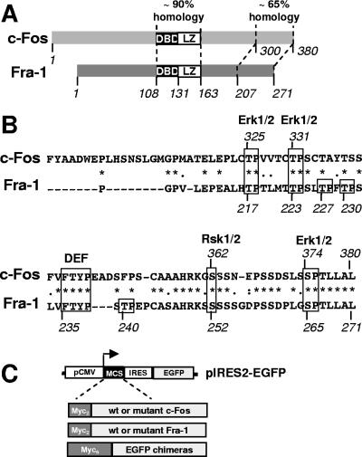
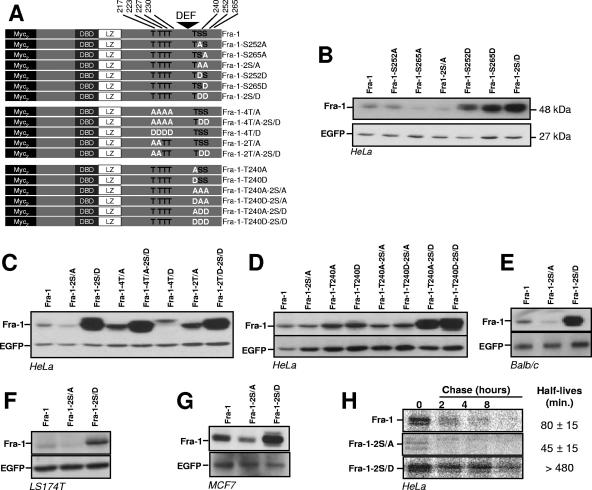
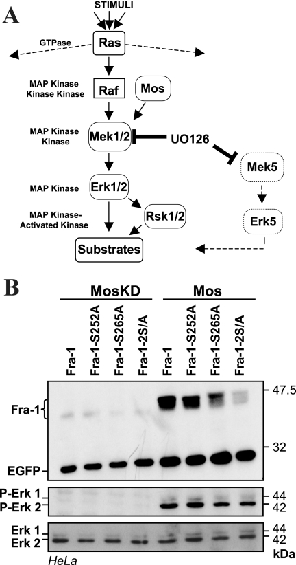
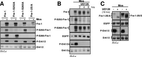
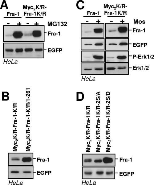
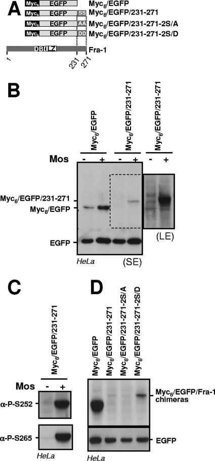
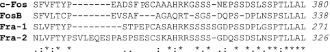
Fra-1, a transcription factor that is phylogenetically and functionally related to the proto-oncoprotein c-Fos, controls many essential cell functions. It is expressed in many cell types, albeit with differing kinetics and abundances. In cells reentering the cell cycle, Fra-1 expression is transiently stimulated albeit later than that of c-Fos and for a longer time. Moreover, Fra-1 overexpression is found in cancer cells displaying high Erk1/2 activity and has been linked to tumorigenesis. One crucial point of regulation of Fra-1 levels is controlled protein degradation, the mechanism of which remains poorly characterized. Here, we have combined genetic, pharmacological, and signaling studies to investigate this process in nontransformed cells and to elucidate how it is altered in cancer cells. We report that the intrinsic instability of Fra-1 depends on a single destabilizer contained within the C-terminal 30 to 40 amino acids. Two serines therein, S252 and S265, are phosphorylated by kinases of the Erk1/2 pathway, which compromises protein destruction upon both normal physiological induction and tumorigenic constitutive activation of this cascade. Our data also indicate that Fra-1, like c-Fos, belongs to a small group of proteins that may, under certain circumstances, undergo ubiquitin-independent degradation by the proteasome. Our work reveals both similitudes and differences between Fra-1 and c-Fos degradation mechanisms. In particular, the presence of a single destabilizer within Fra-1, instead of two that are differentially regulated in c-Fos, explains the much faster turnover of the latter when cells traverse the G(0)/G(1)-to-S-phase transition. Finally, our study offers further insights into the signaling-regulated expression of the other Fos family proteins.


Figures

FIG. 1.
FIG. 1.
Comparison of Fra-1 and c-Fos and analysis of mutants. (A) Structures of c-Fos and Fra-1. The bZIP domain is central. It is the most conserved region between c-Fos and Fra-1. The second region of high homology is the C-terminal domain of both proteins. DBD, DNA-binding domain. (B) Comparison of c-Fos and Fra-1 C-terminal domains. The C-terminal 80 amino acids of human c-Fos were compared with the homologous region in human Fra-1 using the Clustal W program. Identical amino acids are indicated with *, conserved ones are indicated with •, and semiconserved ones are indicated with •. The Erk1/2 and Rsk1/2 target sites and the DEF domain of c-Fos are boxed together with their conserved equivalents in Fra-1. The other putative Erk1/2 target sites of Fra-1 are also boxed. (C) Structure of bicistronic expression plasmids and principle of the immunoblotting assay. pIRES2-EGFP is a CMV promoter-based eukaryotic expression vector. Wild-type (wt) and mutant Fra-1 proteins and EGFP chimeras are cloned in a multicloning site (MCS) linker situated upstream of an encephalomyocarditis virus internal ribosome entry site (IRES)-EGFP expression cassette. Asynchronous cell cultures were transfected in parallel with pIRES2-EGFP-based plasmids. Total cell extracts were prepared 16 to 24 h later, which is a time that is sufficient to reach equilibrium in protein accumulation. They were subsequently analyzed by immunoblotting using specific antibodies against EGFP and the protein to be analyzed (most often the 9E10 anti-Myc tag monoclonal antibody). Note that for EGFP chimeras, a Myc6 tag entails electrophoretic retardation (approximately 10 kDa) that allows easy discrimination with EGFP used as an internal reference. Protein decay can, in the first approximation, be considered to be exponential, which implies that the relative steady-state levels of different proteins synthetized at the same rate are (nearly) proportional to their half-lives. The comparison of protein-to-be-analyzed/EGFP ratios between samples, which can be determined by densitometry scanning of luminograms or direct chemiluminescence signal quantification with a camera-based system, therefore gives the relative stabilities of compared proteins. Importantly, using EGFP as an internal standard and comparing such ratios intrinsically corrects for variations in protein synthesis rates, whether those are due to slight differences in transfection efficiencies or to variations in CMV promoter activity resulting from an alteration of intracellular signaling (see Results).
FIG. 2.
FIG. 2.
Relative stabilities of C-terminal-domain serine and threonine mutants of Fra-1. pIRES2-EGFP-based expression plasmids of the various Fra-1 mutants were transfected in asynchronously growing cells. Immunoblotting experiments were carried out 24 h later using total cell extracts. Fra-1 proteins and EGFP were visualized using the 9E10 anti-Myc tag monoclonal antibody and an anti-EGFP antiserum, respectively. (A) Structure of Fra-1 mutants. DBD, DNA-binding domain. (B) Analysis of serine 252 and serine 265 mutants in HeLa cells. (C) Analysis of threonine 217, 223, 227, and 230 mutants in HeLa cells. (D) Analysis of threonine 240 mutants in HeLa cells. (E) Analysis of Fra-1 serine mutants in BALB/c 3T3 fibroblasts. (F) Analysis of Fra-1 serine mutants in LS174T cells. (G) Analysis of Fra-1 serine mutants in MCF7 cells. (H) Pulse-chase analysis of Fra-1, Fra-1-2S/A, and Fra-1-2S/D in HeLa cells. HeLa cells were transfected to express Fra-1, Fra-1-2S/A, and Fra-1-2S/D, and pulse-chase analyses were carried out as described in Materials and Methods. All data presented are representative of at least three independent experiments.
FIG. 3.
FIG. 3.
S252 and S265 phosphorylation-dependent stabilization of Fra-1 upon Erk1/2 pathway activation. (A) Erk1/2 pathway. Erk1 and Erk2 MAPKs (MAP kinase) (Erk1/2) are activated upon phosphorylation by the Mek1 and Mek2 MAPK kinases (Mek1/2). The UO126 drug can reversibly inhibit the latter as well as Mek5. Mek1/2 is itself activated upon phosphorylation by kinases such as Raf and Mos. The activation of Raf is controlled by Ras small GTPases, which also control other pathways. Erk1/2 phosphorylates its substrates at S/T-P motifs. It activates the Rsk1 and Rsk2 MAPK-activated kinases. Raf and Mos specifically control the Erk1/2 pathway. (B) Effect of Mos on the S252 and S265 mutants of Fra-1. HeLa cells were transfected to express the indicated proteins (Fig. 2A) in the presence or in the absence of vectors for the wild type (Mos) or a kinase-dead mutant (MosKD) of Mos. Note that EGFP levels were higher in the presence of Mos due to the stimulation of the CMV promoter. Taking this point into account, chemiluminescent quantification indicated the absence of Fra-1-2S/A stabilization. The level and the activation of Erk1/2 were assayed using anti-Erk1/2 and anti-phospho-Erk1/2 antisera. The data presented are representative of three independent experiments.
FIG. 4.
FIG. 4.
Erk1/2 pathway-induced phosphorylations of S252 and S265 and Fra-1 stabilization. (A) Erk1/2 pathway-induced phosphorylations of Fra-1 S252 and S265. HeLa cells were transfected to express the indicated proteins (Fig. 2A) in the presence or in the absence of Mos. Total cell extracts were probed with the various rabbit antisera designated for the detection of total Fra-1 (Fra-1), S252-phosphoylated Fra-1 (P-S252-Fra-1), and S265-phosphorylated Fra-1 (P-S265-Fra-1) as well as total Erk1/2 and phosphorylated Erk1/2 (P-Erk1/2). The differences in Fra-1-2S/A abundances in the presence and in the absence of Mos result largely from differences in the transcriptional activity of the expression vector as shown in Fig. 3. (B) UO126 chase of HeLa cells coexpressing Fra-1 and Mos. The UO126 chase was started 16 h after cotransfection of asynchronous HeLa cells transfected with Fra-1 and Mos expression vectors. Immunoblotting experiments were conducted with extracts from cells taken at various time points. (C) UO126 chase in HeLa cells coexpressing Mos with either Fra-1-2S/A or Fra-1-2S/D. The experiment was carried out as in B. + corresponds to 8 h in the presence of UO126. The data presented are representative of three independent experiments.
FIG. 5.
FIG. 5.
Stabilization of Fra-1 in HCT116 colon cancer cells. (A) Relative Erk1/2 activities in HCT116 and LS174T cells. Cell extract immunoblots were probed with antibodies specific for the indicated proteins. (B) Phosphorylation of endogenous Fra-1 on S252 and S265. Extracts from nontreated cells and cells treated with UO126 for 16 h were analyzed by immunoblotting for visualization of the indicated proteins. (C) Destabilization of overexpressed ectopic Fra-1 in HCT116 cells upon Erk1/2 pathway inactivation. HCT116 cells were transiently transfected in the presence or in the absence of UO126 for 16 h to inhibit the phosphorylation of Fra-1. Levels of ectopic total Fra-1 were assayed with the 9E10 antibody, and S252-phosphorylated Fra-1 and S265-phosphorylated Fra-1 were assayed with the relevant anti-phosphoserine antiserum. Transfected Fra-1 was expressed well over the level of endogenous Fra-1, which avoided interference with endogenous Fra-1 when probing with the latter two antisera. The efficiency of UO126 was demonstrated by assaying the levels of phosphorylated and nonphosphorylated Erk1/2. (D) Relative stabilities of Fra-1, Fra-1-2S/A, and Fra-1-2S/D. Transfection experiments were performed as described in the legend of Fig. 2.
FIG. 6.
FIG. 6.
Wild-type and mutant Fra-1 stability during a G0/G1-to-S-phase transition. (A) Phosphorylation of Fra-1 in serum-stimulated cells. BALB/c 3T3 fibroblasts were brought in G0 phase by serum deprivation for 36 h. They were then stimulated for growth by the readdition of culture medium containing 20% serum. Immunoblotting experiments were conducted with extracts from cells stimulated for various periods of time with the indicated antibodies. GAPDH (not shown) and Erk1/2 were used as an invariant internal standards. (B) Fate and phosphorylation of Fra-1 in serum-stimulated cells treated with UO126. In the left panel, cells were treated as described above (A), except that UO126 was added 2 h after stimulation with serum. The right panel corresponds to control cells treated in parallel with no UO126 addition and allows the visualization of the faster Fra-1 decay in the presence of UO126. (C) Structure of transient expression vectors. Fra-1, Fra-1-2S/A, and Fra-1-2S/D open reading frames were cloned in the PM302 vector after the removal of its original c-Fos insert (1). They were stably transfected in BALB/c 3T3 fibroblasts. UTR, untranslated region; SRE, serum-responsive element. (D) Design of the synchronization experiment. Deprivation of and stimulation by serum of the various cells stably transfected with the plasmids described in C were performed as described in A. When required, UO126 was added 1 h poststimulation, which gives sufficient time for Fra-1 accumulation. Ectopic mRNA levels peaked by 45 to 60 min and were back to the basal level by 90 to 120 min after serum addition (1). (E) Immunoblotting assays. Immunoblotting experiments were carried out as described above (A) on the cells stably transfected with the various PM302-based vectors. The data presented are representative of at least three independent experiments.
FIG. 7.
FIG. 7.
Delineation of the Fra-1 destabilizer. (A) Analysis of C-terminal-truncation Fra-1 mutants. Various C-terminal-truncation mutants were cloned in the pIRES2-EGFP expression vectors, and their relative accumulation in transiently transfected HeLa cells was assayed as described in the legend of Fig. 2. DBD, DNA-binding domain. (B) Analysis of EGFP-Fra-1 chimera in asynchronous HeLa cells. The various chimeras were cloned in the pIRES2-EGFP vector for transfection analysis in HeLa cells as described above (A). Immunodetections of Fra-1 proteins and EGFP were performed together with an appropriate combination of anti-Myc tag and anti-EGFP antisera. MCS, multiple cloning site; IRES, internal ribosome entry site. (C) Analysis of Fra-1 mutants during the G0/G1-to-S-phase transition. BALB/c 3T3 fibroblasts, stably transfected to express the indicated proteins from PM302-based plasmids (Fig. 6C), were serum synchronized and analyzed as described in the legend of Fig. 6. Fra-1 was immunodetected with anti-Myc monoclonal antibodies, and GAPDH was used as an internal invariant control. The data presented are representative of at least three independent experiments. UTR, untranslated region.
FIG. 8.
FIG. 8.
Proteasomal degradation of Fra-1. (A) Proteasome-dependent degradation of Fra-1 in asynchronous cells. HeLa cells were transfected with pIRES2-EGFP expression vectors to express the indicated proteins. Twenty-four hours later, they were treated with MG132 or epoxomycin for 8 h. Immunoblotting experiments were carried out as described in the legend of Fig. 2. (B) Inhibition of endogenous Fra-1 decay in HCT116 cells treated with UO126. HCT116 cells were treated with UO126, as described in the legend of Fig. 5B, in the presence of MG132 before immunoblotting analysis. (C) Inhibition of endogenous Fra-1 decay by MG132 in serum-stimulated fibroblasts treated with UO126. BALB/c 3T3 cells were serum stimulated as described in the legend of Fig. 6B. UO126 and MG132 were added 2 h poststimulation. (D) Proteasomal degradation of Fra-1-2S/A during the G0-to-G1-phase transition. BALB/c cells stably transfected to express Fra-1-2S/A from a PM302-based vector (Fig. 6C) were serum deprived for 36 h and then stimulated by the addition of 20% serum. MG132 or epoxomycin was added 1 h later. Immunoblotting assays were performed as described in the legend of Fig. 6 by using GAPDH as an internal control. The data presented are representative of at least three independent experiments. (E) Stabilization of unstable Fra-1 mutants by MG132. HeLa cells were transfected with pIRES2-derived expression vectors coding for the indicated proteins for 24 h and treated for another 8 h in the presence of MG132 before immunoblotting analysis. Normalization of luminograms with EGFP in three independent experiments indicates comparable levels of protein accumulation in the presence of MG132.
FIG. 9.
FIG. 9.
Prior ubiquitylation is not necessary for proteasomal degradation of Fra-1. (A) Proteasomal degradation of Myc2K/R-Fra-1K/R. HeLa cells were transiently transfected to express Fra-1 and Myc2K/R-Fra-1K/R from pIRES2-EGFP-based vectors. MG132 was added 24 h later for a period of 8 h before immunoblotting analysis. (B) Relative stabilities of Myc2K/R-Fra-1K/R and Myc2K/R-Fra-1K/R/1-261. Experiments were carried out in HeLa cells with pIRES2-EGFP-based vectors for the indicated proteins as described above (A). (C) Stabilization of Myc2K/R-Fra-1K/R upon Mos expression. HeLa cells were transfected with pIRES2-EGFP-based vectors for the indicated proteins in the presence of the Mos expression vector. Immunoblotting experiments were conducted 16 h later. (D) Relative stabilities of S252 and S265 mutants of MycK/R-Fra-1K/R. S252 and S265 of Myc2K/R-Fra-1K/R were mutated either in A or in D, and the resulting pIRES2-EGFP-based plasmids were transfected in parallel with that for Myc2K/R-Fra-1K/R. Immunoblotting analysis of the various Fra-1 proteins was performed 16 h later.
FIG. 10.
FIG. 10.
Phosphorylation-driven antagonization of the Fra-1 destabilizer. (A) Structure of EGFP-Fra-1 chimeras. DBD, DNA-binding domain. (B) Mos-driven inhibition of Fra-1 destabilizer activity. Asynchronous HeLa cells were transfected to express the indicated proteins from a pIRES2-EGFP-based plasmid in the presence or in the absence of the Mos expression vector. The immunoblotting analysis was carried out 24 h later with anti-EGFP antibodies. A short-exposure luminogram (SE) is presented in the left panel. Because of the signal saturation for EGFP on this luminogram, transcriptional activation of pIRES2-EGFP is barely appreciable but is similar to that shown in Fig. 3B. For better visualization of Myc6-EGFP/231-271, a longer-exposure luminogram (LE) of the relevant part of the left panel is presented in the right panel. (C) Phosphorylation of Myc6-EGFP/231-271 in the presence of Mos. The protein extracts from cells transfected with a pIRES2-EGFP-based vector for Myc6-EGFP/231-271 in the presence or in the absence of Mos were probed with anti-phospho-S252 and anti-phospho-S265 antisera. (D) Comparison of EGFP chimeras made with wild-type and mutated Fra-1 destabilizer. Asynchronous HeLa cells were transfected with pIRES2-based plasmids for the indicated proteins, and immunoblotting analysis was carried out 24 h later. All data presented are representative of at least three independent experiments.
FIG. 11.
FIG. 11.
Alignments of Fos protein C termini. The domains of the four human Fos proteins corresponding to the C-terminal 40 amino acids of Fra-1 were aligned using the Clustal W multiple sequence alignment program. *:, and • correspond to identical, conserved, and semiconserved amino acids, respectively. The residues corresponding to Fra-1-S252 and Fra-1-S265 are boxed.

References

    1. Acquaviva, C., F. Brockly, P. Ferrara, G. Bossis, C. Salvat, I. Jariel-Encontre, and M. Piechaczyk. 2001. Identification of a C-terminal tripeptide motif involved in the control of rapid proteasomal degradation of c-Fos proto-oncoprotein during the G(0)-to-S phase transition. Oncogene 20:7563-7572. - PubMed
    1. Adiseshaiah, P., S. Peddakama, Q. Zhang, D. V. Kalvakolanu, and S. P. Reddy. 2005. Mitogen regulated induction of FRA-1 proto-oncogene is controlled by the transcription factors binding to both serum and TPA response elements. Oncogene 24:4193-4205. - PubMed
    1. Asher, G., Z. Bercovich, P. Tsvetkov, Y. Shaul, and C. Kahana. 2005. 20S proteasomal degradation of ornithine decarboxylase is regulated by NQO1. Mol. Cell 17:645-655. - PubMed
    1. Asher, G., and Y. Shaul. 2005. p53 proteasomal degradation: poly-ubiquitination is not the whole story. Cell Cycle 4:1015-1018. - PubMed
    1. Asher, G., P. Tsvetkov, C. Kahana, and Y. Shaul. 2005. A mechanism of ubiquitin-independent proteasomal degradation of the tumor suppressors p53 and p73. Genes Dev. 19:316-321. - PMC - PubMed

Publication types

MeSH terms

LinkOut - more resources