. 2007 Jul 1;110(1):201-10.
doi: 10.1182/blood-2006-11-056168. Epub 2007 Mar 19.

Activation-induced expression of CD137 permits detection, isolation, and expansion of the full repertoire of CD8+ T cells responding to antigen without requiring knowledge of epitope specificities

Affiliations

Activation-induced expression of CD137 permits detection, isolation, and expansion of the full repertoire of CD8+ T cells responding to antigen without requiring knowledge of epitope specificities

Matthias Wolfl et al. Blood. .

Abstract

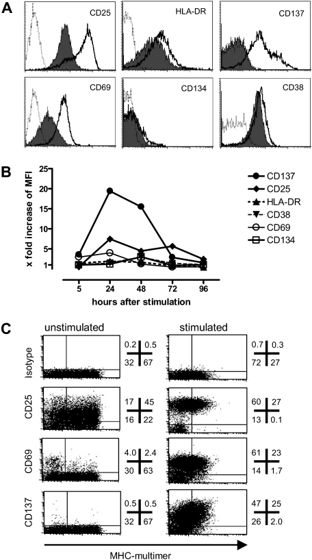
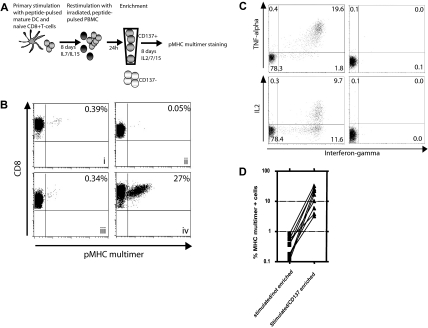
CD137 is a member of the TNFR-family with costimulatory function. Here we show that it also has many favorable characteristics as a surrogate marker for antigen-specific activation of human CD8(+) T cells. Although undetectable on unstimulated CD8(+) T cells, it is uniformly up-regulated 24 hours after stimulation on virtually all responding cells regardless of differentiation stage or profile of cytokine secretion, which circumvents limitations of current surrogate markers for defining the repertoire of responding cells based on only individual functions. Antibody-labeled responding CD137(+) cells can be easily and efficiently isolated by flow sorting or magnetic beads to substantially enrich antigen-specific T cells. To test this approach for epitope discovery, we examined in vitro priming of naive T cells from healthy donors to Wilms tumor antigen 1 (WT1), a protein overexpressed in various malignancies. Two overlapping pentadecamers were identified as immunogenic, and further analysis defined WT1((286-293)) as the minimal amino acid sequence and HLA-Cw07 as the HLA restriction element. In conclusion, this approach appears to be an efficient and sensitive in vitro technique to rapidly identify and isolate antigen-specific CD8(+) T cells present at low frequencies and displaying heterogeneous functional profiles, and does not require prior knowledge of the specific epitopes recognized or the HLA-restricting elements.


Figures

Figure 1
Figure 1
Expression level and kinetics of activation markers after antigen-specific activation. To evaluate a panel of activation markers, a T-cell clone specific for the HLA-A*0201–restricted epitope of Melan-A(26-35L) was stimulated with peptide-pulsed T2 cells, and assessed for (A) antigen-specific up-regulation of denoted markers 24 hours after stimulation (thick lines) compared with control peptide (filled) or isotype-matched antibodies (dotted line); and (B) expression kinetics of the panel of markers in the same experiment (x-fold increase: MFI of the specifically stimulated sample/MFI of the sample stimulated with the irrelevant control peptide). (C) A polyclonal Melan-A–specific T-cell line generated by 2 in vitro stimulations was assessed for baseline expression and specific up-regulation 24 hours after stimulation of the 3 most informative activation markers (numbers indicate the percentage of viable CD8+ T cells in each quadrant).
Figure 2
Figure 2
CD137 up-regulation as a surrogate marker for specifically activated CD8+ T cells in vitro and ex vivo. The activation threshold for several markers of response was compared in responding T-cell clones (A), T-cell lines (B), or memory T cells directly ex vivo (C). (A) Two HLA-A*0201–restricted T-cell clones, specific for either Melan-A(26-35L) or WT1(126-134), were stimulated with titrating amounts of peptides. IFNγ (squares) and TNFα (triangles) production was assessed after 5 hours of stimulation in the presence of brefeldin A, and CD137 expression (circles) was determined 24 hours after stimulation (note: these reflect optimal time points for detecting each of these events). (B) Six separate T-cell lines specific for the NS3(1406-1415)-epitope of HCV were generated, and assessed for cytokine production and degranulation after 5 hours of stimulation, and CD137 expression after 24 hours and pMHC-multimer staining of the unstimulated sample. Pearson correlation (2-tailed) was calculated, and the P value and the coefficient of determination (r2) appear in each plot. (C) PBMCs from 7 CMV+/HLA-A*0201+ donors were thawed, rested overnight, and analyzed for pMHC multimer staining, cytokine production, and degranulation after 5 hours of stimulation and CD137 expression at 24 hours. P value; r2 appear in each plot.
Figure 3
Figure 3
Use of CD137 expression for enrichment of antigen-specific T cells. (A) Experimental outline: Naive CD45ROCD8+ T cells were stimulated with peptide-pulsed DCs and cultured for 1 week with IL-7 and IL-15 added on day 4. On day 8, the cells were restimulated with irradiated, autologous, peptide-pulsed PBMCs. Twenty-four hours later, CD137+ cells were isolated with magnetic beads and cultured for an additional 8 days in medium containing IL-2, IL-7, and IL-15 to allow both expansion and re-expression of the TCR. The cells were then analyzed for specificity using pMHC multimers. (B) Enrichment of an HCV/NS3(1406-1415)-specific T-cell line by selection of CD137+ cells. Eight days after primary stimulation of naive T cells, an aliquot was stained with an NS3(1406-1415)/MHC multimer to determine the frequency of antigen-specific cells before restimulation and/or enrichment (i). These cells were then split and either cultured in IL-2–, IL-7–, and IL-15–containing medium without any restimulation (ii), restimulated and cultured but not enriched (iii), or restimulated and enriched for CD137+ cells 24 hours later and then cultured for an additional 8 days (iv), and then stained with anti-CD8 antibody and the pMHC multimer. (C) Parallel to the pMHC-multimer staining, an aliquot of the enriched T-cell population was also restimulated with peptide-pulsed T2 cells and cytokine production was assessed after 5 hours (left). T cells incubated with unloaded T2 cells served as control (right). (D) Nine different HCV/NS3-specific T-cell lines were generated and enriched 24 hours after the second stimulation as above, and the number of pMHC multimer+ T cells assessed 8 days later and compared with lines not enriched (2-tailed, paired t test: P = .002).
Figure 4
Figure 4
CD137 enrichment is effective with memory and naive T-cell populations. (A) PBMCs from a CMV+/HLA-B7+ donor were used to compare the CD137 enrichment method versus the IFNγ-secretion assay (all plots, except the ex vivo staining, show groups after an additional 8 days of culture): (i) CMVpp65(417-426)-pMHC multimer+ cells ex vivo; (ii) stimulation with peptide, no enrichment; (iii) stimulation with peptide and enrichment with IFNγ-secretion assay; (iv) stimulation with peptide and CD137 enrichment; (v) IFN group (flowthrough/depleted); (vi) CD137 cells (flowthrough/depleted). (B) PBMCs from a healthy donor were stimulated with Melan-A(26-35L) peptide directly ex vivo for 24 hours and then either cultured for an additional week in medium containing IL-7 and IL-15 (left), or enriched for CD137+ cells and cultured for one week (right). The enriched sample was then restimulated a second time (day 8 of culture) and assessed for (C) up-regulation of CD137 at 24 hours after peptide-specific stimulation (left, black line) or stimulation with irrelevant control peptide (filled). In parallel, the pattern of cytokine responses was determined by intracellular cytokine staining (right, 5-hour stimulation period, % responsive CD8+ cells noted).
Figure 5
Figure 5
CD137 enrichment includes tumor-lytic T cells. (A) A Melan-A–specific T-cell line was generated by 2 rounds of stimulation (i). The cells were restimulated with peptide-pulsed autologous PBMCs, and 24 hours later CD137+ cells were positively selected using antibody/bead complexes (ii). Selected T cells were cultured for additional 8 days until pMHC multimer staining was performed (iii). This enriched line was then coincubated for 24 hours (ratio 1:1) with the HLA-A2+ Melan-A melanoma cell line A325 (iv) or with the HLA-A2+ Melan-A+ melanoma cell line Mel526 (v), before staining for CD137. (B) The same enriched line (filled symbols) and the CD137 flowthrough (open symbols) were also tested in a 4-hour chromium release assay using the melanoma cell lines as targets (circles indicate targets; Melan-A+, Mel526 melanoma cells; and triangles, Melan A A325 melanoma cells).
Figure 6
Figure 6
Implementing CD137 enrichment for epitope discovery. (A) Experimental outline: After 3 rounds of stimulation with a pool of 110 overlapping 15-mers, CD137+ cells were enriched and further expanded. The immunogenic peptides were subsequently identified by step-wise testing of smaller peptide pools. (Bi) An enriched T-cell line was tested for IFNγ production in response to autologous EBV-transformed B lymphoblastoid cell lines (B-LCLs) pulsed with pools of peptides (10 or 11 peptides/pool). (ii) Peptides derived from pools 3, 4 (clearly positive), and 7 (potentially positive) were tested in smaller pools containing 2 to 5 peptides, in which the peptides were rearranged by selecting partially overlapping peptide sequences. (iii) Identification of peptides 71 and 72 as the stimulating peptides. (C) Fine mapping of the minimal essential amino acid sequence was performed using a T-cell line generated from a second HLA-Cw07+ donor after 3 stimulations with the immunogenic peptide. A panel of peptides was synthesized and tested for recognition of the minimal essential amino acid (see also Table 4) using peptide-pulsed, autologous B-LCLs as stimulators.

References

    1. Altman JD, Moss PA, Goulder PJ, et al. Phenotypic analysis of antigen-specific T lymphocytes. Science. 1996;274:94–96. - PubMed
    1. Manz R, Assenmacher M, Pfluger E, Miltenyi S, Radbruch A. Analysis and sorting of live cells according to secreted molecules, relocated to a cell-surface affinity matrix. Proc Natl Acad Sci U S A. 1995;92:1921–1925. - PMC - PubMed
    1. Betts MR, Brenchley JM, Price DA, et al. Sensitive and viable identification of antigen-specific CD8+ T cells by a flow cytometric assay for degranulation. J Immunol Methods. 2003;281:65–78. - PubMed
    1. Mosmann TR, Li L, Sad S. Functions of CD8 T-cell subsets secreting different cytokine patterns. Semin Immunol. 1997;9:87–92. - PubMed
    1. Sallusto F, Geginat J, Lanzavecchia A. Central memory and effector memory T cell subsets: function, generation, and maintenance. Annu Rev Immunol. 2004;22:745–763. - PubMed

Publication types

MeSH terms

Substances