. 2007 Apr 1;178(7):4304-14.
doi: 10.4049/jimmunol.178.7.4304.

Distinct cytokine-driven responses of activated blood gammadelta T cells: insights into unconventional T cell pleiotropy

Affiliations

Distinct cytokine-driven responses of activated blood gammadelta T cells: insights into unconventional T cell pleiotropy

David Vermijlen et al. J Immunol. .

Abstract

Human Vgamma9/Vdelta2 T cells comprise a small population of peripheral blood T cells that in many infectious diseases respond to the microbial metabolite, (E)-4-hydroxy-3-methyl-but-2-enyl pyrophosphate (HMB-PP), expanding to up to 50% of CD3(+) cells. This "transitional response," occurring temporally between the rapid innate and slower adaptive response, is widely viewed as proinflammatory and/or cytolytic. However, increasing evidence that different cytokines drive widely different effector functions in alphabeta T cells provoked us to apply cDNA microarrays to explore the potential pleiotropy of HMB-PP-activated Vgamma9/Vdelta2 T cells. The data and accompanying validations show that the related cytokines, IL-2, IL-4, or IL-21, each drive proliferation and comparable CD69 up-regulation but induce distinct effector responses that differ from prototypic alphabeta T cell responses. For example, the Th1-like response to IL-2 also includes expression of IL-5 and IL-13 that conversely are not induced by IL-4. The data identify specific molecules that may mediate gammadelta T cell effects. Thus, IL-21 induces a lymphoid-homing phenotype and high, unexpected expression of the follicular B cell-attracting chemokine CXCL13/BCA-1, suggesting a novel follicular B-helper-like T cell that may play a hitherto underappreciated role in humoral immunity early in infection. Such broad plasticity emphasizes the capacity of gammadelta T cells to influence the nature of the immune response to different challenges and has implications for the ongoing clinical application of cytokines together with Vgamma9/Vdelta2 TCR agonists.


Figures
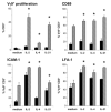
FIGURE 1
FIGURE 1
Flow cytometric analysis of activated γδ T cells. PBMC were cultured in the absence (∎) or presence (formula image) of HMB-PP, together with the cytokines indicated, and analyzed by flow cytometry. Cell proliferation was assessed by expansion of Vγ9+CD3+ cells within the CD3+ lymphocyte population (n = 12). Expression of CD69 on Vγ9+ CD3+ cells was determined after 18 h (n = 8), expression of ICAM-1 (n = 6), and LFA-1 (n = 4) after 6 days.
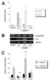
FIGURE 2
FIGURE 2
IL-2-dependent gene expression. Expression of IFN-γ (n = 3–6) and TNF-α (n = 6) in Vγ9+ CD3+ cells was analyzed by intracellular flow cytometry after 72 h. GM-CSF (n = 4), CCL3 (n = 3), and IL-13 (n = 4) were detected in 72 h culture supernatants, IL-5 (n = 2) after 6 days. Solid and hatched bars as in Fig. 1 legend.
FIGURE 3
FIGURE 3
IL-4-dependent gene expression. A, Expression of IL-4 and LT-α mRNAs by γδ T cells purified from PBMC after 72 h, as analyzed by real-time RT-PCR. Expression levels were normalized to values in medium controls and represent mean values and SDs from duplicate experiments. B, Expression of IL-4 and GATA-3 mRNAs by γδ T cells purified from PBMC after 72 h. Semiquantitative RT-PCR data shown are representative of three blood donors. C, Surface expression of CD27 (n = 6–7) as analyzed by flow cytometry on day 6.
FIGURE 4
FIGURE 4
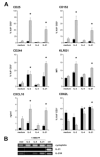
IL-21-dependent gene expression. A, Surface expression of CD25 (n = 6), intracellular expression of CD152 (n = 6), and CXCL10 secretion into the culture supernatants (n = 4–9) were detected after 72 h. Surface expression of CD244 (n = 8–11), KLRG1 (n = 3), and CD62L (n = 5) was measured after 6 days (note that KLRG1 expression is depicted as mean fluorescence values as at baseline Vγ9/Vδ2 T cells were already ~80% KLRG1+). Solid and hatched bars as in Fig. 1 legend. B, Expression of IL-21 and IL-21R mRNAs by γδ T cells purified from PBMC after 72 h in culture was analyzed by semiquantitative RT-PCR; data shown are representative of three individual blood donors (ctrl, whole PBMC stimulated with PHA).
FIGURE 5
FIGURE 5. IL-21-dependent expression of CXCL13
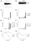
A, M/A plot of IL-21-stimulated vs IL-2-stimulated Vγ9/Vδ2 T cells.The locations of the IL-2-induced genes encoding for GM-CSF and LT-α and the IL-21-induced gene encoding for CXCL13 are indicated. B and C, Expression of CXCL13 mRNA by γδ T cells purified from PBMC cultured for 72 h in medium alone or with HMB-PP and the cytokines indicated. Semiquantitative RT-PCR data shown are representative of three blood donors; real-time data are mean values and SDs from duplicate experiments. D, Detection of CXCL13 protein in the supernatant of PBMC cultured for 72 h with and without HMB-PP, in the presence of the cytokines indicated (n = 6–9). E, CXCL13 production by PBMC cultured for 72 h in medium alone or with HMB-PP plus IL-21 after depletion of γδ T cells, monocytes, or B cells (n = 3). F, CXL13 production by purified γδ T cells in the absence or presence of monocytes as feeder cells after 72 h (n = 4). G, Inhibition of the IL-21-induced γδ T cell proliferation by IFN-β. PBMC were cultured for 6 days with HMB-PP plus IL-2 or HMB-PP plus IL-21 in the absence or presence of IFN-β (n = 3). H, Inhibition of the IL-21-dependent CXCL13 expression by IFN-β. PBMC were cultured for 72 h with HMB-PP plus IL-2 or HMB-PP plus IL-21 in the absence or presence of IFN-β (n = 5).
FIGURE 6
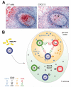
FIGURE 6. A role for γδ T cells in regulating follicular B cell maturation
A, Immunohistochemical analysis of serial sections of an active gut follicle from a patient with Yersinia ileitis, with γδ T cells and CXCL13, respectively, stained in red. Colocalization of CXCL13-producing cells and γδ T cells is indicated by arrows. Note that a similar picture of the same section stained for γδ T cells was already published earlier (23 ), but has been included here for the sake of optimum comparison. B, Proposed interaction between γδ T cells, B cells, and TFH cells in secondary lymphoid tissue. Expression of CCR7 by naive and central memory αβ T cells permits entry to the LNs, and subsequent colocalization with CCR7+ DC in the T zone that is defined by the CCR7 ligands CCL19 and CCL21. T cell priming results in the generation of TFH cells expressing CXCR5, thereby conferring responsiveness to the B zone-specific chemokine CXCL13. After relocalization of TFH cells and CXCR5+ B cells to the follicles, mutual interaction through ICOS and CD40 with their respective ligands induces plasma cell maturation on one hand, and gives rise to effector αβ T cells on the other hand (see also Refs. 2 and 60 ). Upon infection with microbial pathogens, peripheral γδ T cells recognize HMB-PP at sites of inflammation and contribute to the local immune response by secretion of proinflammatory cytokines and chemokines, and lysis of infected cells. Alternatively or additionally, they acquire a LN homing phenotype by up-regulation of CCR7 and CD62L (Ref. and this study), and HMB-PP released in the periphery may reach local LN through tissue drainage and act on LN resident γδ T cells. Data presented here show that γδ T cells moving into the GCs will respond to TFH-derived IL-21 by producing CXCL13 and CXCL10, thus contributing to the molecular definition of the B zone and aiding further recruitment of CXCR5+ B cells, TFH cells, and monocytes. HMB-PP triggered γδ T cells may also directly present microbial Ags to TFH cells at an early stage of infection (65 ).

Similar articles

Cited by

References

    1. Stockinger B, Bourgeois C, Kassiotis G. CD4+ memory T cells: functional differentiation and homeostasis. Immunol. Rev. 2006;211:39–48. - PubMed
    1. Vinuesa CG, Tangye SG, Moser B, Mackay CR. Follicular B helper T cells in antibody responses and autoimmunity. Nat. Rev. Immunol. 2005;5:853–865. - PubMed
    1. Pennington DJ, Vermijlen D, Wise EL, Clarke SL, Tigelaar RE, Hayday AC. The integration of conventional and unconventional T cells that characterizes cell-mediated responses. Adv. Immunol. 2005;87:27–59. - PubMed
    1. Hayday AC. γδ cells: a right time and a right place for a conserved third way of protection. Annu. Rev. Immunol. 2000;18:975–1026. - PubMed
    1. Shires J, Theodoridis E, Hayday AC. Biological insights into TCRγδ+ and TCRαβ+ intraepithelial lymphocytes provided by serial analysis of gene expression (SAGE) Immunity. 2001;15:419–434. - PubMed

Publication types