. 2007 Apr 1;178(7):4424-35.
doi: 10.4049/jimmunol.178.7.4424.

The role of antibody polyspecificity and lipid reactivity in binding of broadly neutralizing anti-HIV-1 envelope human monoclonal antibodies 2F5 and 4E10 to glycoprotein 41 membrane proximal envelope epitopes

Affiliations

The role of antibody polyspecificity and lipid reactivity in binding of broadly neutralizing anti-HIV-1 envelope human monoclonal antibodies 2F5 and 4E10 to glycoprotein 41 membrane proximal envelope epitopes

S Munir Alam et al. J Immunol. .

Abstract

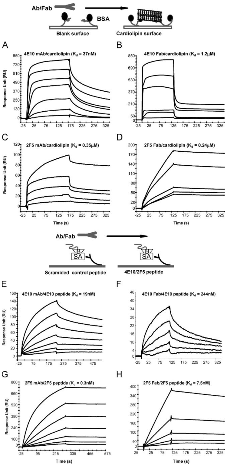
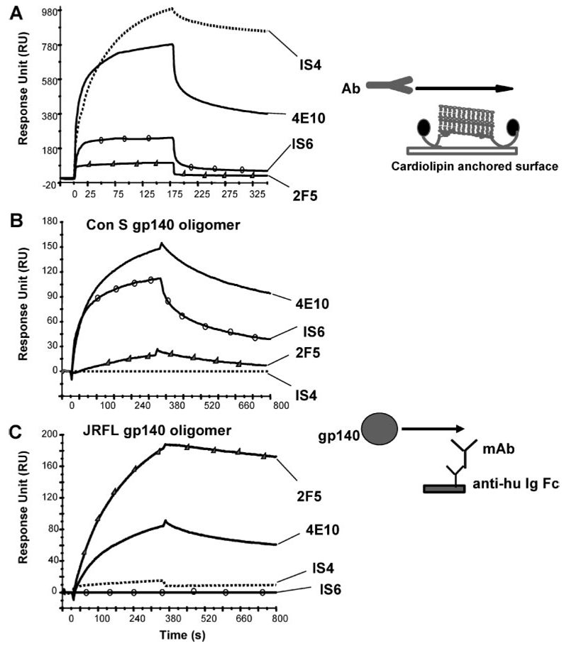
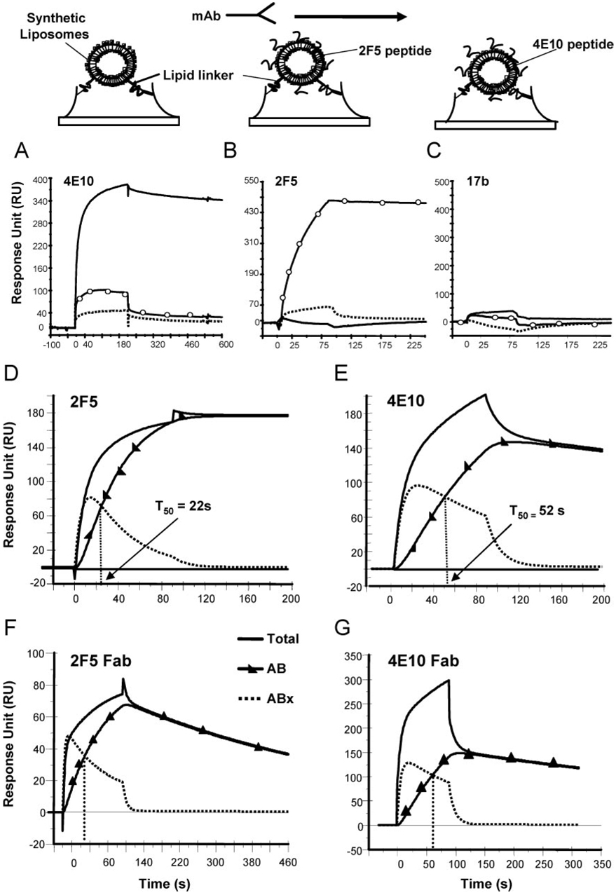
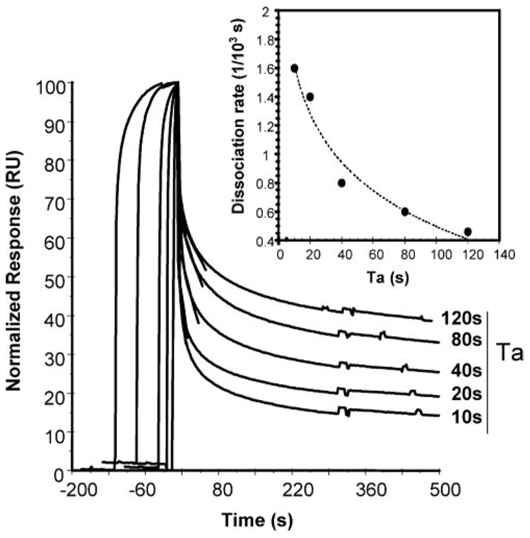
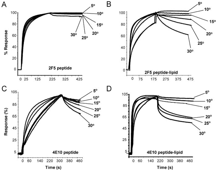
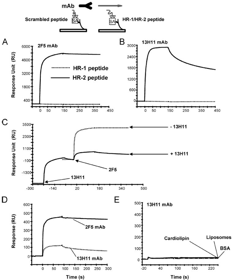
Two neutralizing human mAbs, 2F5 and 4E10, that react with the HIV-1 envelope gp41 membrane proximal region are also polyspecific autoantibodies that bind to anionic phospholipids. To determine the autoantibody nature of these Abs, we have compared their reactivities with human anti-cardiolipin mAbs derived from a primary antiphospholipid syndrome patient. To define the role of lipid polyreactivity in binding of 2F5 and 4E10 mAbs to HIV-1 envelope membrane proximal epitopes, we determined the kinetics of binding of mAbs 2F5 and 4E10 to their nominal gp41 epitopes vs liposome-gp41 peptide conjugates. Both anti-HIV-1 mAbs 2F5 and 4E10 bound to cardiolipin with K(d) values similar to those of autoimmune anti-cardiolipin Abs, IS4 and IS6. Binding kinetics studies revealed that mAb 2F5 and 4E10 binding to their respective gp41 peptide-lipid conjugates could best be defined by a two-step (encounter-docking) conformational change model. In contrast, binding of 2F5 and 4E10 mAbs to linear peptide epitopes followed a simple Langmuir model. A mouse mAb, 13H11, that cross-blocks mAb 2F5 binding to the gp41 epitope did not cross-react with lipids nor did it neutralize HIV-1 viruses. Taken together, these data demonstrate the similarity of 2F5 and 4E10 mAbs to known anti-cardiolipin Abs and support the model that mAb 2F5 and 4E10 binding to HIV-1 involves both viral lipid membrane and gp41 membrane proximal epitopes.


Conflict of interest statement

Disclosures The authors have no financial conflict of interest.

Figures

FIGURE 1
FIGURE 1
Binding of 4E10 and 2F5 mAb and Fab to cardiolipin and peptide epitopes. Approximately 400–600 RU of cardiolipin in an aqueous suspension was coupled to the BIAcore L1 sensor, on which ~3000 RU of BSA was immobilized previously. Whole mAb (A, C, E, and G) or Fab (B, D, F, and H) of either 2F5 or 4E10 was then injected at varying concentrations. The Ab concentration range used was as follows: 4E10 mAb (1.66–333 nM); 4E10 Fab (27.3–1800 nM); 2F5 mAb (46.6–1800 nM); 2F5 Fab (120–2000 nM). Due to the biphasic nature of binding, steady-state KD values were derived for 4E10 mAb and 4E10 Fab and 2F5 mAb, whereas Kd values for 2F5 Fab were derived from kinetics rate constants. Data shown are specific binding of each mAb or Fab after subtraction of nonspecific signals over a BSA immobilized surface. For binding to peptide epitopes, µ150–200 RU of biotinylated peptides were immobilized to streptavidin that was coupled to a BIAcore sensor chip (SA). Whole mAb or Fab were then injected at varying concentrations, which were at 11–714 nM (4E10), and 0.156–5 nM (2F5) for whole mAb and 125–2000 nM (4E10) and 0.62–9.95 nM (2F5) for Fab.
FIGURE 2
FIGURE 2
Interactions of anti-MPER mAb and anti-cardiolipin mAbs to cardiolipin and rHIV-1 Env gp140. A, Binding of anti-cardiolipin mAbs (IS4, IS6) and anti-HIV-1 MPER mAbs (2F5, 4E10) to anchored cardiolipin. A total of 100 µg/ml of each mAb was injected over 500 RU of cardiolipin anchored on a L1 sensor chip. Binding of mAb IS6 to Con S gp140 but not to JRFL gp140. Each of the mAb was captured at ~1,000 RU on a surface immobilized with anti-human Ig Fc (13,000 RU). rEnv gp140, Con S (B), or JRFL (C), was injected over each surface anchored with either mAb 4E10 (solid line), 2F5 (▲), IS4 (dotted line), or IS6 (●). Binding kinetics data are given in Table I. CDRH3 sequences of each mAb is given and the hydrophobic loop is underlined. 2F5 and 4E10 are from Protein Data Bank code 1TJI and ITZG, respectively. IS6 IgG was sequenced as described in Materials and Methods and the sequence for IS4 mAb were from Zhu et al. (29). IS4 GRRDVRGVLWRGRHD; IS6 DRSGRRQRWGMGY; 2F5 RRGPTTLFGVPIARGPVNAMDV; 4E10 EGTTGWGWLGKPIGAFAH.
FIGURE 3
FIGURE 3
mAbs 4E10 and 2F5 binding to peptide-liposome conjugates. Approximately 600 RU of either synthetic liposomes (broken line); 2F5 peptide-liposomes (○); or 4E10 peptide-liposomes were coupled on to a BIAcore L1 sensor chip. mAbs 4E10 (A), 2F5 (B), or 17b (C) was then injected at 100 µg/ml. Both 4E10 and 2F5 bound to peptide-liposomes in a peptide-dependent manner. DG, The binding of mAbs or Fab of 4E10 and 2F5 to their respective epitope peptide-liposome conjugates follows the two-step conformational change model. Curve-fitting analysis of data generated from binding of mAb 4E10 and 2F5 to their respective peptide-liposome conjugates. The two-step conformational change model was used to derive the rate constants for the encounter complex (●) and the docked complex (▲) and is given in Table I. In each of the overlay, the binding data is shown in solid line and represents the observed total binding response. The component curves for the encounter complex (●) and the docked complex (▲) were simulated from the experimentally determined rate constants. T50 defines the time required for half of the encounter complex to be converted to docked complex. Both mAbs bound to their peptide-liposomes with an overall high apparent Kd (Table I).
FIGURE 4
FIGURE 4
Effect of varying contact time on dissociation rates. 4E10 mAb (100 µg/ml) was injected with varying contact time (Ta) in seconds (10, 20, 40, 80, and 120) over 600 RU of 4E10 peptide-liposomes immobilized surface. Binding data were normalized to 100 RU and the end of injection point (start of dissociation) was aligned on the x-axis at time zero. Dissociation rate constants were measured during the first 60 s of the dissociation phase and were plotted against Ta (inset). An inverse relationship between early dissociation rates and Ta shows that the two steps (encounter and docking) of the binding of the mAb are linked and sequential.
FIGURE 5
FIGURE 5
Effect of temperature on binding kinetics of 2F5 and 4E10 mAb to peptide and peptide-lipid conjugates. Binding kinetics of 2F5 (A and B) and 4E10 (C and D) was measured at temperatures ranging from 5 to 30°C for their respective epitope peptide (A and C) or peptide-lipid conjugate (B and D). Specific binding signal was recorded in reference to either control peptide or synthetic liposomes as described in Materials and Methods. Data shown are expressed as the percent response and was derived following normalization of binding responses recorded at each temperature.
FIGURE 6
FIGURE 6
Binding of a non-neutralizing mAb, 13H11, to MPER peptides. A and B, Binding of MPER mAb 2F5 (A), 13H11 mAb (B) to the full-length HR-2 peptide (YTSLIHSLIEESQNQQEKNEQELLELDKWASLWNF; solid line), and to the control HR-1 peptide (dotted line). C, Blocking of 2F5 mAb binding to the full-length HR-2 peptide, DP178, by 13H11 mAb. 13H11 mAb was bound to saturation and then mAb 2F5 was injected at the time indicated with arrows. The binding of mAb 25F in the absence (dotted line) or presence (solid line) of prebound 13H11 is shown. D, Comparison of 13H11 mAb (dotted line) to 2F5 mAb binding to 2F5 peptide-lipid conjugates. E, Binding of 13H11 mAb (100 µg/ml) to cardiolipin, synthetic liposomes, or BSA-immobilized surfaces.

Similar articles

Cited by

References

    1. Ofek G, Tang M, Sambor A, Katinger H, Mascola JR, Wyatt R, Kwong PD. Structure and mechanistic analysis of the anti-human immunodeficiency virus type 1 antibody 2F5 in complex with its gp41 epitope. J. Virol. 2004;78:10724–10737. - PMC - PubMed
    1. Cardoso RM, Zwick MB, Stanfield RL, Kunert R, Binley JM, Katinger H, Burton DR, Wilson IA. Broadly neutralizing anti-HIV antibody 4E10 recognizes a helical conformation of a highly conserved fusion-associated motif in gp41. Immunity. 2005;22:163–173. - PubMed
    1. Stiegler G, Kunert R, Purtscher M, Wolbank S, Voglauer R, Steindl F, Katinger H. A potent cross-clade neutralizing human monoclonal antibody against a novel epitope on gp41 of human immunodeficiency virus type 1. AIDS Res. Hum. Retroviruses. 2001;17:1757–1765. - PubMed
    1. D’Souza MP, Livnat D, Bradac JA, Bridges SH. Evaluation of monoclonal antibodies to human immunodeficiency virus type 1 primary isolates by neutralization assays: performance criteria for selecting candidate antibodies for clinical trials. AIDS Clinical Trials Group Antibody Selection Working Group. J. Infect. Dis. 1997;175:1056–1062. - PubMed
    1. Barbato GE, Baianchi P, Inagallinella P, Hurni WH, Miller MD, Ciliberto R, Cortrese R, Bazzo R, Shiver JW, Pessi A. Structural analysis of the epitope of the anti-HIV antibody 2F5 sheds light into its mechanism of neutralization and HIV fusion. J. Mol. Biol. 2003;330:1101–1115. - PubMed

Publication types